Other Sites:

Bibliography Options Menu

Robert J. Robbins is a biologist, an educator, a science administrator, a publisher, an information technologist, and an IT leader and manager who specializes in advancing biomedical knowledge and supporting education through the application of information technology. More About: RJR | OUR TEAM | OUR SERVICES | THIS WEBSITE
RJR: Recommended Bibliography 30 Jul 2026 at 02:01 Created:
N-Acetyl-Cysteine: Wonder Drug?
Wikipedia: Acetylcysteine,
also known as N-acetylcysteine (NAC), is a medication that is used to treat paracetamol overdose and to loosen thick mucus in individuals with chronic bronchopulmonary disorders like pneumonia and bronchitis. It has been used to treat lactobezoar in infants. It can be taken intravenously, by mouth, or inhaled as a mist. Some people use it as a dietary supplement.
Common side effects include nausea and vomiting when taken by mouth. The skin may occasionally become red and itchy with any route of administration. A non-immune type of anaphylaxis may also occur. It appears to be safe in pregnancy. For paracetamol overdose, it works by increasing the level of glutathione, an antioxidant that can neutralise the toxic breakdown products of paracetamol. When inhaled, it acts as a mucolytic by decreasing the thickness of mucus.
NAC, as a commercially available dietary supplement, is touted as A potent antioxidant that supports comprehensive wellness, including lung, liver, kidney and immune function.
Is NAC a life-extending wonder drug? What does the scientific literature say?
Created with PubMed® Query: nac acetylcysteine OR "acetyl-cysteine" NOT pmcbook NOT ispreviousversion
Citations The Papers (from PubMed®)
RevDate: 2026-07-29
CmpDate: 2026-07-29
Betaine Attenuates Hyperhomocysteinemia-Induced Cognitive Impairment by Suppressing Oxidative Stress and Activating the PI3K/AKT/GSK-3β Pathway.
Antioxidants (Basel, Switzerland), 15(7):.
High homocysteine levels are a key risk factor for cognitive impairment, a major public health concern in aging societies. Although betaine is known to reduce Hcy levels, its effects on hyperhomocysteinemia (hHcy)-induced cognitive impairment and the underlying mechanisms remain unclear. Here, we established an hHcy-induced cognitive impairment mouse model by feeding mice a high-methionine diet for 8 weeks, followed by betaine supplementation for 14 days. Betaine treatment attenuated hHcy-induced cognitive impairment. This improvement was accompanied by alleviation of neuropathological alterations and enhancement of antioxidant capacity. Notably, betaine suppressed reactive oxygen species (ROS) accumulation, neuronal apoptosis, and Tau hyperphosphorylation at Ser396 and Thr231 in both mouse hippocampus and HT-22 cells. Mechanistically, betaine-induced activation of the PI3K/AKT/GSK-3β pathway was effectively blocked by the PI3K inhibitor LY294002. Notably, treatment with the ROS scavenger N-acetylcysteine (NAC) alone phenocopied this activation, suggesting that ROS functions as an upstream regulator of this signaling cascade. Collectively, our data demonstrate that betaine attenuates hHcy-induced cognitive impairment by suppressing oxidative stress-driven apoptosis and Tau pathology through modulation of the PI3K/AKT/GSK-3β signaling pathway. These findings suggest that betaine may hold promise for further preclinical and clinical studies, although long-term efficacy and safety evaluations remain necessary.
Additional Links: PMID-42510538
PubMed:
Citation:
show bibtex listing
hide bibtex listing
@article {pmid42510538,
year = {2026},
author = {Gu, X and Fu, Y and Zhao, Y and Liu, Z and Yang, Y and Xie, Q and Ma, P and Peng, Z and Liu, Z and Li, J and Xie, J},
title = {Betaine Attenuates Hyperhomocysteinemia-Induced Cognitive Impairment by Suppressing Oxidative Stress and Activating the PI3K/AKT/GSK-3β Pathway.},
journal = {Antioxidants (Basel, Switzerland)},
volume = {15},
number = {7},
pages = {},
pmid = {42510538},
issn = {2076-3921},
support = {82201841//National Natural Science Foundation of China/ ; U23A20420//National Natural Science Foundation of China/ ; 202203021212369//Shanxi Basic Research Program/ ; 202403021221156//Shanxi Basic Research Program/ ; XD2114//Science Research Start⁃up Fund for Doctor of Shanxi Medical University/ ; BYBLD//Shanxi Province Higher Education "Billion Project" Science and Technology Guidance Project/ ; },
abstract = {High homocysteine levels are a key risk factor for cognitive impairment, a major public health concern in aging societies. Although betaine is known to reduce Hcy levels, its effects on hyperhomocysteinemia (hHcy)-induced cognitive impairment and the underlying mechanisms remain unclear. Here, we established an hHcy-induced cognitive impairment mouse model by feeding mice a high-methionine diet for 8 weeks, followed by betaine supplementation for 14 days. Betaine treatment attenuated hHcy-induced cognitive impairment. This improvement was accompanied by alleviation of neuropathological alterations and enhancement of antioxidant capacity. Notably, betaine suppressed reactive oxygen species (ROS) accumulation, neuronal apoptosis, and Tau hyperphosphorylation at Ser396 and Thr231 in both mouse hippocampus and HT-22 cells. Mechanistically, betaine-induced activation of the PI3K/AKT/GSK-3β pathway was effectively blocked by the PI3K inhibitor LY294002. Notably, treatment with the ROS scavenger N-acetylcysteine (NAC) alone phenocopied this activation, suggesting that ROS functions as an upstream regulator of this signaling cascade. Collectively, our data demonstrate that betaine attenuates hHcy-induced cognitive impairment by suppressing oxidative stress-driven apoptosis and Tau pathology through modulation of the PI3K/AKT/GSK-3β signaling pathway. These findings suggest that betaine may hold promise for further preclinical and clinical studies, although long-term efficacy and safety evaluations remain necessary.},
}
RevDate: 2026-07-29
CmpDate: 2026-07-29
N-Acetylcysteine Attenuates Oxidative Stress and Preserves Red Blood Cell Quality During Whole Blood Storage.
Antioxidants (Basel, Switzerland), 15(7):.
Whole blood (WB) storage induces biochemical and biomechanical alterations that may compromise red blood cell (RBC) quality. Since oxidative stress is a major driver of storage lesions, we investigated whether N-acetylcysteine (NAC) could attenuate these changes during refrigerated storage. WB from healthy donors was stored at 4 °C for 42 days with or without NAC, added either once at baseline or every 10 days. Plasma albumin proteoforms were assessed by liquid chromatography-mass spectrometry, free hemoglobin species by spectrophotometry, plasma proteomic changes by proximity extension assay, and RBC hemorheological properties by LORRCA analysis. Storage decreased reduced albumin (HSA-SH) and increased oxidized albumin (HSA-Cys), indicating plasma oxidation. Free oxyhemoglobin, deoxyhemoglobin, and methemoglobin increased, consistent with hemoglobin oxidation and hemolysis. Storage also induced plasma proteomic alterations and impaired RBC osmotic and deformability parameters. NAC preserved albumin redox status, limited free hemoglobin accumulation, and attenuated storage-induced proteomic changes. Moreover, NAC partially preserved RBC osmotic and rheological properties, particularly parameters related to osmotic fragility and hydration. No clear advantage of 20 mM over 10 mM NAC was observed. Overall, NAC attenuated oxidative and functional alterations associated with refrigerated whole blood storage, supporting further investigation of antioxidant supplementation as a strategy to mitigate storage lesions under ex vivo conditions.
Additional Links: PMID-42510589
PubMed:
Citation:
show bibtex listing
hide bibtex listing
@article {pmid42510589,
year = {2026},
author = {Eligini, S and Brocca, L and Mallia, A and Valeriano, A and Gianazza, E and Banfi, C},
title = {N-Acetylcysteine Attenuates Oxidative Stress and Preserves Red Blood Cell Quality During Whole Blood Storage.},
journal = {Antioxidants (Basel, Switzerland)},
volume = {15},
number = {7},
pages = {},
pmid = {42510589},
issn = {2076-3921},
support = {PNC-E3-2022-23683268//Ministero della Salute/ ; },
abstract = {Whole blood (WB) storage induces biochemical and biomechanical alterations that may compromise red blood cell (RBC) quality. Since oxidative stress is a major driver of storage lesions, we investigated whether N-acetylcysteine (NAC) could attenuate these changes during refrigerated storage. WB from healthy donors was stored at 4 °C for 42 days with or without NAC, added either once at baseline or every 10 days. Plasma albumin proteoforms were assessed by liquid chromatography-mass spectrometry, free hemoglobin species by spectrophotometry, plasma proteomic changes by proximity extension assay, and RBC hemorheological properties by LORRCA analysis. Storage decreased reduced albumin (HSA-SH) and increased oxidized albumin (HSA-Cys), indicating plasma oxidation. Free oxyhemoglobin, deoxyhemoglobin, and methemoglobin increased, consistent with hemoglobin oxidation and hemolysis. Storage also induced plasma proteomic alterations and impaired RBC osmotic and deformability parameters. NAC preserved albumin redox status, limited free hemoglobin accumulation, and attenuated storage-induced proteomic changes. Moreover, NAC partially preserved RBC osmotic and rheological properties, particularly parameters related to osmotic fragility and hydration. No clear advantage of 20 mM over 10 mM NAC was observed. Overall, NAC attenuated oxidative and functional alterations associated with refrigerated whole blood storage, supporting further investigation of antioxidant supplementation as a strategy to mitigate storage lesions under ex vivo conditions.},
}
RevDate: 2026-07-25
Testicular damage from electromagnetic radiation in rats and evaluation of protective agents.
Urologia [Epub ahead of print].
BACKGROUND: Male infertility has been associated with various environmental, physiological, and genetic factors. In recent years, the widespread use of mobile phones has raised concerns regarding exposure to electromagnetic radiation (EMR). EMR emitted from mobile devices may adversely affect male reproductive function by inducing oxidative stress and impairing spermatogenesis.
OBJECTIVE: This study aimed to evaluate the potential protective effects of vitamin E and N-acetylcysteine (NAC) against EMR-induced testicular damage in rats.
METHODS: A total of 35 adult male Wistar rats were randomly divided into five groups (n = 7 per group): control, EMR exposure, EMR + NAC, EMR + vitamin E, and EMR + NAC + vitamin E. Rats were exposed to EMR generated by a mobile phone operating in the GSM frequency band (900/1800 MHz) in active call mode, positioned at a fixed distance of 15 cm from the cages, for 3 h daily over 28 days. The specific absorption rate (SAR) was based on manufacturer-reported values. Biochemical analyses were performed to assess total antioxidant capacity (TAC), glutathione peroxidase (GPX), superoxide dismutase (SOD), and malondialdehyde (MDA) levels. Data distribution was evaluated using the Shapiro-Wilk test, and group comparisons were conducted using the Kruskal-Wallis test with appropriate post-hoc analyses.
RESULTS: Significant differences were observed among groups in terms of total antioxidant capacity (TAC) (p < 0.001). TAC levels were reduced in the EMR-only group compared to controls, whereas antioxidant supplementation (NAC and/or vitamin E) resulted in increased TAC levels. Post-hoc analyses demonstrated significant improvements in TAC in treatment groups compared to both control and EMR-only groups. However, no statistically significant differences were observed among groups for GPX, SOD, and MDA levels (p > 0.05).
CONCLUSION: N-acetylcysteine (NAC) and vitamin E may exert partial protective effects against EMR-induced oxidative alterations in rat testes, particularly as reflected by improvements in total antioxidant capacity. However, given that other oxidative stress markers did not demonstrate statistically significant differences, these findings should be interpreted with caution. Further experimental and clinical studies with larger sample sizes and detailed histopathological evaluation are required to better elucidate the potential therapeutic role and clinical relevance of these antioxidant agents.
Additional Links: PMID-42499284
Publisher:
PubMed:
Citation:
show bibtex listing
hide bibtex listing
@article {pmid42499284,
year = {2026},
author = {Gözüküçük, A and Çakıroğlu, B and Uyanik, BS and Kılıç, HH and Çelik, İS},
title = {Testicular damage from electromagnetic radiation in rats and evaluation of protective agents.},
journal = {Urologia},
volume = {},
number = {},
pages = {3915603261448981},
doi = {10.1177/03915603261448981},
pmid = {42499284},
issn = {1724-6075},
abstract = {BACKGROUND: Male infertility has been associated with various environmental, physiological, and genetic factors. In recent years, the widespread use of mobile phones has raised concerns regarding exposure to electromagnetic radiation (EMR). EMR emitted from mobile devices may adversely affect male reproductive function by inducing oxidative stress and impairing spermatogenesis.
OBJECTIVE: This study aimed to evaluate the potential protective effects of vitamin E and N-acetylcysteine (NAC) against EMR-induced testicular damage in rats.
METHODS: A total of 35 adult male Wistar rats were randomly divided into five groups (n = 7 per group): control, EMR exposure, EMR + NAC, EMR + vitamin E, and EMR + NAC + vitamin E. Rats were exposed to EMR generated by a mobile phone operating in the GSM frequency band (900/1800 MHz) in active call mode, positioned at a fixed distance of 15 cm from the cages, for 3 h daily over 28 days. The specific absorption rate (SAR) was based on manufacturer-reported values. Biochemical analyses were performed to assess total antioxidant capacity (TAC), glutathione peroxidase (GPX), superoxide dismutase (SOD), and malondialdehyde (MDA) levels. Data distribution was evaluated using the Shapiro-Wilk test, and group comparisons were conducted using the Kruskal-Wallis test with appropriate post-hoc analyses.
RESULTS: Significant differences were observed among groups in terms of total antioxidant capacity (TAC) (p < 0.001). TAC levels were reduced in the EMR-only group compared to controls, whereas antioxidant supplementation (NAC and/or vitamin E) resulted in increased TAC levels. Post-hoc analyses demonstrated significant improvements in TAC in treatment groups compared to both control and EMR-only groups. However, no statistically significant differences were observed among groups for GPX, SOD, and MDA levels (p > 0.05).
CONCLUSION: N-acetylcysteine (NAC) and vitamin E may exert partial protective effects against EMR-induced oxidative alterations in rat testes, particularly as reflected by improvements in total antioxidant capacity. However, given that other oxidative stress markers did not demonstrate statistically significant differences, these findings should be interpreted with caution. Further experimental and clinical studies with larger sample sizes and detailed histopathological evaluation are required to better elucidate the potential therapeutic role and clinical relevance of these antioxidant agents.},
}
RevDate: 2026-07-27
CmpDate: 2026-07-27
The Effects of Short-Term N-Acetylcysteine Supplementation on Biochemical Parameters in Endurance-Trained Adults: A Randomized Clinical Trial.
Metabolites, 16(7): pii:metabo16070505.
Background: The main aim of this study was to assess the effects of short-term N-acetylcysteine (NAC) supplementation on concentrations of homocysteine (Hcy) and reduced glutathione (rGSH), blood lipid profile and liver enzyme activities in endurance-trained adults, and to determine whether these effects are modified by methylenetetrahydrofolate reductase (MTHFR) C677T and glutathione S-transferase Pi 1 (GSTP1) A313G. Methods: A total of 56 males and 21 females completed a randomized, double-blind, placebo-controlled crossover trial. Participants received 1200 mg of NAC or a placebo for seven days in a crossover design. Serum Hcy and plasma rGSH concentrations were assessed using dedicated biochemical assays, while blood lipid profile and liver enzyme activities were measured using the biochemical analyzer Konelab 20i. Genotyping was conducted using TaqMan probes. A series of within-subject/between-subject repeated-measures analysis of variance (ANOVA) within a general linear model framework were performed to compare Hcy, rGSH, blood lipid profile and liver enzymes activities before and after the intervention. Results: Hcy concentrations significantly decreased following NAC supplementation (18.58 ± 5.45 µmol/L vs. 16.51 ± 4.97 µmol/L; p = 0.009), although subgroup analysis indicated that the decrease was significant only among females (15.40 ± 4.96 µmol/L vs. 13.60 ± 3.68 µmol/L; p = 0.002) without any significant effect among males. We did not observe any significant changes in rGSH, lipid profile, or liver enzyme activities. There was no interaction between NAC supplementation, MTHFR and GSTP1 genotypes and the changes noted in the parameters we analyzed. Conclusions: In conclusion, short-term NAC supplementation may reduce circulating Hcy concentrations in endurance-trained adults, particularly in females. No consistent effects were observed for rGSH, lipid profile, or liver enzyme activities.
Additional Links: PMID-42506456
Publisher:
PubMed:
Citation:
show bibtex listing
hide bibtex listing
@article {pmid42506456,
year = {2026},
author = {Sadowski, M and Zawieja, E and Muzsik-Kazimierska, A and Bulczak, E and Chmurzynska, A},
title = {The Effects of Short-Term N-Acetylcysteine Supplementation on Biochemical Parameters in Endurance-Trained Adults: A Randomized Clinical Trial.},
journal = {Metabolites},
volume = {16},
number = {7},
pages = {},
doi = {10.3390/metabo16070505},
pmid = {42506456},
issn = {2218-1989},
abstract = {Background: The main aim of this study was to assess the effects of short-term N-acetylcysteine (NAC) supplementation on concentrations of homocysteine (Hcy) and reduced glutathione (rGSH), blood lipid profile and liver enzyme activities in endurance-trained adults, and to determine whether these effects are modified by methylenetetrahydrofolate reductase (MTHFR) C677T and glutathione S-transferase Pi 1 (GSTP1) A313G. Methods: A total of 56 males and 21 females completed a randomized, double-blind, placebo-controlled crossover trial. Participants received 1200 mg of NAC or a placebo for seven days in a crossover design. Serum Hcy and plasma rGSH concentrations were assessed using dedicated biochemical assays, while blood lipid profile and liver enzyme activities were measured using the biochemical analyzer Konelab 20i. Genotyping was conducted using TaqMan probes. A series of within-subject/between-subject repeated-measures analysis of variance (ANOVA) within a general linear model framework were performed to compare Hcy, rGSH, blood lipid profile and liver enzymes activities before and after the intervention. Results: Hcy concentrations significantly decreased following NAC supplementation (18.58 ± 5.45 µmol/L vs. 16.51 ± 4.97 µmol/L; p = 0.009), although subgroup analysis indicated that the decrease was significant only among females (15.40 ± 4.96 µmol/L vs. 13.60 ± 3.68 µmol/L; p = 0.002) without any significant effect among males. We did not observe any significant changes in rGSH, lipid profile, or liver enzyme activities. There was no interaction between NAC supplementation, MTHFR and GSTP1 genotypes and the changes noted in the parameters we analyzed. Conclusions: In conclusion, short-term NAC supplementation may reduce circulating Hcy concentrations in endurance-trained adults, particularly in females. No consistent effects were observed for rGSH, lipid profile, or liver enzyme activities.},
}
RevDate: 2026-07-27
Beyond Antioxidant Activity, Towards Redox Modulation: N-Acetylcysteine (NAC) in Endometriosis and Uterine Leiomyomas.
Free radical research [Epub ahead of print].
Endometriosis and uterine leiomyomas are common chronic gynecologic diseases associated with immune dysregulation, inflammation and aberrant cellular proliferation, processes that contribute to increased oxidative stress. Current management relies largely on surgical and hormonal interventions, highlighting the need for effective long-term nonhormonal therapeutic strategies. N-acetylcysteine (NAC), a glutathione precursor, exhibits antioxidant and anti-inflammatory properties that may target key mechanisms underlying both conditions. NAC functions as both a direct reactive oxygen species scavenger and regulator of intracellular redox homeostasis through replenishment of glutathione stores. These findings suggest that NAC may represent a potential adjunctive therapy warranting further investigation and may influence disease-associated biological pathways. Future clinical and translational studies are needed to define its efficacy, optimal dosing, and role in redox-based management of endometriosis and uterine leiomyomas.
Additional Links: PMID-42506968
Publisher:
PubMed:
Citation:
show bibtex listing
hide bibtex listing
@article {pmid42506968,
year = {2026},
author = {Mohebbi, L and Sanz Maset, A and Macri, V and Jospeh, E and Segars, J and Singh, B},
title = {Beyond Antioxidant Activity, Towards Redox Modulation: N-Acetylcysteine (NAC) in Endometriosis and Uterine Leiomyomas.},
journal = {Free radical research},
volume = {},
number = {},
pages = {1-19},
doi = {10.1080/10715762.2026.2710249},
pmid = {42506968},
issn = {1029-2470},
abstract = {Endometriosis and uterine leiomyomas are common chronic gynecologic diseases associated with immune dysregulation, inflammation and aberrant cellular proliferation, processes that contribute to increased oxidative stress. Current management relies largely on surgical and hormonal interventions, highlighting the need for effective long-term nonhormonal therapeutic strategies. N-acetylcysteine (NAC), a glutathione precursor, exhibits antioxidant and anti-inflammatory properties that may target key mechanisms underlying both conditions. NAC functions as both a direct reactive oxygen species scavenger and regulator of intracellular redox homeostasis through replenishment of glutathione stores. These findings suggest that NAC may represent a potential adjunctive therapy warranting further investigation and may influence disease-associated biological pathways. Future clinical and translational studies are needed to define its efficacy, optimal dosing, and role in redox-based management of endometriosis and uterine leiomyomas.},
}
RevDate: 2026-07-27
Escitalopram and N-acetylcysteine alleviate major depressive disorder with cognitive impairment through YTHDC1/ATF4/CHOP-mediated pyroptosis.
Behavioural brain research pii:S0166-4328(26)00379-7 [Epub ahead of print].
BACKGROUND: Cognitive impairment (CI) is an important factor contributing to poor prognosis in major depressive disorder (MDD). Endoplasmic reticulum stress and microglial inflammatory response are closely associated with the occurrence and development of MDD with CI. Escitalopram (ESC) and N-acetylcysteine (NAC) are two drugs used to improve depression. However, the therapeutic efficacy and underlying molecular mechanisms of ESC and NAC in treating MDD with CI are still not fully understood.
METHODS: The depression rat was established using the chronic unpredictable mild stress (CUMS) protocol. The antidepressant effects of ESC and NAC were evaluated through behavioral tests including the open field test, sucrose preference test, and Morris water maze test and. The underlying molecular mechanisms were investigated using ELISA and immunofluorescence techniques.
RESULTS: The CUMS group of rats showed decreased responsiveness and memory capacity, which were significantly improved following intervention with ESC and NAC. Treatment with ESC and NAC alleviated hippocampal injury in the CUMS group of rats. The expression of microglial activation markers CD68 and IBA1 in hippocampal tissue was significantly reduced. The levels of NLRP3 inflammasome and pro-inflammatory cytokines IL-6, IL-18, and IL-1β in the hippocampus were significantly decreased. Mechanistically, ESC and NAC downregulate ATF4/CHOP expression by inhibiting the m6A reader YTHDC1, thereby reducing hippocampal cell pyroptosis.
CONCLUSION: This study demonstrates that ESC and NAC alleviate hippocampal tissue damage by modulating YTHDC1/ATF4/CHOP-mediated pyroptosis, thereby improving MDD with CI. These findings provide experimental evidence supporting the therapeutic potential of targeting YTHDC1/ATF4/CHOP-mediated pyroptosis in MDD-associated CI.
Additional Links: PMID-42508696
Publisher:
PubMed:
Citation:
show bibtex listing
hide bibtex listing
@article {pmid42508696,
year = {2026},
author = {Zhou, Y and Zhang, Y and Yang, L},
title = {Escitalopram and N-acetylcysteine alleviate major depressive disorder with cognitive impairment through YTHDC1/ATF4/CHOP-mediated pyroptosis.},
journal = {Behavioural brain research},
volume = {},
number = {},
pages = {116403},
doi = {10.1016/j.bbr.2026.116403},
pmid = {42508696},
issn = {1872-7549},
abstract = {BACKGROUND: Cognitive impairment (CI) is an important factor contributing to poor prognosis in major depressive disorder (MDD). Endoplasmic reticulum stress and microglial inflammatory response are closely associated with the occurrence and development of MDD with CI. Escitalopram (ESC) and N-acetylcysteine (NAC) are two drugs used to improve depression. However, the therapeutic efficacy and underlying molecular mechanisms of ESC and NAC in treating MDD with CI are still not fully understood.
METHODS: The depression rat was established using the chronic unpredictable mild stress (CUMS) protocol. The antidepressant effects of ESC and NAC were evaluated through behavioral tests including the open field test, sucrose preference test, and Morris water maze test and. The underlying molecular mechanisms were investigated using ELISA and immunofluorescence techniques.
RESULTS: The CUMS group of rats showed decreased responsiveness and memory capacity, which were significantly improved following intervention with ESC and NAC. Treatment with ESC and NAC alleviated hippocampal injury in the CUMS group of rats. The expression of microglial activation markers CD68 and IBA1 in hippocampal tissue was significantly reduced. The levels of NLRP3 inflammasome and pro-inflammatory cytokines IL-6, IL-18, and IL-1β in the hippocampus were significantly decreased. Mechanistically, ESC and NAC downregulate ATF4/CHOP expression by inhibiting the m6A reader YTHDC1, thereby reducing hippocampal cell pyroptosis.
CONCLUSION: This study demonstrates that ESC and NAC alleviate hippocampal tissue damage by modulating YTHDC1/ATF4/CHOP-mediated pyroptosis, thereby improving MDD with CI. These findings provide experimental evidence supporting the therapeutic potential of targeting YTHDC1/ATF4/CHOP-mediated pyroptosis in MDD-associated CI.},
}
RevDate: 2026-07-27
In vivo Activity of Combination of N-acetyl Cysteine and Levofloxacin against Colonization and Catheter-Associated Infection.
Current microbiology, 83(9):.
Despite many recent advancements and improvements to infection control measures and practices in healthcare facilities, infections associated with intravascular catheters remain problematic and even fatal. Since these infections are generally biofilm-based, effective antibiofilm strategies combined with antimicrobial approaches that can inhibit pathogenic colonization are important in preventing catheter-associated infections. We evaluated in vivo the activities of intravascular catheters impregnated with a combination of N-acetyl cysteine, an antibiofilm agent, and levofloxacin (NACLEV) compared to chlorhexidine/silver sulfadiazine-treated catheters (CHSS) against bacterial colonization and device-associated infection with representatives of both Gram-positive and Gram-negative bacteria including Staphylococcus aureus, vancomycin-resistant enterococcus (VRE), Pseudomonas aeruginosa, or Klebsiella pneumoniae. NACLEV catheters inoculated with S. aureus, VRE, or K. pneumoniae were generally as effective as inoculated CHSS catheters against device colonization (75%, 70%, or 80% vs. 65%, 55%, or 85% colonization reduction, respectively) and device-associated infection (65%, 65%, or 60% vs. 50%,3%, or 50% device-associated infection reduction, respectively). However, NACLEV catheters inoculated with P. aeruginosa were significantly more effective compared to CHSS catheters against both colonization (80% vs. 10% colonization reduction, P < 0.001) and device-associated infection (55% vs. 5% device-associated infection reduction, P = 0.001). Furthermore, impregnation of catheters with the N-acetyl cysteine and levofloxacin combination did not affect the mechanical integrity of the catheters since the mean force at break of 30.91 N for the impregnated catheters with the combination was well above the threshold of 15 N as recommended by the International Organization for Standardization BS EN ISO 10555-1 and 10555-3.
Additional Links: PMID-42509467
PubMed:
Citation:
show bibtex listing
hide bibtex listing
@article {pmid42509467,
year = {2026},
author = {Mansouri, MD and Mansouri, DL and Hull, RA},
title = {In vivo Activity of Combination of N-acetyl Cysteine and Levofloxacin against Colonization and Catheter-Associated Infection.},
journal = {Current microbiology},
volume = {83},
number = {9},
pages = {},
pmid = {42509467},
issn = {1432-0991},
abstract = {Despite many recent advancements and improvements to infection control measures and practices in healthcare facilities, infections associated with intravascular catheters remain problematic and even fatal. Since these infections are generally biofilm-based, effective antibiofilm strategies combined with antimicrobial approaches that can inhibit pathogenic colonization are important in preventing catheter-associated infections. We evaluated in vivo the activities of intravascular catheters impregnated with a combination of N-acetyl cysteine, an antibiofilm agent, and levofloxacin (NACLEV) compared to chlorhexidine/silver sulfadiazine-treated catheters (CHSS) against bacterial colonization and device-associated infection with representatives of both Gram-positive and Gram-negative bacteria including Staphylococcus aureus, vancomycin-resistant enterococcus (VRE), Pseudomonas aeruginosa, or Klebsiella pneumoniae. NACLEV catheters inoculated with S. aureus, VRE, or K. pneumoniae were generally as effective as inoculated CHSS catheters against device colonization (75%, 70%, or 80% vs. 65%, 55%, or 85% colonization reduction, respectively) and device-associated infection (65%, 65%, or 60% vs. 50%,3%, or 50% device-associated infection reduction, respectively). However, NACLEV catheters inoculated with P. aeruginosa were significantly more effective compared to CHSS catheters against both colonization (80% vs. 10% colonization reduction, P < 0.001) and device-associated infection (55% vs. 5% device-associated infection reduction, P = 0.001). Furthermore, impregnation of catheters with the N-acetyl cysteine and levofloxacin combination did not affect the mechanical integrity of the catheters since the mean force at break of 30.91 N for the impregnated catheters with the combination was well above the threshold of 15 N as recommended by the International Organization for Standardization BS EN ISO 10555-1 and 10555-3.},
}
RevDate: 2026-07-22
CmpDate: 2026-07-22
Re-exploration of the pharmacotherapeutic strategy for drug-induced liver injury: a narrative review.
Frontiers in medicine, 13:1880114.
The incidence of drug-induced liver injury (DILI) is on the rise, and discontinuation of suspected causative drugs is the core therapeutic principle. However, some patients may progress to moderate-to-severe liver injury, liver failure, or chronicity; thus, pharmacotherapy maybe considered a potentially valuable supportive treatment option in appropriate clinical contexts. At present, there is no unified consensus on the therapeutic indications and drug selection for DILI worldwide, and irrational practices such as prophylactic medication, indication-free medication and polypharmacy exist in China. This review proposes formulating therapeutic indications based on the severity, type of liver injury and Hy's Law, identifying TBIL≥2 × ULN, cholestatic and mixed liver injury, acute liver failure (ALF), and high risk of ALF as indications for pharmacotherapy. For drug selection, N-acetylcysteine (NAC) is recommended by all domestic and international guidelines, Glucocorticoid (GC) may be used in selected cases, and hepatoprotective drugs such as magnesium isoglycyrrhizinate (MgIG) and bicyclol are widely prescribed in China. Pharmacotherapeutic regimens can be tailored according to the type and severity of liver injury.
Additional Links: PMID-42482898
PubMed:
Citation:
show bibtex listing
hide bibtex listing
@article {pmid42482898,
year = {2026},
author = {Liu, Y and Guo, Y and Xu, H and Han, Y and Feng, H and Fang, Y and Pan, F},
title = {Re-exploration of the pharmacotherapeutic strategy for drug-induced liver injury: a narrative review.},
journal = {Frontiers in medicine},
volume = {13},
number = {},
pages = {1880114},
pmid = {42482898},
issn = {2296-858X},
abstract = {The incidence of drug-induced liver injury (DILI) is on the rise, and discontinuation of suspected causative drugs is the core therapeutic principle. However, some patients may progress to moderate-to-severe liver injury, liver failure, or chronicity; thus, pharmacotherapy maybe considered a potentially valuable supportive treatment option in appropriate clinical contexts. At present, there is no unified consensus on the therapeutic indications and drug selection for DILI worldwide, and irrational practices such as prophylactic medication, indication-free medication and polypharmacy exist in China. This review proposes formulating therapeutic indications based on the severity, type of liver injury and Hy's Law, identifying TBIL≥2 × ULN, cholestatic and mixed liver injury, acute liver failure (ALF), and high risk of ALF as indications for pharmacotherapy. For drug selection, N-acetylcysteine (NAC) is recommended by all domestic and international guidelines, Glucocorticoid (GC) may be used in selected cases, and hepatoprotective drugs such as magnesium isoglycyrrhizinate (MgIG) and bicyclol are widely prescribed in China. Pharmacotherapeutic regimens can be tailored according to the type and severity of liver injury.},
}
RevDate: 2026-07-27
A Multi-Analyte-Responsive, Multi-Organ-Imaging, Dual-NIR-Emissive Fluorescent Probe for Visualizing Pyroptosis-Mediated Hg[2+]-Induced Hepatorenal Dysfunction.
Analytical chemistry [Epub ahead of print].
Mercury ions (Hg[2+]) are highly toxic heavy metals that are widely distributed in the environment and pose a serious threat to both ecosystems and human health. After entering the body, mercury preferentially accumulates in the liver and kidneys, underscoring the importance of early detection of acute and chronic hepatorenal injuries. Although fluorescent probes have been widely explored for disease diagnosis, most existing systems cannot achieve simultaneous in vivo imaging of the liver and kidneys, nor can they monitor multiple pathological parameters during hepatorenal injury, thus limiting their utility in evaluating Hg[2+]-induced multiorgan damage. In this study, we developed a multifunctional fluorescent probe, VPO, with high sensitivity and good biocompatibility, which can be used to monitor polarity, viscosity, and ONOO[-] levels in cells. Using VPO, we observed, for the first time, a significant increase in ONOO[-] during pyroptosis, accompanied by an elevated mitochondrial viscosity and reduced polarity. In addition, VPO successfully enabled the real-time visual monitoring of these changes in mercury-exposed cells, zebrafish, and mice. Moreover, its multiresponsive property enabled the evaluation of the therapeutic efficacy of N-acetylcysteine (NAC) against Hg[2+]-induced hepatorenal injury. NAC alleviated hepatic and renal mitochondrial oxidative stress, as well as pyroptosis-related inflammatory signaling, by reversing Hg[2+]-induced biomarker abnormalities. In summary, these findings demonstrate that VPO provides a comprehensive platform for the early diagnosis, therapeutic evaluation, and mechanistic investigation of mercury poisoning.
Additional Links: PMID-42483836
Publisher:
PubMed:
Citation:
show bibtex listing
hide bibtex listing
@article {pmid42483836,
year = {2026},
author = {Zhai, Z and Tao, L and Huang, J and Zhang, H and Wang, X and Deng, M and Wen, Q and Cheng, D and He, L and Li, S},
title = {A Multi-Analyte-Responsive, Multi-Organ-Imaging, Dual-NIR-Emissive Fluorescent Probe for Visualizing Pyroptosis-Mediated Hg[2+]-Induced Hepatorenal Dysfunction.},
journal = {Analytical chemistry},
volume = {},
number = {},
pages = {},
doi = {10.1021/acs.analchem.6c02189},
pmid = {42483836},
issn = {1520-6882},
abstract = {Mercury ions (Hg[2+]) are highly toxic heavy metals that are widely distributed in the environment and pose a serious threat to both ecosystems and human health. After entering the body, mercury preferentially accumulates in the liver and kidneys, underscoring the importance of early detection of acute and chronic hepatorenal injuries. Although fluorescent probes have been widely explored for disease diagnosis, most existing systems cannot achieve simultaneous in vivo imaging of the liver and kidneys, nor can they monitor multiple pathological parameters during hepatorenal injury, thus limiting their utility in evaluating Hg[2+]-induced multiorgan damage. In this study, we developed a multifunctional fluorescent probe, VPO, with high sensitivity and good biocompatibility, which can be used to monitor polarity, viscosity, and ONOO[-] levels in cells. Using VPO, we observed, for the first time, a significant increase in ONOO[-] during pyroptosis, accompanied by an elevated mitochondrial viscosity and reduced polarity. In addition, VPO successfully enabled the real-time visual monitoring of these changes in mercury-exposed cells, zebrafish, and mice. Moreover, its multiresponsive property enabled the evaluation of the therapeutic efficacy of N-acetylcysteine (NAC) against Hg[2+]-induced hepatorenal injury. NAC alleviated hepatic and renal mitochondrial oxidative stress, as well as pyroptosis-related inflammatory signaling, by reversing Hg[2+]-induced biomarker abnormalities. In summary, these findings demonstrate that VPO provides a comprehensive platform for the early diagnosis, therapeutic evaluation, and mechanistic investigation of mercury poisoning.},
}
RevDate: 2026-07-23
Targeting Aβ25-35-Induced Neuronal Senescence-Like Features Using Curcumin-Loaded Solid Self-Emulsifying Drug Delivery Systems.
Current aging science pii:CAS-EPUB-157035 [Epub ahead of print].
INTRODUCTION: Curcumin is widely recognized for its anti-neurodegenerative properties. However, its clinical utility is hindered by poor aqueous solubility and low bioavailability. To address these limitations, Curcumin-loaded Solid Self-Emulsifying Drug Delivery Systems (C-SSEDDS) have been formulated to improve pharmacokinetics and neuronal targeting. Despite these advancements, the molecular mechanisms underlying curcumin in Alzheimer's Disease (AD)-related neuronal senescence remain poorly understood. Therefore, this study provides novel molecular insights into the therapeutic potential of C-SSEDDS for targeting neuronal senescence- like features in the AD context.
METHODS: An in vitro model of neuronal senescence-like features was established by exposing SK-N-SH cells to 0.075 μM Aβ25-35 for 24 hours, resulting in a 20-30% decline in cell viability. Subsequently, cells were post-treated with C-SSEDDS at optimized non-toxic concentrations (0.1, 1, and 10 μg/mL curcumin equivalent) and 10 μg/mL commercial curcumin for 24, 48, and 72 hours in Aβ25-35-free media. The study comprehensively assessed cell viability, morphological alterations, and key biomarkers associated with neuronal senescence-like features, including senescence- associated beta-galactosidase (SA-β-gal) positive cells, Reactive Oxygen Species (ROS) levels, NLRP3, Sirt-1, and synaptophysin expression.
RESULTS: C-SSEDDS post-treatment significantly improved cell viability, reduced apoptotic cells, and decreased SA-β-gal positive cells in Aβ25-35-induced senescence-like SK-N-SH cells compared to the untreated Aβ25-35 group. It also effectively decreased intracellular ROS level and NLRP3 expression, and markedly upregulated Sirt-1 and synaptophysin expression. Notably, CSSEDDS demonstrated therapeutic efficacy superior to commercial curcumin and comparable to N-acetylcysteine (NAC).
DISCUSSION: In SK-N-SH cells exhibiting senescence-like features, the C-SSEDDS formulation effectively counteracts Aβ25-35-induced damage. It mitigates key drivers of cellular stress, including oxidative stress, inflammation-related markers, and apoptosis, while also enhancing Sirt-1 activity and promoting synaptic plasticity.
CONCLUSION: C-SSEDDS demonstrates significant potential as a promising therapeutic strategy against Aβ25-35 -induced neuronal stress and senescence-like changes in AD.
Additional Links: PMID-42487594
Publisher:
PubMed:
Citation:
show bibtex listing
hide bibtex listing
@article {pmid42487594,
year = {2026},
author = {Lonlab, K and Nitjapol, A and Thummayot, S and Tiyaboonchai, W and Jumnongprakhon, P},
title = {Targeting Aβ25-35-Induced Neuronal Senescence-Like Features Using Curcumin-Loaded Solid Self-Emulsifying Drug Delivery Systems.},
journal = {Current aging science},
volume = {},
number = {},
pages = {},
doi = {10.2174/0118746098440081260430205824},
pmid = {42487594},
issn = {1874-6128},
abstract = {INTRODUCTION: Curcumin is widely recognized for its anti-neurodegenerative properties. However, its clinical utility is hindered by poor aqueous solubility and low bioavailability. To address these limitations, Curcumin-loaded Solid Self-Emulsifying Drug Delivery Systems (C-SSEDDS) have been formulated to improve pharmacokinetics and neuronal targeting. Despite these advancements, the molecular mechanisms underlying curcumin in Alzheimer's Disease (AD)-related neuronal senescence remain poorly understood. Therefore, this study provides novel molecular insights into the therapeutic potential of C-SSEDDS for targeting neuronal senescence- like features in the AD context.
METHODS: An in vitro model of neuronal senescence-like features was established by exposing SK-N-SH cells to 0.075 μM Aβ25-35 for 24 hours, resulting in a 20-30% decline in cell viability. Subsequently, cells were post-treated with C-SSEDDS at optimized non-toxic concentrations (0.1, 1, and 10 μg/mL curcumin equivalent) and 10 μg/mL commercial curcumin for 24, 48, and 72 hours in Aβ25-35-free media. The study comprehensively assessed cell viability, morphological alterations, and key biomarkers associated with neuronal senescence-like features, including senescence- associated beta-galactosidase (SA-β-gal) positive cells, Reactive Oxygen Species (ROS) levels, NLRP3, Sirt-1, and synaptophysin expression.
RESULTS: C-SSEDDS post-treatment significantly improved cell viability, reduced apoptotic cells, and decreased SA-β-gal positive cells in Aβ25-35-induced senescence-like SK-N-SH cells compared to the untreated Aβ25-35 group. It also effectively decreased intracellular ROS level and NLRP3 expression, and markedly upregulated Sirt-1 and synaptophysin expression. Notably, CSSEDDS demonstrated therapeutic efficacy superior to commercial curcumin and comparable to N-acetylcysteine (NAC).
DISCUSSION: In SK-N-SH cells exhibiting senescence-like features, the C-SSEDDS formulation effectively counteracts Aβ25-35-induced damage. It mitigates key drivers of cellular stress, including oxidative stress, inflammation-related markers, and apoptosis, while also enhancing Sirt-1 activity and promoting synaptic plasticity.
CONCLUSION: C-SSEDDS demonstrates significant potential as a promising therapeutic strategy against Aβ25-35 -induced neuronal stress and senescence-like changes in AD.},
}
RevDate: 2026-07-23
CmpDate: 2026-07-23
Efficacy of N-acetylcysteine in enhancing paracetamol's analgesic effect and inhibiting hepatotoxicity: a preclinical mouse model.
Pain reports, 11(4):e1461.
INTRODUCTION: Paracetamol (PCM) is extensively administered in clinical settings to alleviate mild to moderate pain and/or decrease fever. However, noncompliance with prescribed dosages can lead to severe and irreversible liver damage from PCM overdose. Although N-acetylcysteine (NAC) serves as the standard antidote for PCM toxicity, no commercially available combination of PCM and NAC has been marketed to mitigate its toxic effects. Objectives: This study aims to evaluate both the antinociceptive and hepatoprotective effects of the PCM and NAC combination in a murine model.
METHODS: Male and female C57Bl6/J mice were assigned to 4 groups. Group A received oral PCM exclusively, administered every 6 hours (4 doses within 24 hours), whereas groups B-D were administered PCM alone, a combination of PCM/L-NAC (N-acetyl-l-cysteine), or PCM/D-NAC (N-acetyl-d-cysteine) every 4 hours (6 doses within 24 hours).
RESULTS: Both PCM/L-NAC and PCM/D-NAC combinations yielded superior analgesia compared with the standard regimen. Furthermore, L-NAC administration mitigated the increase of glutamate pyruvate transaminase, a key hepatic marker associated with PCM hepatotoxicity. In addition, a protective effect from D-NAC in conjunction with PCM on glutathione levels was suggested. Our findings suggest that NAC enhances the analgesic properties of PCM while concurrently reducing its hepatotoxic potential in mice. Moreover, data indicate that L-NAC may represent a more effective candidate than D-NAC for this purpose, particularly regarding hepatoprotection.
CONCLUSION: This study presents novel insights into the synergistic effects of the PCM/NAC combination on pain relief and hepatic safeguarding, warranting consideration for the development of novel therapeutic strategies targeting pain and fever management in patients.
Additional Links: PMID-42491779
PubMed:
Citation:
show bibtex listing
hide bibtex listing
@article {pmid42491779,
year = {2026},
author = {Chrétien, B and Goy, R and Guillaume, M and Bazin, G and Collet, M and Dutheil, JJ and Baugé, C and Vivien, D and Agin, V and Dolladille, C},
title = {Efficacy of N-acetylcysteine in enhancing paracetamol's analgesic effect and inhibiting hepatotoxicity: a preclinical mouse model.},
journal = {Pain reports},
volume = {11},
number = {4},
pages = {e1461},
pmid = {42491779},
issn = {2471-2531},
abstract = {INTRODUCTION: Paracetamol (PCM) is extensively administered in clinical settings to alleviate mild to moderate pain and/or decrease fever. However, noncompliance with prescribed dosages can lead to severe and irreversible liver damage from PCM overdose. Although N-acetylcysteine (NAC) serves as the standard antidote for PCM toxicity, no commercially available combination of PCM and NAC has been marketed to mitigate its toxic effects. Objectives: This study aims to evaluate both the antinociceptive and hepatoprotective effects of the PCM and NAC combination in a murine model.
METHODS: Male and female C57Bl6/J mice were assigned to 4 groups. Group A received oral PCM exclusively, administered every 6 hours (4 doses within 24 hours), whereas groups B-D were administered PCM alone, a combination of PCM/L-NAC (N-acetyl-l-cysteine), or PCM/D-NAC (N-acetyl-d-cysteine) every 4 hours (6 doses within 24 hours).
RESULTS: Both PCM/L-NAC and PCM/D-NAC combinations yielded superior analgesia compared with the standard regimen. Furthermore, L-NAC administration mitigated the increase of glutamate pyruvate transaminase, a key hepatic marker associated with PCM hepatotoxicity. In addition, a protective effect from D-NAC in conjunction with PCM on glutathione levels was suggested. Our findings suggest that NAC enhances the analgesic properties of PCM while concurrently reducing its hepatotoxic potential in mice. Moreover, data indicate that L-NAC may represent a more effective candidate than D-NAC for this purpose, particularly regarding hepatoprotection.
CONCLUSION: This study presents novel insights into the synergistic effects of the PCM/NAC combination on pain relief and hepatic safeguarding, warranting consideration for the development of novel therapeutic strategies targeting pain and fever management in patients.},
}
RevDate: 2026-07-21
The KAT5-TSP-1 Axis Maintains Ovarian Redox Homeostasis and Is Impaired in Polycystic Ovary Syndrome.
Reproductive sciences (Thousand Oaks, Calif.) [Epub ahead of print].
Polycystic ovary syndrome (PCOS) is characterized by oxidative stress (OS). This study reveals a novel epigenetic mechanism linking lysine acetyltransferase 5 (KAT5) and thrombospondin-1 (TSP-1) in PCOS pathogenesis. A meta-analysis of 50 clinical studies demonstrated elevated systemic oxidative markers (total oxidant status, nitric oxide, malondialdehyde) and reduced paraoxonase-1 levels in PCOS patients, alongside compromised total antioxidant capacity in follicular fluid. Bioinformatic analysis identified TSP-1 as a key downregulated gene in PCOS, and the hTFtarget database predicted KAT5 as an upstream regulator of TSP-1. In vitro, H2O2-induced oxidative stress in KGN granulosa cells suppressed TSP-1, KAT5, and H2AK5ac levels, which were restored by N-acetylcysteine (NAC). Mechanistically, KAT5 was found to regulate TSP-1 expression via H2AK5 acetylation at its promoter. Knockdown of KAT5 exacerbated oxidative stress, apoptosis, and senescence, while TSP-1 overexpression counteracted these effects. In a letrozole-induced PCOS rat model, ovarian tissues exhibited decreased H2AK5 acetylation, reduced KAT5 and TSP-1 expression, and impaired redox homeostasis. Interventions with N-acetylcysteine or TSP-1 ameliorated ovarian pathological changes, hormonal imbalances, oxidative stress, apoptosis, and cellular senescence. Collectively, these findings establish the KAT5-TSP-1 axis as a critical regulator of ovarian redox homeostasis, unveiling a novel epigenetic mechanism and proposing a dual-target therapeutic strategy for PCOS.
Additional Links: PMID-42481896
PubMed:
Citation:
show bibtex listing
hide bibtex listing
@article {pmid42481896,
year = {2026},
author = {Jin, Q and Yang, W and Wang, C and Liu, Y and Wang, R and Liu, L and Zhang, X and Yan, J and Liu, M},
title = {The KAT5-TSP-1 Axis Maintains Ovarian Redox Homeostasis and Is Impaired in Polycystic Ovary Syndrome.},
journal = {Reproductive sciences (Thousand Oaks, Calif.)},
volume = {},
number = {},
pages = {},
pmid = {42481896},
issn = {1933-7205},
support = {LH2019H070//Natural Science Foundation of Heilongjiang Province/ ; KYCX2018-03//College Youth Innovation Foundation/ ; PL2025H115//Cultivation Project of the Joint Fund of Heilongjiang Provincial Natural Science Foundation/ ; },
abstract = {Polycystic ovary syndrome (PCOS) is characterized by oxidative stress (OS). This study reveals a novel epigenetic mechanism linking lysine acetyltransferase 5 (KAT5) and thrombospondin-1 (TSP-1) in PCOS pathogenesis. A meta-analysis of 50 clinical studies demonstrated elevated systemic oxidative markers (total oxidant status, nitric oxide, malondialdehyde) and reduced paraoxonase-1 levels in PCOS patients, alongside compromised total antioxidant capacity in follicular fluid. Bioinformatic analysis identified TSP-1 as a key downregulated gene in PCOS, and the hTFtarget database predicted KAT5 as an upstream regulator of TSP-1. In vitro, H2O2-induced oxidative stress in KGN granulosa cells suppressed TSP-1, KAT5, and H2AK5ac levels, which were restored by N-acetylcysteine (NAC). Mechanistically, KAT5 was found to regulate TSP-1 expression via H2AK5 acetylation at its promoter. Knockdown of KAT5 exacerbated oxidative stress, apoptosis, and senescence, while TSP-1 overexpression counteracted these effects. In a letrozole-induced PCOS rat model, ovarian tissues exhibited decreased H2AK5 acetylation, reduced KAT5 and TSP-1 expression, and impaired redox homeostasis. Interventions with N-acetylcysteine or TSP-1 ameliorated ovarian pathological changes, hormonal imbalances, oxidative stress, apoptosis, and cellular senescence. Collectively, these findings establish the KAT5-TSP-1 axis as a critical regulator of ovarian redox homeostasis, unveiling a novel epigenetic mechanism and proposing a dual-target therapeutic strategy for PCOS.},
}
RevDate: 2026-07-21
Investigation of Payloads and Related Impurities in the cysteine-linked Antibody-Drug Conjugate Using Mass Spectrometry Techniques.
Applied biochemistry and biotechnology [Epub ahead of print].
Loncastuximab tesirine is a cysteine-linked antibody-drug conjugate composed of an anti-CD19 monoclonal antibody linked to the PBD dimer payload SG3249 via a protease-cleavable linker, enabling targeted release of a highly cytotoxic DNA cross-linking agent. NAC-SG3249, a thiol adduct formed with N-acetylcysteine, serves as an important indicator of payload release and ADC stability. In this study, mass spectrometry techniques were employed to systematically investigate the stability and related small-molecule impurities of this ADC. Quantitative analysis of the free payload showed good linearity over 0.05-100 ng/mL (R[2] ≥ 0.991), with an LLOQ of 0.05 ng/mL, accuracy of 80%-120%, and precision below 5%, and the results were consistent between HRMS and TQMS, both methods can achieve effective quantification. Potential payload-related impurities were further characterized based on predicted hydrolysis pathways using targeted MRM analysis. Forced degradation studies under acidic, alkaline, photolytic, and thermal conditions revealed multiple degradation products using a highly sensitive nanoLC-HRMS method, with alkaline conditions producing the greatest number of impurities. Clinical trial number: not applicable.
Additional Links: PMID-42479388
PubMed:
Citation:
show bibtex listing
hide bibtex listing
@article {pmid42479388,
year = {2026},
author = {Wu, G and Xu, G and Zhao, Y and Gao, T and Ni, Y and Lv, X and Chen, H and Li, M and Du, J and Yu, C},
title = {Investigation of Payloads and Related Impurities in the cysteine-linked Antibody-Drug Conjugate Using Mass Spectrometry Techniques.},
journal = {Applied biochemistry and biotechnology},
volume = {},
number = {},
pages = {},
pmid = {42479388},
issn = {1559-0291},
support = {Z251100004625003//Beijing Municipal Science and Technology Commission/ ; 2023YFC3404004//National Key Research and Development Program of China/ ; 2025SKLDRS0336//State Key Laboratory of Drug Regulatory science/ ; BZ2025137//Standardization Improvement Project of Chinese Pharmacopeiae/ ; },
abstract = {Loncastuximab tesirine is a cysteine-linked antibody-drug conjugate composed of an anti-CD19 monoclonal antibody linked to the PBD dimer payload SG3249 via a protease-cleavable linker, enabling targeted release of a highly cytotoxic DNA cross-linking agent. NAC-SG3249, a thiol adduct formed with N-acetylcysteine, serves as an important indicator of payload release and ADC stability. In this study, mass spectrometry techniques were employed to systematically investigate the stability and related small-molecule impurities of this ADC. Quantitative analysis of the free payload showed good linearity over 0.05-100 ng/mL (R[2] ≥ 0.991), with an LLOQ of 0.05 ng/mL, accuracy of 80%-120%, and precision below 5%, and the results were consistent between HRMS and TQMS, both methods can achieve effective quantification. Potential payload-related impurities were further characterized based on predicted hydrolysis pathways using targeted MRM analysis. Forced degradation studies under acidic, alkaline, photolytic, and thermal conditions revealed multiple degradation products using a highly sensitive nanoLC-HRMS method, with alkaline conditions producing the greatest number of impurities. Clinical trial number: not applicable.},
}
RevDate: 2026-07-21
Cisplatin-Induced Hearing Loss Prevention With Intratympanic Therapy Systematic Review and Meta-Analysis.
Otology & neurotology : official publication of the American Otological Society, American Neurotology Society [and] European Academy of Otology and Neurotology pii:00129492-990000000-01260 [Epub ahead of print].
INTRODUCTION: Cisplatin-induced hearing loss (CIHL) is a well-described, long-term consequence of cisplatin treatment for malignancy. Intratympanic (IT) injections have been trialed to prevent CIHL in humans. To provide clarity on which agents have been studied through IT injection and to review their efficacy for hearing loss prevention, we performed a systematic review and meta-analysis.
DATA SOURCES: OVID Medline, Embase, Web of Science, and Cochrane Library were queried.
METHODS: Databases were searched in accordance with the Preferred Reporting Items for Systematic Reviews and Meta-Analysis guidelines. Prospective randomized trials were included, and a systematic review was performed for all studies. Demographic, audiometric, and therapeutic data were collected. Random-effects models were used to compare across studies, and subgroup analyses were performed for each IT agent.
RESULTS: The initial database search yielded 1017 articles, which were screened according to inclusion and exclusion criteria. Ten studies were identified, involving a total of 284 patients. Studies included data on IT dexamethasone, IT N-acetylcysteine (NAC), and IT sodium thiosulfate (STS). Pooled analysis across all agents and frequencies did not reveal a significant difference in hearing thresholds between treatment and control ears [prediction interval [-3.77, 3.20], negative favors treatment). Subgroup analysis of IT dexamethasone [-1.74, 3.80] and IT NAC [-1.02, 4.64] also did not demonstrate significant differences. STS data were not amenable to pooled analysis; however, one study demonstrated a significant decrease in ASHA-defined ototoxicity (40% vs. 85%, P=0.0027).
CONCLUSIONS: To date, no IT agent has consistently prevented CIHL, although limited data suggest that IT STS may decrease ototoxicity. More trials are necessary to fully elucidate these effects.
Additional Links: PMID-42480209
Publisher:
PubMed:
Citation:
show bibtex listing
hide bibtex listing
@article {pmid42480209,
year = {2026},
author = {Kons, ZA and Kersbergen, CJ and Goss, D and Remenschneider, AK},
title = {Cisplatin-Induced Hearing Loss Prevention With Intratympanic Therapy Systematic Review and Meta-Analysis.},
journal = {Otology & neurotology : official publication of the American Otological Society, American Neurotology Society [and] European Academy of Otology and Neurotology},
volume = {},
number = {},
pages = {},
doi = {10.1097/MAO.0000000000005011},
pmid = {42480209},
issn = {1537-4505},
abstract = {INTRODUCTION: Cisplatin-induced hearing loss (CIHL) is a well-described, long-term consequence of cisplatin treatment for malignancy. Intratympanic (IT) injections have been trialed to prevent CIHL in humans. To provide clarity on which agents have been studied through IT injection and to review their efficacy for hearing loss prevention, we performed a systematic review and meta-analysis.
DATA SOURCES: OVID Medline, Embase, Web of Science, and Cochrane Library were queried.
METHODS: Databases were searched in accordance with the Preferred Reporting Items for Systematic Reviews and Meta-Analysis guidelines. Prospective randomized trials were included, and a systematic review was performed for all studies. Demographic, audiometric, and therapeutic data were collected. Random-effects models were used to compare across studies, and subgroup analyses were performed for each IT agent.
RESULTS: The initial database search yielded 1017 articles, which were screened according to inclusion and exclusion criteria. Ten studies were identified, involving a total of 284 patients. Studies included data on IT dexamethasone, IT N-acetylcysteine (NAC), and IT sodium thiosulfate (STS). Pooled analysis across all agents and frequencies did not reveal a significant difference in hearing thresholds between treatment and control ears [prediction interval [-3.77, 3.20], negative favors treatment). Subgroup analysis of IT dexamethasone [-1.74, 3.80] and IT NAC [-1.02, 4.64] also did not demonstrate significant differences. STS data were not amenable to pooled analysis; however, one study demonstrated a significant decrease in ASHA-defined ototoxicity (40% vs. 85%, P=0.0027).
CONCLUSIONS: To date, no IT agent has consistently prevented CIHL, although limited data suggest that IT STS may decrease ototoxicity. More trials are necessary to fully elucidate these effects.},
}
RevDate: 2026-07-18
Bilobalide alleviates ischemia-reperfusion injury during liver transplantation by regulating macrophage polarization through inhibiting NF-κB pathway.
Hepatobiliary & pancreatic diseases international : HBPD INT pii:S1499-3872(26)00113-X [Epub ahead of print].
BACKGROUND: Hepatic ischemia-reperfusion injury (IRI) causes donor graft dysfunction and increases mortality after liver transplantation (LT). Bilobalide (BB), a natural sesquiterpene lactone extracted from Ginkgo biloba leaves, exhibits anti-inflammatory and anti-necrotic bioactivities. However, there is no study currently on the use of bilobalide in IRI therapy for liver transplant. This study aimed to investigate whether bilobalide protects grafted livers from IRI and to explore the underlying mechanisms.
METHODS: Donor livers preserved in histidine-tryptophan-ketoglutarate (HTK) solution for 5 h underwent orthotopic liver transplantation in Sprague-Dawley rats. Following LT, rats were administered intraperitoneal injections of bilobalide (30 mg/kg, LT + BB group), saline (LT group), or N-acetylcysteine (150 mg/kg, LT + NAC group) at two-time points: immediately after surgery and again at 12 h post-transplant. Serum liver enzymes levels at 24 h after transplantation were measured. Liver tissues underwent hematoxylin and eosin (H&E) staining to confirm the injury. To examine hepatocyte apoptosis, both TUNEL staining and Western blot analysis were employed. Inflammatory responses in liver tissue were examined using immunohistochemistry, enzyme-linked immunosorbent assay (ELISA), and reverse transcription-quantitative polymerase chain reaction (RT-qPCR). Potential signaling pathways were identified through transcriptome sequencing (RNA-seq) and subsequently validated by Western blot analysis. In addition, macrophage polarization was evaluated using immunofluorescence and Western blot.
RESULTS: The LT + BB group exhibited significantly milder liver injury and hepatocyte apoptosis compared to the LT group. The LT + NAC group also showed reduced liver injury and apoptosis, although the protective effects were less pronounced than those observed in the LT + BB group. Bilobalide treatment significantly reduced inflammatory cell infiltration and suppressed pro-inflammatory cytokine expression. RNA-seq and Western blot analyses demonstrated that bilobalide inhibited the activation of the nuclear factor Kappa B (NF-κB) signaling pathway in transplanted liver tissue. Immunofluorescence and Western blot results further revealed that bilobalide suppressed macrophage polarization toward the M1 phenotype.
CONCLUSIONS: Bilobalide protects the transplanted liver from IRI by regulating macrophage polarization through NF-κB signaling pathway.
Additional Links: PMID-42471283
Publisher:
PubMed:
Citation:
show bibtex listing
hide bibtex listing
@article {pmid42471283,
year = {2026},
author = {Tang, YX and Zhong, XY and Shi, D and Wu, ZY and Wu, ZS and Lu, D and Wang, K and Chen, J and Xu, H and Liu, ZK and Xu, X and Wei, Q},
title = {Bilobalide alleviates ischemia-reperfusion injury during liver transplantation by regulating macrophage polarization through inhibiting NF-κB pathway.},
journal = {Hepatobiliary & pancreatic diseases international : HBPD INT},
volume = {},
number = {},
pages = {},
doi = {10.1016/j.hbpd.2026.07.003},
pmid = {42471283},
issn = {1499-3872},
abstract = {BACKGROUND: Hepatic ischemia-reperfusion injury (IRI) causes donor graft dysfunction and increases mortality after liver transplantation (LT). Bilobalide (BB), a natural sesquiterpene lactone extracted from Ginkgo biloba leaves, exhibits anti-inflammatory and anti-necrotic bioactivities. However, there is no study currently on the use of bilobalide in IRI therapy for liver transplant. This study aimed to investigate whether bilobalide protects grafted livers from IRI and to explore the underlying mechanisms.
METHODS: Donor livers preserved in histidine-tryptophan-ketoglutarate (HTK) solution for 5 h underwent orthotopic liver transplantation in Sprague-Dawley rats. Following LT, rats were administered intraperitoneal injections of bilobalide (30 mg/kg, LT + BB group), saline (LT group), or N-acetylcysteine (150 mg/kg, LT + NAC group) at two-time points: immediately after surgery and again at 12 h post-transplant. Serum liver enzymes levels at 24 h after transplantation were measured. Liver tissues underwent hematoxylin and eosin (H&E) staining to confirm the injury. To examine hepatocyte apoptosis, both TUNEL staining and Western blot analysis were employed. Inflammatory responses in liver tissue were examined using immunohistochemistry, enzyme-linked immunosorbent assay (ELISA), and reverse transcription-quantitative polymerase chain reaction (RT-qPCR). Potential signaling pathways were identified through transcriptome sequencing (RNA-seq) and subsequently validated by Western blot analysis. In addition, macrophage polarization was evaluated using immunofluorescence and Western blot.
RESULTS: The LT + BB group exhibited significantly milder liver injury and hepatocyte apoptosis compared to the LT group. The LT + NAC group also showed reduced liver injury and apoptosis, although the protective effects were less pronounced than those observed in the LT + BB group. Bilobalide treatment significantly reduced inflammatory cell infiltration and suppressed pro-inflammatory cytokine expression. RNA-seq and Western blot analyses demonstrated that bilobalide inhibited the activation of the nuclear factor Kappa B (NF-κB) signaling pathway in transplanted liver tissue. Immunofluorescence and Western blot results further revealed that bilobalide suppressed macrophage polarization toward the M1 phenotype.
CONCLUSIONS: Bilobalide protects the transplanted liver from IRI by regulating macrophage polarization through NF-κB signaling pathway.},
}
RevDate: 2026-07-19
ADSL drives fumarate mediated scrib-rictor complex formation to promote metastasis dissemination in triple-negative breast cancer.
Molecular cancer pii:10.1186/s12943-026-02742-2 [Epub ahead of print].
BACKGROUND: Triple-negative breast cancer (TNBC) is characterized by aggressive metastatic behavior and limited therapeutic options. Although metabolic reprogramming is increasingly recognized as a hallmark of TNBC, the mechanisms by which specific metabolic enzymes and intermediates drive metastasis remain poorly defined.
METHODS: We performed untargeted metabolomic profiling on TNBC tumors and matched normal tissues to identify dysregulated metabolic pathways. Functional assays, chemoproteomic succination profiling, molecular interaction analyses, and in vivo cancer metastasis models were used to define the mechanistic and biological consequences of altered metabolism. In vitro experiments validated the effects of N-acetylcysteine (NAC) in TNBC cell lines.
RESULTS: Metabolomic analyses revealed aberrant activation of the alanine-aspartate-glutamate axis and upregulation of adenylosuccinate lyase (ADSL) in TNBC. ADSL promoted tumor cell proliferation and metastasis by generating fumarate, which accumulated primarily through covalent protein succination. Chemoproteomic profiling identified the cell polarity regulator SCRIB as a critical fumarate target. Fumarate-mediated succination of SCRIB impaired its membrane localization, promoted epithelial-mesenchymal transition, and facilitated aberrant interaction with the mTORC2 component RICTOR, leading to activation of AKT/mTOR signaling. Genetic disruption of SCRIB succination abrogated these effects. Importantly, pharmacological perturbation of fumarate using NAC reduced SCRIB succination and attenuated the malignant phenotypes of TNBC cells in vitro.
CONCLUSIONS: These findings identify an ADSL-fumarate-SCRIB signaling axis that links metabolic reprogramming to the loss of cell polarity and activation of pro-metastatic signaling in TNBC. Targeting fumarate-mediated protein succination represents a previously unrecognized and experimental vulnerability in TNBC.
Additional Links: PMID-42472815
Publisher:
PubMed:
Citation:
show bibtex listing
hide bibtex listing
@article {pmid42472815,
year = {2026},
author = {Fan, Y and He, X and Deng, W and Sun, J and Qin, J and Zou, Y and Zhu, X and Xie, X and Tang, H and Yu, P and Bao, W and Ye, F},
title = {ADSL drives fumarate mediated scrib-rictor complex formation to promote metastasis dissemination in triple-negative breast cancer.},
journal = {Molecular cancer},
volume = {},
number = {},
pages = {},
doi = {10.1186/s12943-026-02742-2},
pmid = {42472815},
issn = {1476-4598},
support = {2024A1515012207//Natural Science Foundation of Guangdong Province/ ; 2023A1515030040//Natural Science Foundation of Guangdong Province/ ; 25JRRA1295//Science and Technology Plan Projects of Gansu Province/ ; 82303738//National Natural Science Foundation of China/ ; },
abstract = {BACKGROUND: Triple-negative breast cancer (TNBC) is characterized by aggressive metastatic behavior and limited therapeutic options. Although metabolic reprogramming is increasingly recognized as a hallmark of TNBC, the mechanisms by which specific metabolic enzymes and intermediates drive metastasis remain poorly defined.
METHODS: We performed untargeted metabolomic profiling on TNBC tumors and matched normal tissues to identify dysregulated metabolic pathways. Functional assays, chemoproteomic succination profiling, molecular interaction analyses, and in vivo cancer metastasis models were used to define the mechanistic and biological consequences of altered metabolism. In vitro experiments validated the effects of N-acetylcysteine (NAC) in TNBC cell lines.
RESULTS: Metabolomic analyses revealed aberrant activation of the alanine-aspartate-glutamate axis and upregulation of adenylosuccinate lyase (ADSL) in TNBC. ADSL promoted tumor cell proliferation and metastasis by generating fumarate, which accumulated primarily through covalent protein succination. Chemoproteomic profiling identified the cell polarity regulator SCRIB as a critical fumarate target. Fumarate-mediated succination of SCRIB impaired its membrane localization, promoted epithelial-mesenchymal transition, and facilitated aberrant interaction with the mTORC2 component RICTOR, leading to activation of AKT/mTOR signaling. Genetic disruption of SCRIB succination abrogated these effects. Importantly, pharmacological perturbation of fumarate using NAC reduced SCRIB succination and attenuated the malignant phenotypes of TNBC cells in vitro.
CONCLUSIONS: These findings identify an ADSL-fumarate-SCRIB signaling axis that links metabolic reprogramming to the loss of cell polarity and activation of pro-metastatic signaling in TNBC. Targeting fumarate-mediated protein succination represents a previously unrecognized and experimental vulnerability in TNBC.},
}
RevDate: 2026-07-20
CmpDate: 2026-07-20
The efficacy of N-acetylcysteine in the management of chronic obstructive pulmonary disease: a systematic review and meta-analysis.
PeerJ, 14:e21448.
INTRODUCTION: This study aimed to evaluate the efficacy and safety of N-acetylcysteine (NAC) in patients with chronic obstructive pulmonary disease (COPD).
METHODS: This systematic review was registered in PROSPERO (CRD42024597263). In February 2026, a systematic search of PubMed, Embase, Web of Science, and the Cochrane Library was conducted to identify eligible published studies. The methodological quality of the included studies was assessed using the Cochrane Collaboration's risk-of-bias tool. Statistical analyses, including the generation of forest plots, were performed using Review Manager software.
RESULTS: Fourteen randomized controlled trials involving 2,856 patients were included in the final analysis. The primary analysis of acute exacerbations included 2,609 participants, with 1,295 patients in the NAC group and 1,314 in the control group. Compared with the control group, NAC was associated with a reduced incidence of acute exacerbations in patients with COPD (risk ratio (RR) = 0.87, 95% confidence interval (CI) [0.79-0.96], P = 0.006). Subgroup analysis by NAC dosage showed that low-dose NAC (≤600 mg/day) was associated with a lower risk of acute exacerbations (RR = 0.84, 95% CI [0.73-0.97], P = 0.02), whereas high-dose NAC (>600 mg/day) showed no statistically significant benefit (RR = 0.88, 95% CI [0.73-1.06], P = 0.19). In addition, NAC did not significantly improve pulmonary function, including forced expiratory volume in one second (FEV1) and forced vital capacity (FVC), St. George's Respiratory Questionnaire scores, or glutathione (GSH) levels. NAC was also not associated with an increased risk of adverse events compared with the control group.
CONCLUSIONS: These findings suggest that NAC may reduce the risk of acute exacerbations in patients with COPD without increasing drug-related adverse events. However, the available evidence does not indicate clear benefits for pulmonary ventilation, quality of life, or GSH levels. Further high-quality, large-scale, and rigorously designed randomized controlled trials are needed to confirm these findings.
Additional Links: PMID-42473447
PubMed:
Citation:
show bibtex listing
hide bibtex listing
@article {pmid42473447,
year = {2026},
author = {Cai, X and Peng, S and Mao, S and Zhong, Y and Yu, X},
title = {The efficacy of N-acetylcysteine in the management of chronic obstructive pulmonary disease: a systematic review and meta-analysis.},
journal = {PeerJ},
volume = {14},
number = {},
pages = {e21448},
pmid = {42473447},
issn = {2167-8359},
mesh = {*Acetylcysteine/therapeutic use/administration & dosage/adverse effects ; Humans ; *Pulmonary Disease, Chronic Obstructive/drug therapy/physiopathology ; Randomized Controlled Trials as Topic ; Treatment Outcome ; },
abstract = {INTRODUCTION: This study aimed to evaluate the efficacy and safety of N-acetylcysteine (NAC) in patients with chronic obstructive pulmonary disease (COPD).
METHODS: This systematic review was registered in PROSPERO (CRD42024597263). In February 2026, a systematic search of PubMed, Embase, Web of Science, and the Cochrane Library was conducted to identify eligible published studies. The methodological quality of the included studies was assessed using the Cochrane Collaboration's risk-of-bias tool. Statistical analyses, including the generation of forest plots, were performed using Review Manager software.
RESULTS: Fourteen randomized controlled trials involving 2,856 patients were included in the final analysis. The primary analysis of acute exacerbations included 2,609 participants, with 1,295 patients in the NAC group and 1,314 in the control group. Compared with the control group, NAC was associated with a reduced incidence of acute exacerbations in patients with COPD (risk ratio (RR) = 0.87, 95% confidence interval (CI) [0.79-0.96], P = 0.006). Subgroup analysis by NAC dosage showed that low-dose NAC (≤600 mg/day) was associated with a lower risk of acute exacerbations (RR = 0.84, 95% CI [0.73-0.97], P = 0.02), whereas high-dose NAC (>600 mg/day) showed no statistically significant benefit (RR = 0.88, 95% CI [0.73-1.06], P = 0.19). In addition, NAC did not significantly improve pulmonary function, including forced expiratory volume in one second (FEV1) and forced vital capacity (FVC), St. George's Respiratory Questionnaire scores, or glutathione (GSH) levels. NAC was also not associated with an increased risk of adverse events compared with the control group.
CONCLUSIONS: These findings suggest that NAC may reduce the risk of acute exacerbations in patients with COPD without increasing drug-related adverse events. However, the available evidence does not indicate clear benefits for pulmonary ventilation, quality of life, or GSH levels. Further high-quality, large-scale, and rigorously designed randomized controlled trials are needed to confirm these findings.},
}
MeSH Terms:
show MeSH Terms
hide MeSH Terms
*Acetylcysteine/therapeutic use/administration & dosage/adverse effects
Humans
*Pulmonary Disease, Chronic Obstructive/drug therapy/physiopathology
Randomized Controlled Trials as Topic
Treatment Outcome
RevDate: 2026-07-21
Myeloperoxidase-Mediated Metabolic Activation of Aristolochic Acid I Aggravates Liver Injury Under Inflammatory Conditions.
Journal of agricultural and food chemistry [Epub ahead of print].
This study aimed to characterize the roles of inflammation and myeloperoxidase (MPO) in aristolochic acid I (AAI)-induced liver injury and to identify the resulting reactive metabolites. We found that acetaminophen-induced inflammation exacerbated AAI hepatotoxicity in mice and increased hepatic AAI-related protein adduction. While Mpo-knockout mice exhibited attenuated liver injury and fewer AAI-protein adduction. Further investigation revealed that MPO mediates the metabolic activation of AAI, specifically its metabolite aristolactam I (ALI). An ALI-chloride (ALI-Cl) intermediate was identified in both ALI/leukocyte incubations and ALI/hypochlorous acid incubations. ALI-Cl exhibited chemical reactivity toward N-acetylcysteine (NAC) and cysteine, forming ALI-NAC or ALI-cysteine conjugates when ALI was incubated with leukocytes. Additionally, ALI-Cl also reacted with sulfhydryl groups on cysteine residues of cellular protein in leukocytes. After enzymatic hydrolysis, the resultant ALI-cysteine adduct matched that identified in the liver digests of AAI-exposed mice. This study facilitates the understanding of the mechanisms underlying AAI toxicity under inflammatory conditions.
Additional Links: PMID-42478876
Publisher:
PubMed:
Citation:
show bibtex listing
hide bibtex listing
@article {pmid42478876,
year = {2026},
author = {Jia, Y and Zhao, Y and Deng, X and Guo, Y and Liu, M and Li, X and Li, W and Hu, Z and Luan, Y and Zhang, X and Peng, Y and Li, H and Zheng, J},
title = {Myeloperoxidase-Mediated Metabolic Activation of Aristolochic Acid I Aggravates Liver Injury Under Inflammatory Conditions.},
journal = {Journal of agricultural and food chemistry},
volume = {},
number = {},
pages = {},
doi = {10.1021/acs.jafc.6c06815},
pmid = {42478876},
issn = {1520-5118},
abstract = {This study aimed to characterize the roles of inflammation and myeloperoxidase (MPO) in aristolochic acid I (AAI)-induced liver injury and to identify the resulting reactive metabolites. We found that acetaminophen-induced inflammation exacerbated AAI hepatotoxicity in mice and increased hepatic AAI-related protein adduction. While Mpo-knockout mice exhibited attenuated liver injury and fewer AAI-protein adduction. Further investigation revealed that MPO mediates the metabolic activation of AAI, specifically its metabolite aristolactam I (ALI). An ALI-chloride (ALI-Cl) intermediate was identified in both ALI/leukocyte incubations and ALI/hypochlorous acid incubations. ALI-Cl exhibited chemical reactivity toward N-acetylcysteine (NAC) and cysteine, forming ALI-NAC or ALI-cysteine conjugates when ALI was incubated with leukocytes. Additionally, ALI-Cl also reacted with sulfhydryl groups on cysteine residues of cellular protein in leukocytes. After enzymatic hydrolysis, the resultant ALI-cysteine adduct matched that identified in the liver digests of AAI-exposed mice. This study facilitates the understanding of the mechanisms underlying AAI toxicity under inflammatory conditions.},
}
RevDate: 2026-07-17
CmpDate: 2026-07-17
N-Acetylcysteine Reduces Tryptophan-induced Abnormalities in People with Schizophrenia.
medRxiv : the preprint server for health sciences pii:2026.06.25.26356572.
The current study assessed whether N-acetylcysteine (NAC), which inhibits the kynurenic acid (KYNA)-synthesizing enzyme kynurenine aminotransferase (KAT) II, affects tryptophan (TRYP)-induced peripheral formation of the kynurenine pathway metabolites kynurenine and KYNA and improves selected functional outcome measures in people with schizophrenia. Fifty-eight participants with DSM-5 schizophrenia or schizoaffective disorder entered a double-blind, placebo-controlled, randomized cross-over challenge study, in which they were pretreated with either NAC (up to a maximum of 15 g) or placebo, then received TRYP, 6 g. Prior to and after receiving the study medications, participants underwent laboratory (serum kynurenine and KYNA), symptom (BPRS, SANS, and CDS), cognitive (6 MCCB tests) and brain MRI (ASL, DTI, [1] H-MRS) assessments. In contrast to placebo pre-treatment, NAC significantly reduced the TRYP-induced increase in peripheral serum levels of kynurenine (t=-2.02; p<0.05) and KYNA (t=-3.21; p=0.002). NAC pre-treatment was associated with significantly smaller increases in total white matter (WM) cerebral blood flow (CBF) (t=-2.15; p=0.04) and a trend for smaller increases in total gray matter (GM) CBF (t=-1.81; p=0.08). NAC pre-treatment significantly reduced the TRYP-induced decrease in MCCB composite score (t=2.07; p=0.04). There was no differential treatment effect on DTI or [1] H-MRS or symptom measures. The observation that NAC attenuated the de novo formation of KYNA, reduced WM CBF elevations, tended to decrease GM CBF, and blocked the worsening of cognitive performance in participants following TRYP administration, supports the concept that KAT II inhibition is a promising novel strategy for the treatment of cognitive impairments in people with schizophrenia.
Additional Links: PMID-42465956
Full Text:
Publisher:
PubMed:
Citation:
show bibtex listing
hide bibtex listing
@article {pmid42465956,
year = {2026},
author = {Hare, SM and Kelly, DL and Pan, Y and Chen, S and Blatt, F and Gorelick, DA and Gold, JM and Sathyasaikumar, KV and Adhikari, BM and Kochunov, P and Wijtenburg, SA and Rowland, LM and Schwarcz, R and Buchanan, RW},
title = {N-Acetylcysteine Reduces Tryptophan-induced Abnormalities in People with Schizophrenia.},
journal = {medRxiv : the preprint server for health sciences},
volume = {},
number = {},
pages = {},
doi = {10.64898/2026.06.25.26356572},
pmid = {42465956},
abstract = {The current study assessed whether N-acetylcysteine (NAC), which inhibits the kynurenic acid (KYNA)-synthesizing enzyme kynurenine aminotransferase (KAT) II, affects tryptophan (TRYP)-induced peripheral formation of the kynurenine pathway metabolites kynurenine and KYNA and improves selected functional outcome measures in people with schizophrenia. Fifty-eight participants with DSM-5 schizophrenia or schizoaffective disorder entered a double-blind, placebo-controlled, randomized cross-over challenge study, in which they were pretreated with either NAC (up to a maximum of 15 g) or placebo, then received TRYP, 6 g. Prior to and after receiving the study medications, participants underwent laboratory (serum kynurenine and KYNA), symptom (BPRS, SANS, and CDS), cognitive (6 MCCB tests) and brain MRI (ASL, DTI, [1] H-MRS) assessments. In contrast to placebo pre-treatment, NAC significantly reduced the TRYP-induced increase in peripheral serum levels of kynurenine (t=-2.02; p<0.05) and KYNA (t=-3.21; p=0.002). NAC pre-treatment was associated with significantly smaller increases in total white matter (WM) cerebral blood flow (CBF) (t=-2.15; p=0.04) and a trend for smaller increases in total gray matter (GM) CBF (t=-1.81; p=0.08). NAC pre-treatment significantly reduced the TRYP-induced decrease in MCCB composite score (t=2.07; p=0.04). There was no differential treatment effect on DTI or [1] H-MRS or symptom measures. The observation that NAC attenuated the de novo formation of KYNA, reduced WM CBF elevations, tended to decrease GM CBF, and blocked the worsening of cognitive performance in participants following TRYP administration, supports the concept that KAT II inhibition is a promising novel strategy for the treatment of cognitive impairments in people with schizophrenia.},
}
RevDate: 2026-07-18
Potential of N-acetylcysteine as an adjuvant to amoxicillin against mastitis-associated Staphylococcus aureus and Escherichia coli.
Microbial pathogenesis, 219:108708 pii:S0882-4010(26)00434-1 [Epub ahead of print].
Bovine mastitis is a major health and economic problem in dairy animals, primarily caused by bacterial pathogens such as Staphylococcus aureus, Streptococcus spp. and Escherichia coli. The rise of antimicrobial resistance has reduced the effectiveness of conventional antibiotics, emphasizing the need for alternative or adjunct treatment strategies. The present study evaluated the in vitro synergistic antimicrobial activity of amoxicillin in combination with N-acetylcysteine (NAC) against virulent isolates of S. aureus and E. coli recovered from clinical bovine mastitis cases. Antimicrobial susceptibility test was conducted using disc diffusion test, and the interaction between amoxicillin and NAC was assessed through a qualitative minimum inhibitory concentration test strip method and quantitative checkerboard assay. The combination of amoxicillin and NAC exhibited enhanced antibacterial activity compared to amoxicillin alone, indicating a synergistic effect against the virulent S. aureus and E. coli. Qualitative analysis revealed an increased zone of inhibition with the combination treatment relative to individual agents. Quantitative findings showed a 32-fold reduction in MIC for S. aureus and E. coli. The improved antimicrobial efficacy attributed to the antibiofilm and mucolytic properties of NAC, which can disrupt biofilm architecture and enhance antibiotic penetration. Overall, the findings suggest that NAC serve as a potential adjuvant to conventional antibiotic therapy for improved management of mastitis caused by resistant bacterial pathogens.
Additional Links: PMID-42468749
Publisher:
PubMed:
Citation:
show bibtex listing
hide bibtex listing
@article {pmid42468749,
year = {2026},
author = {Mounica, V and Chauhan, M and Nayakvadi, S and Vaid, RK and Dhali, A and Kumar, V and Dodamani, P},
title = {Potential of N-acetylcysteine as an adjuvant to amoxicillin against mastitis-associated Staphylococcus aureus and Escherichia coli.},
journal = {Microbial pathogenesis},
volume = {219},
number = {},
pages = {108708},
doi = {10.1016/j.micpath.2026.108708},
pmid = {42468749},
issn = {1096-1208},
abstract = {Bovine mastitis is a major health and economic problem in dairy animals, primarily caused by bacterial pathogens such as Staphylococcus aureus, Streptococcus spp. and Escherichia coli. The rise of antimicrobial resistance has reduced the effectiveness of conventional antibiotics, emphasizing the need for alternative or adjunct treatment strategies. The present study evaluated the in vitro synergistic antimicrobial activity of amoxicillin in combination with N-acetylcysteine (NAC) against virulent isolates of S. aureus and E. coli recovered from clinical bovine mastitis cases. Antimicrobial susceptibility test was conducted using disc diffusion test, and the interaction between amoxicillin and NAC was assessed through a qualitative minimum inhibitory concentration test strip method and quantitative checkerboard assay. The combination of amoxicillin and NAC exhibited enhanced antibacterial activity compared to amoxicillin alone, indicating a synergistic effect against the virulent S. aureus and E. coli. Qualitative analysis revealed an increased zone of inhibition with the combination treatment relative to individual agents. Quantitative findings showed a 32-fold reduction in MIC for S. aureus and E. coli. The improved antimicrobial efficacy attributed to the antibiofilm and mucolytic properties of NAC, which can disrupt biofilm architecture and enhance antibiotic penetration. Overall, the findings suggest that NAC serve as a potential adjuvant to conventional antibiotic therapy for improved management of mastitis caused by resistant bacterial pathogens.},
}
RevDate: 2026-07-17
CmpDate: 2026-07-17
Targeting ferroptosis in gefitinib-resistant HCC827 NSCLC cells using erastin.
American journal of cancer research, 16(6):2342-2354.
Overcoming drug resistance remains essential for improving clinical outcomes in non-small cell lung cancer (NSCLC). This study explores the differential susceptibility of drug-resistant cancer cells, utilizing gefitinib (GEF)-resistant HCC827GR NSCLC cells as a model system. We observed that HCC827GR cells inherently display elevated levels of reactive oxygen species (ROS) and increased expression of ferroptosis-related markers when compared with GEF-sensitive cells. Accordingly, the HCC827GR lineage showed heightened responsiveness to erastin-mediated cytotoxicity, as determined by MTT and soft-agar assays. Our mechanistic investigations established that erastin treatment resulted in non-apoptotic cell death, accompanied by mitochondrial dysfunction and G2/M phase arrest in the cell cycle. In-depth analysis demonstrated that erastin notably raised intracellular iron and ROS concentrations and induced lipid peroxidation. Pretreatment with N-acetyl cysteine (NAC) validated the crucial involvement of ROS, and the use of ferrostatin-1 conclusively verified ferroptosis as the predominant mode of cell death. In summary, GEF-resistant HCC827GR cells exhibit increased sensitivity to erastin-induced ferroptosis compared with the parental line. This distinct vulnerability should be further studied as a potential therapeutic approach for targeting drug-resistant NSCLC.
Additional Links: PMID-42465009
PubMed:
Citation:
show bibtex listing
hide bibtex listing
@article {pmid42465009,
year = {2026},
author = {Nam, AY and Joo, SH and Lee, NY and Yoon, G and Park, JW and Shim, JH},
title = {Targeting ferroptosis in gefitinib-resistant HCC827 NSCLC cells using erastin.},
journal = {American journal of cancer research},
volume = {16},
number = {6},
pages = {2342-2354},
pmid = {42465009},
issn = {2156-6976},
abstract = {Overcoming drug resistance remains essential for improving clinical outcomes in non-small cell lung cancer (NSCLC). This study explores the differential susceptibility of drug-resistant cancer cells, utilizing gefitinib (GEF)-resistant HCC827GR NSCLC cells as a model system. We observed that HCC827GR cells inherently display elevated levels of reactive oxygen species (ROS) and increased expression of ferroptosis-related markers when compared with GEF-sensitive cells. Accordingly, the HCC827GR lineage showed heightened responsiveness to erastin-mediated cytotoxicity, as determined by MTT and soft-agar assays. Our mechanistic investigations established that erastin treatment resulted in non-apoptotic cell death, accompanied by mitochondrial dysfunction and G2/M phase arrest in the cell cycle. In-depth analysis demonstrated that erastin notably raised intracellular iron and ROS concentrations and induced lipid peroxidation. Pretreatment with N-acetyl cysteine (NAC) validated the crucial involvement of ROS, and the use of ferrostatin-1 conclusively verified ferroptosis as the predominant mode of cell death. In summary, GEF-resistant HCC827GR cells exhibit increased sensitivity to erastin-induced ferroptosis compared with the parental line. This distinct vulnerability should be further studied as a potential therapeutic approach for targeting drug-resistant NSCLC.},
}
RevDate: 2026-07-17
Cysteine derivatives coordinate photosynthetic performance and osmotic adjustment to enhance drought tolerance in vegetable soybean.
BMC plant biology pii:10.1186/s12870-026-09484-9 [Epub ahead of print].
BACKGROUND: Drought stress severely constrains photosynthetic efficiency in vegetable soybean. However, the role of cysteine-based biostimulants in coordinating photosynthetic regulation and osmotic adjustment remains poorly understood. This study evaluated how N-acetylcysteine (NAC) and N-acetylcysteine trisulfide (NAC-TS) modulate photosynthetic function, photoprotection, and osmotic adjustment to enhance growth and reproductive performance under drought in two vegetable soybean (Glycine max L. Merr.) cultivars with different yielding capabilities and drought sensitivities (UVE17 and AGS429).
RESULTS: Both cysteine derivatives slightly increased stomatal conductance under drought. N-acetylcysteine enhanced photosystem II (PSII) functionality in UVE17, as evidenced by increased performance index (PIabs), increased energy absorption and trapping per reaction centre, reduced energy dissipation, and improved pigment stability. In contrast, NAC-TS elicited stronger photochemical and energy flux responses in AGS429, while promoting higher chlorophyll and carotenoid accumulation in UVE17. These responses were closely associated with cultivar-dependent osmotic adjustment, where NAC increased relative water content and soluble sugars in UVE17, whereas NAC-TS enhanced proline and soluble sugars in AGS429. The coordinated improvement in water status, pigment stability, and osmotic balance helped sustain photosynthetic activity and protect the photosynthetic apparatus under drought stress. Consequently, these physiological adjustments translated into improved growth and reproductive performance, which were also strongly cultivar and treatment-dependent. NAC enhanced shoot biomass, leaf area, and shoot length (notably in UVE17), while NAC-TS increased shoot length and leaf area in both cultivars. At the reproductive level, NAC-TS showed improved reproductive development in AGS429 by increasing pod number.
CONCLUSIONS: NAC and NAC-TS enhance drought resilience in vegetable soybean through coordinated regulation of photosynthesis, photoprotection, osmotic adjustment, and water relations, ultimately improving growth and reproductive performance. Their effects are strongly cultivar- and trait-dependent, highlighting the importance of genotype-specific optimisation of cysteine derivative applications for effective drought mitigation.
Additional Links: PMID-42464118
Publisher:
PubMed:
Citation:
show bibtex listing
hide bibtex listing
@article {pmid42464118,
year = {2026},
author = {Khoza, BM and Bowden, NB and Moloi, MJ},
title = {Cysteine derivatives coordinate photosynthetic performance and osmotic adjustment to enhance drought tolerance in vegetable soybean.},
journal = {BMC plant biology},
volume = {},
number = {},
pages = {},
doi = {10.1186/s12870-026-09484-9},
pmid = {42464118},
issn = {1471-2229},
support = {PhD bursary//National Research Foundation/ ; TTK210329591284//National Research Foundation/ ; },
abstract = {BACKGROUND: Drought stress severely constrains photosynthetic efficiency in vegetable soybean. However, the role of cysteine-based biostimulants in coordinating photosynthetic regulation and osmotic adjustment remains poorly understood. This study evaluated how N-acetylcysteine (NAC) and N-acetylcysteine trisulfide (NAC-TS) modulate photosynthetic function, photoprotection, and osmotic adjustment to enhance growth and reproductive performance under drought in two vegetable soybean (Glycine max L. Merr.) cultivars with different yielding capabilities and drought sensitivities (UVE17 and AGS429).
RESULTS: Both cysteine derivatives slightly increased stomatal conductance under drought. N-acetylcysteine enhanced photosystem II (PSII) functionality in UVE17, as evidenced by increased performance index (PIabs), increased energy absorption and trapping per reaction centre, reduced energy dissipation, and improved pigment stability. In contrast, NAC-TS elicited stronger photochemical and energy flux responses in AGS429, while promoting higher chlorophyll and carotenoid accumulation in UVE17. These responses were closely associated with cultivar-dependent osmotic adjustment, where NAC increased relative water content and soluble sugars in UVE17, whereas NAC-TS enhanced proline and soluble sugars in AGS429. The coordinated improvement in water status, pigment stability, and osmotic balance helped sustain photosynthetic activity and protect the photosynthetic apparatus under drought stress. Consequently, these physiological adjustments translated into improved growth and reproductive performance, which were also strongly cultivar and treatment-dependent. NAC enhanced shoot biomass, leaf area, and shoot length (notably in UVE17), while NAC-TS increased shoot length and leaf area in both cultivars. At the reproductive level, NAC-TS showed improved reproductive development in AGS429 by increasing pod number.
CONCLUSIONS: NAC and NAC-TS enhance drought resilience in vegetable soybean through coordinated regulation of photosynthesis, photoprotection, osmotic adjustment, and water relations, ultimately improving growth and reproductive performance. Their effects are strongly cultivar- and trait-dependent, highlighting the importance of genotype-specific optimisation of cysteine derivative applications for effective drought mitigation.},
}
RevDate: 2026-07-16
A randomized trial of omega-3 fatty acids plus inositol versus N-acetylcysteine for the treatment of depression and mania in emotionally dysregulated youth age 5-17 with and without autism traits.
Journal of affective disorders pii:S0165-0327(26)01130-4 [Epub ahead of print].
The aim of this study was to assess the effectiveness of the nutraceutical treatments combined omega-3 fatty acid plus inositol (O3I) versus N-acetylcysteine (NAC) in children and adolescents with emotional dysregulation and the impact of the co-occurrence of autism traits (AT). Participants were male and female children (5-17) with emotional dysregulation and the presence/absence of AT, as defined by Child Behavior Checklist score, randomized to receive open-label O3I (1020 mg EPA and 1000-2000 mg Inositol) or NAC (1800-2700 mg) daily for 6 weeks. Clinicians measured severity and improvement of depression and mania with the NIMH Clinical Global Improvement Scale (CGI). Parents completed Parent-Youth Mania Rating Scale (P-YMRS) and Children's Depression Inventory (CDI). Both O3I and NAC resulted in modest improvements in mania and depression and did not differ significantly in between group comparison in their effectiveness. Participants taking O3I, but not NAC, demonstrated statistically significant within group improvement in depression per CDI. Participants taking NAC had a greater decrease in P-YMRS scores versus O3I, but the difference did not reach statistical significance. In the presence of AT, NAC was more effective than O3I for mania but not for depression. Further, participants taking NAC, but not O3I, demonstrated significant within group improvement in SRS and BRIEF scores. These results suggest that O3I and NAC may be beneficial for mania and depression in youth, with trends towards O3I more effective for depression and NAC more effective for mania. The presence of AT in youth with emotional dysregulation may moderate the impact of NAC.
Additional Links: PMID-42462928
Publisher:
PubMed:
Citation:
show bibtex listing
hide bibtex listing
@article {pmid42462928,
year = {2026},
author = {Wozniak, J and Iorini, M and DiSalvo, M and O'Connor, H and Uchida, M and Kandel, H and Ceranoglu, AT and O'Connor, M and Faraone, S and Joshi, G},
title = {A randomized trial of omega-3 fatty acids plus inositol versus N-acetylcysteine for the treatment of depression and mania in emotionally dysregulated youth age 5-17 with and without autism traits.},
journal = {Journal of affective disorders},
volume = {},
number = {},
pages = {122277},
doi = {10.1016/j.jad.2026.122277},
pmid = {42462928},
issn = {1573-2517},
abstract = {The aim of this study was to assess the effectiveness of the nutraceutical treatments combined omega-3 fatty acid plus inositol (O3I) versus N-acetylcysteine (NAC) in children and adolescents with emotional dysregulation and the impact of the co-occurrence of autism traits (AT). Participants were male and female children (5-17) with emotional dysregulation and the presence/absence of AT, as defined by Child Behavior Checklist score, randomized to receive open-label O3I (1020 mg EPA and 1000-2000 mg Inositol) or NAC (1800-2700 mg) daily for 6 weeks. Clinicians measured severity and improvement of depression and mania with the NIMH Clinical Global Improvement Scale (CGI). Parents completed Parent-Youth Mania Rating Scale (P-YMRS) and Children's Depression Inventory (CDI). Both O3I and NAC resulted in modest improvements in mania and depression and did not differ significantly in between group comparison in their effectiveness. Participants taking O3I, but not NAC, demonstrated statistically significant within group improvement in depression per CDI. Participants taking NAC had a greater decrease in P-YMRS scores versus O3I, but the difference did not reach statistical significance. In the presence of AT, NAC was more effective than O3I for mania but not for depression. Further, participants taking NAC, but not O3I, demonstrated significant within group improvement in SRS and BRIEF scores. These results suggest that O3I and NAC may be beneficial for mania and depression in youth, with trends towards O3I more effective for depression and NAC more effective for mania. The presence of AT in youth with emotional dysregulation may moderate the impact of NAC.},
}
RevDate: 2026-07-15
CmpDate: 2026-07-15
Effects of Rosmarinic Acid and Doxorubicin Combination in Breast Cancer Cells.
Biology, 15(13):.
Rosmarinic acid (RA), a naturally occurring polyphenolic compound, has demonstrated promising anticancer activity; however, its combinatorial potential with conventional chemotherapeutic agents remains incompletely characterized. This study investigated the cytotoxic, pro-apoptotic, oxidative stress-associated, and cytokine-associated effects of RA alone and in combination with doxorubicin (DOX) in 4T1 murine breast cancer cells and HaCaT human keratinocyte cells as a non-cancerous control model. Cellular viability, apoptosis, cell cycle progression, oxidative stress, mitochondrial function, cytokine responses, and apoptosis-associated molecular alterations were evaluated using complementary cellular and molecular approaches. In addition, three-dimensional (3D) tumor spheroid experiments were performed to assess treatment responses under physiologically relevant tumor-like conditions. Results demonstrated that RA synergistically enhanced DOX-induced cytotoxicity in 4T1 cells while exhibiting comparatively lower toxicity toward HaCaT cells. Combination treatment significantly increased apoptotic cell death, mitochondrial depolarization, intracellular reactive oxygen species (ROS) accumulation, apoptotic DNA fragmentation, and G2/M-phase accumulation. N-acetylcysteine (NAC)-mediated rescue experiments partially reversed ROS elevation and treatment-associated cytotoxicity in both monolayer and 3D spheroid models. Furthermore, the RA+DOX combination markedly disrupted spheroid integrity and reduced spheroid viability compared with monotherapies. Collectively, these findings indicate that RA enhances the anticancer activity of DOX and support further investigation of this combination strategy in breast cancer models.
Additional Links: PMID-42450570
PubMed:
Citation:
show bibtex listing
hide bibtex listing
@article {pmid42450570,
year = {2026},
author = {Orhaner, C and Orhaner, A and Tuncer, MC and Özdemir, İ},
title = {Effects of Rosmarinic Acid and Doxorubicin Combination in Breast Cancer Cells.},
journal = {Biology},
volume = {15},
number = {13},
pages = {},
pmid = {42450570},
issn = {2079-7737},
abstract = {Rosmarinic acid (RA), a naturally occurring polyphenolic compound, has demonstrated promising anticancer activity; however, its combinatorial potential with conventional chemotherapeutic agents remains incompletely characterized. This study investigated the cytotoxic, pro-apoptotic, oxidative stress-associated, and cytokine-associated effects of RA alone and in combination with doxorubicin (DOX) in 4T1 murine breast cancer cells and HaCaT human keratinocyte cells as a non-cancerous control model. Cellular viability, apoptosis, cell cycle progression, oxidative stress, mitochondrial function, cytokine responses, and apoptosis-associated molecular alterations were evaluated using complementary cellular and molecular approaches. In addition, three-dimensional (3D) tumor spheroid experiments were performed to assess treatment responses under physiologically relevant tumor-like conditions. Results demonstrated that RA synergistically enhanced DOX-induced cytotoxicity in 4T1 cells while exhibiting comparatively lower toxicity toward HaCaT cells. Combination treatment significantly increased apoptotic cell death, mitochondrial depolarization, intracellular reactive oxygen species (ROS) accumulation, apoptotic DNA fragmentation, and G2/M-phase accumulation. N-acetylcysteine (NAC)-mediated rescue experiments partially reversed ROS elevation and treatment-associated cytotoxicity in both monolayer and 3D spheroid models. Furthermore, the RA+DOX combination markedly disrupted spheroid integrity and reduced spheroid viability compared with monotherapies. Collectively, these findings indicate that RA enhances the anticancer activity of DOX and support further investigation of this combination strategy in breast cancer models.},
}
RevDate: 2026-07-15
CmpDate: 2026-07-15
Efficacy and Safety of Glutathione Supplementation in Type 2 Diabetes & Diabetes Complications.
Nutrients, 18(13):.
Glutathione (GSH), the most abundant intracellular antioxidant, plays a central role in maintaining redox homeostasis, regulating immune responses, and protecting cellular integrity. In chronic diseases such as type 2 diabetes mellitus (T2DM), GSH deficiency is a consistent hallmark, contributing to oxidative stress, mitochondrial dysfunction, inflammation, and progressive organ damage. This review critically examines the efficacy and safety of GSH supplementation and precursor strategies, synthesizing evidence across mechanistic studies, clinical trials, and translational research. In T2DM, GSH augmentation has been linked to improved insulin sensitivity, reduced oxidative damage, and better microvascular outcomes, although findings remain preliminary and heterogeneous. Safety profiles across populations are highly favorable, with gastrointestinal discomfort being the most reported adverse effect and serious toxicities rare. Importantly, both acute and chronic studies reinforce the compatibility of GSH and its precursors with standard antiretroviral and antidiabetic therapies. Despite this encouraging data, significant research gaps remain. Standardization of biomarkers, dose-response mapping, and long-term outcomes are urgently needed to move from proof-of-concept to clinical trials. Future directions include integrating mechanistic endpoints such as mitochondrial function and multi-omic profiling, exploring targeted delivery systems, and embedding implementation science to ensure real-world feasibility and equity. Collectively, the emerging evidence supports GSH-centered strategies as promising adjuncts for oxidative stress-driven chronic disease. Rigorous, well-designed trials are now required to define their definitive role in clinical care.
Additional Links: PMID-42451135
PubMed:
Citation:
show bibtex listing
hide bibtex listing
@article {pmid42451135,
year = {2026},
author = {Au, S and Dawi, J and Affa, S and Misakyan, Y and Gonzalez, E and Chorbajian, A and Hammi, M and Dave, P and Qumsieh, K and Venketaraman, V},
title = {Efficacy and Safety of Glutathione Supplementation in Type 2 Diabetes & Diabetes Complications.},
journal = {Nutrients},
volume = {18},
number = {13},
pages = {},
pmid = {42451135},
issn = {2072-6643},
mesh = {Humans ; *Diabetes Mellitus, Type 2/drug therapy/complications ; *Glutathione/adverse effects/administration & dosage/therapeutic use ; Oxidative Stress/drug effects ; *Dietary Supplements/adverse effects ; *Diabetes Complications/drug therapy ; *Antioxidants/administration & dosage/adverse effects ; Animals ; },
abstract = {Glutathione (GSH), the most abundant intracellular antioxidant, plays a central role in maintaining redox homeostasis, regulating immune responses, and protecting cellular integrity. In chronic diseases such as type 2 diabetes mellitus (T2DM), GSH deficiency is a consistent hallmark, contributing to oxidative stress, mitochondrial dysfunction, inflammation, and progressive organ damage. This review critically examines the efficacy and safety of GSH supplementation and precursor strategies, synthesizing evidence across mechanistic studies, clinical trials, and translational research. In T2DM, GSH augmentation has been linked to improved insulin sensitivity, reduced oxidative damage, and better microvascular outcomes, although findings remain preliminary and heterogeneous. Safety profiles across populations are highly favorable, with gastrointestinal discomfort being the most reported adverse effect and serious toxicities rare. Importantly, both acute and chronic studies reinforce the compatibility of GSH and its precursors with standard antiretroviral and antidiabetic therapies. Despite this encouraging data, significant research gaps remain. Standardization of biomarkers, dose-response mapping, and long-term outcomes are urgently needed to move from proof-of-concept to clinical trials. Future directions include integrating mechanistic endpoints such as mitochondrial function and multi-omic profiling, exploring targeted delivery systems, and embedding implementation science to ensure real-world feasibility and equity. Collectively, the emerging evidence supports GSH-centered strategies as promising adjuncts for oxidative stress-driven chronic disease. Rigorous, well-designed trials are now required to define their definitive role in clinical care.},
}
MeSH Terms:
show MeSH Terms
hide MeSH Terms
Humans
*Diabetes Mellitus, Type 2/drug therapy/complications
*Glutathione/adverse effects/administration & dosage/therapeutic use
Oxidative Stress/drug effects
*Dietary Supplements/adverse effects
*Diabetes Complications/drug therapy
*Antioxidants/administration & dosage/adverse effects
Animals
RevDate: 2026-07-15
CmpDate: 2026-07-15
N-Acetyl-L-Cysteine as a Potential Adjunctive Strategy in STEC-HUS: Mechanistic Rationale and Current Evidence.
Molecules (Basel, Switzerland), 31(13):.
Shiga toxin-producing Escherichia coli (STEC) infections are a major cause of hemolytic uremic syndrome (HUS), a thrombotic microangiopathy characterized by microangiopathic hemolytic anemia, thrombocytopenia, and acute kidney injury. The pathogenesis of STEC-HUS is primarily driven by Shiga toxins (Stx), which induce endothelial injury, inflammation, platelet activation, and microvascular thrombosis. Hemolysis associated with thrombotic microangiopathy leads to the release of hemoglobin and free heme into the circulation. Free heme, an iron-containing molecule with potent pro-oxidative, pro-inflammatory, and cytotoxic properties, contributes to oxidative stress, endothelial dysfunction, complement activation, and further tissue injury. Oxidative stress plays a crucial role in both host and bacterial cells, influencing disease progression and the expression of bacterial virulence factors, including Shiga toxin. N-acetyl-L-cysteine (NAC), a precursor of glutathione (GSH) and a well-established antioxidant, has attracted attention as a potential adjunctive therapeutic agent due to its antioxidant, anti-inflammatory, antiplatelet, and cytoprotective properties. In addition, NAC may influence iron- and heme-mediated oxidative damage and improve erythrocyte resistance to oxidative stress. This review summarizes current knowledge regarding the roles of oxidative stress and free heme in STEC-HUS and examines the mechanistic rationale and current evidence supporting NAC as a potential adjunctive strategy. The available evidence remains largely indirect and preclinical; therefore, the potential role of NAC in STEC-HUS should be considered hypothesis-generating and requires further investigation in clinical studies.
Additional Links: PMID-42451632
PubMed:
Citation:
show bibtex listing
hide bibtex listing
@article {pmid42451632,
year = {2026},
author = {Wróblewska, J and Wróblewski, M and Woźniak, A},
title = {N-Acetyl-L-Cysteine as a Potential Adjunctive Strategy in STEC-HUS: Mechanistic Rationale and Current Evidence.},
journal = {Molecules (Basel, Switzerland)},
volume = {31},
number = {13},
pages = {},
pmid = {42451632},
issn = {1420-3049},
mesh = {Humans ; *Hemolytic-Uremic Syndrome/drug therapy/microbiology/metabolism ; *Acetylcysteine/therapeutic use/pharmacology ; *Shiga-Toxigenic Escherichia coli/pathogenicity/drug effects ; Oxidative Stress/drug effects ; Antioxidants/pharmacology/therapeutic use ; *Escherichia coli Infections/drug therapy/microbiology ; Animals ; Heme/metabolism ; },
abstract = {Shiga toxin-producing Escherichia coli (STEC) infections are a major cause of hemolytic uremic syndrome (HUS), a thrombotic microangiopathy characterized by microangiopathic hemolytic anemia, thrombocytopenia, and acute kidney injury. The pathogenesis of STEC-HUS is primarily driven by Shiga toxins (Stx), which induce endothelial injury, inflammation, platelet activation, and microvascular thrombosis. Hemolysis associated with thrombotic microangiopathy leads to the release of hemoglobin and free heme into the circulation. Free heme, an iron-containing molecule with potent pro-oxidative, pro-inflammatory, and cytotoxic properties, contributes to oxidative stress, endothelial dysfunction, complement activation, and further tissue injury. Oxidative stress plays a crucial role in both host and bacterial cells, influencing disease progression and the expression of bacterial virulence factors, including Shiga toxin. N-acetyl-L-cysteine (NAC), a precursor of glutathione (GSH) and a well-established antioxidant, has attracted attention as a potential adjunctive therapeutic agent due to its antioxidant, anti-inflammatory, antiplatelet, and cytoprotective properties. In addition, NAC may influence iron- and heme-mediated oxidative damage and improve erythrocyte resistance to oxidative stress. This review summarizes current knowledge regarding the roles of oxidative stress and free heme in STEC-HUS and examines the mechanistic rationale and current evidence supporting NAC as a potential adjunctive strategy. The available evidence remains largely indirect and preclinical; therefore, the potential role of NAC in STEC-HUS should be considered hypothesis-generating and requires further investigation in clinical studies.},
}
MeSH Terms:
show MeSH Terms
hide MeSH Terms
Humans
*Hemolytic-Uremic Syndrome/drug therapy/microbiology/metabolism
*Acetylcysteine/therapeutic use/pharmacology
*Shiga-Toxigenic Escherichia coli/pathogenicity/drug effects
Oxidative Stress/drug effects
Antioxidants/pharmacology/therapeutic use
*Escherichia coli Infections/drug therapy/microbiology
Animals
Heme/metabolism
RevDate: 2026-07-16
Application of Acetylcysteine for Selective Separation of Chalcopyrite and Molybdenite: Flotation Performance and Depression Mechanism.
Langmuir : the ACS journal of surfaces and colloids [Epub ahead of print].
Given the comparable natural floatability of molybdenite and chalcopyrite, together with the environmental concerns associated with conventional inorganic depressants, achieving their flotation separation remains a major challenge. Herein, acetylcysteine (NAC), a low-toxicity organic reagent, was investigated as a selective depressant for the flotation separation of molybdenite and chalcopyrite. Flotation results showed that, at pH 8.0 and 40.0 mg/L NAC, efficient separation of molybdenite from chalcopyrite was achieved, with a molybdenite recovery of 97.69%, a chalcopyrite recovery of 10.02%, and a selectivity index of 19.50. Furthermore, NAC remained highly effective in depressing chalcopyrite flotation under xanthate-containing conditions. To elucidate the interfacial origin of NAC selectivity, zeta-potential, wettability, adsorption capacity, FTIR, XPS, and DFT analyses were integrated to reveal the adsorption behavior and interaction mechanism of NAC on chalcopyrite and molybdenite. The results suggest that the -SH and -C═O groups of NAC participate in interfacial interactions with surface Cu sites on chalcopyrite, thereby contributing to its preferential adsorption, whereas only weak interaction is observed on molybdenite. This preferential adsorption markedly increased the hydrophilicity of chalcopyrite, while molybdenite retained its hydrophobicity. These findings indicate that NAC is a promising organic depressant for the selective flotation separation of molybdenite from chalcopyrite.
Additional Links: PMID-42461130
Publisher:
PubMed:
Citation:
show bibtex listing
hide bibtex listing
@article {pmid42461130,
year = {2026},
author = {Zhang, T and Yin, W and Yao, J and Ban, X and Sun, W and Du, W and Yin, C},
title = {Application of Acetylcysteine for Selective Separation of Chalcopyrite and Molybdenite: Flotation Performance and Depression Mechanism.},
journal = {Langmuir : the ACS journal of surfaces and colloids},
volume = {},
number = {},
pages = {},
doi = {10.1021/acs.langmuir.6c02304},
pmid = {42461130},
issn = {1520-5827},
abstract = {Given the comparable natural floatability of molybdenite and chalcopyrite, together with the environmental concerns associated with conventional inorganic depressants, achieving their flotation separation remains a major challenge. Herein, acetylcysteine (NAC), a low-toxicity organic reagent, was investigated as a selective depressant for the flotation separation of molybdenite and chalcopyrite. Flotation results showed that, at pH 8.0 and 40.0 mg/L NAC, efficient separation of molybdenite from chalcopyrite was achieved, with a molybdenite recovery of 97.69%, a chalcopyrite recovery of 10.02%, and a selectivity index of 19.50. Furthermore, NAC remained highly effective in depressing chalcopyrite flotation under xanthate-containing conditions. To elucidate the interfacial origin of NAC selectivity, zeta-potential, wettability, adsorption capacity, FTIR, XPS, and DFT analyses were integrated to reveal the adsorption behavior and interaction mechanism of NAC on chalcopyrite and molybdenite. The results suggest that the -SH and -C═O groups of NAC participate in interfacial interactions with surface Cu sites on chalcopyrite, thereby contributing to its preferential adsorption, whereas only weak interaction is observed on molybdenite. This preferential adsorption markedly increased the hydrophilicity of chalcopyrite, while molybdenite retained its hydrophobicity. These findings indicate that NAC is a promising organic depressant for the selective flotation separation of molybdenite from chalcopyrite.},
}
RevDate: 2026-07-14
Induction of paraptotic cell death in breast cancer cells by an iron chelator involved activation of UPR/MAPK pathway and inactivation of PI3K/AKT/mTOR.
Scientific reports pii:10.1038/s41598-026-62456-9 [Epub ahead of print].
Breast cancer is the most frequently diagnosed cancer in the world, but its treatment effect is limited, so new treatment strategies are needed. An iron chelator, 2,2'-di-pyridineketone hydrazone dithiocarbamate butyric acid ester (DpdtbA), exerts significant growth inhibition in breast cancer cell lines. Our data showed that DpdtbA treatment significantly promoted extensive cytoplasmic vacuolization derived from the endoplasmic reticulum, mitochondrial swelling, and Alix downregulation in MDA-MB-231 and SK-BR-3 cell lines. Furthermore, cytoplasmic vacuolation could be abolished by cycloheximide (CHX) and antioxidant N-acetylcysteine (NAC), indicating that vacuole formation required new protein synthesis and was ROS-dependent. These results support that DpdtbA is able to induce paraptosis, a programmed cell death pattern. The enhanced cytoplasmic Ca[2+]after DpdtbA treatment or deceased Ca[2+] upon co-treatment with the Ca[2+] chelator (BAPTA-AM) prompted that ROS production may partially correlate with the Ca[2+]-mediated decrease in mitochondrial membrane potential. In addition, DpdtbA triggered ER stress and accumulation of unfolded proteins, leading to the activation of p-PERK/p-EIF2α and p-IRE1/ATF4/chop pathway, inactivation of ATF6 (activating transcription factor 6) that may contribute to the anti-proliferative effect. Furthermore, our data showed that DpdtbA treatment not only led to excessive activation of MAPKs (mitogen activated protein kinases) signaling, but also resulted in inactivation of the PI3K/AKT/mTOR pathway, both of which were involved in the regulation of paraptosis. The results demonstrated that paraptotic cell death induced by an iron chelator in breast cancer cells was partially involved in Ca[2+]-mediated ROS production, UPR(unfolded protein response)/MAPKs activation, and PI3K/AKT/mTOR inactivation.
Additional Links: PMID-42448757
Publisher:
PubMed:
Citation:
show bibtex listing
hide bibtex listing
@article {pmid42448757,
year = {2026},
author = {Yuan, L and Dong, S and Shao, Y and Li, C},
title = {Induction of paraptotic cell death in breast cancer cells by an iron chelator involved activation of UPR/MAPK pathway and inactivation of PI3K/AKT/mTOR.},
journal = {Scientific reports},
volume = {},
number = {},
pages = {},
doi = {10.1038/s41598-026-62456-9},
pmid = {42448757},
issn = {2045-2322},
support = {242102310094//Development Plan of Henan Provincial Department of Science and Technology/ ; },
abstract = {Breast cancer is the most frequently diagnosed cancer in the world, but its treatment effect is limited, so new treatment strategies are needed. An iron chelator, 2,2'-di-pyridineketone hydrazone dithiocarbamate butyric acid ester (DpdtbA), exerts significant growth inhibition in breast cancer cell lines. Our data showed that DpdtbA treatment significantly promoted extensive cytoplasmic vacuolization derived from the endoplasmic reticulum, mitochondrial swelling, and Alix downregulation in MDA-MB-231 and SK-BR-3 cell lines. Furthermore, cytoplasmic vacuolation could be abolished by cycloheximide (CHX) and antioxidant N-acetylcysteine (NAC), indicating that vacuole formation required new protein synthesis and was ROS-dependent. These results support that DpdtbA is able to induce paraptosis, a programmed cell death pattern. The enhanced cytoplasmic Ca[2+]after DpdtbA treatment or deceased Ca[2+] upon co-treatment with the Ca[2+] chelator (BAPTA-AM) prompted that ROS production may partially correlate with the Ca[2+]-mediated decrease in mitochondrial membrane potential. In addition, DpdtbA triggered ER stress and accumulation of unfolded proteins, leading to the activation of p-PERK/p-EIF2α and p-IRE1/ATF4/chop pathway, inactivation of ATF6 (activating transcription factor 6) that may contribute to the anti-proliferative effect. Furthermore, our data showed that DpdtbA treatment not only led to excessive activation of MAPKs (mitogen activated protein kinases) signaling, but also resulted in inactivation of the PI3K/AKT/mTOR pathway, both of which were involved in the regulation of paraptosis. The results demonstrated that paraptotic cell death induced by an iron chelator in breast cancer cells was partially involved in Ca[2+]-mediated ROS production, UPR(unfolded protein response)/MAPKs activation, and PI3K/AKT/mTOR inactivation.},
}
RevDate: 2026-07-15
CmpDate: 2026-07-15
Porcine Reproductive and Respiratory Syndrome Virus (PRRSV)-Induced Reactive Oxygen Species Inhibit Phagocytosis in Alveolar Macrophages.
International journal of molecular sciences, 27(13): pii:ijms27135800.
Porcine reproductive and respiratory syndrome (PRRS) is an immunosuppressive disease caused by PRRS virus (PRRSV). PRRSV infection not only compromises the host immune defenses, but also predisposes the host to secondary infections by other pathogens, of which PRV is one of the common secondary infection pathogens. Porcine alveolar macrophages (PAMs) are the primary target cells of PRRSV, and their phagocytic function is critical for immune defense, homeostasis maintenance, and disease regulation. However, PRRSV disrupts PAMs phagocytosis, impairing the host's ability to combat infection. This study used PRV-pAb complexes as phagocytic indicators, investigated the effect of PRRSV infection on PAMs phagocytosis and its underlying molecular mechanisms. We found that PRRSV infection interfered with phagosome maturation-a process regulated by Rab7 and other regulators, thereby blocking phagocytic degradation and significantly suppressing PAMs phagocytic activity. Further analysis revealed that reactive oxygen species (ROS) play a key role in this process. Elevated ROS levels damaged lysosomal membrane integrity, ultimately inhibiting phagosome-lysosome fusion. Notably, phagocytosis of PRRSV-infected PAMs was partially restored with N-acetylcysteine (NAC) by reducing ROS levels. These findings offer novel insights into PRRSV-induced immunosuppression and secondary infections while providing a theoretical foundation for developing more effective PRRSV prevention and control strategies.
Additional Links: PMID-42450073
Publisher:
PubMed:
Citation:
show bibtex listing
hide bibtex listing
@article {pmid42450073,
year = {2026},
author = {Xia, Y and Li, Y and Wang, J and Zhang, M and Li, J and Yang, Z and Zhao, S and Wu, Y and Chen, J and Zhang, Y and Dai, H and Wang, M},
title = {Porcine Reproductive and Respiratory Syndrome Virus (PRRSV)-Induced Reactive Oxygen Species Inhibit Phagocytosis in Alveolar Macrophages.},
journal = {International journal of molecular sciences},
volume = {27},
number = {13},
pages = {},
doi = {10.3390/ijms27135800},
pmid = {42450073},
issn = {1422-0067},
mesh = {Animals ; *Macrophages, Alveolar/virology/metabolism/immunology ; *Porcine respiratory and reproductive syndrome virus/physiology/immunology ; *Reactive Oxygen Species/metabolism ; *Phagocytosis ; Swine ; *Porcine Reproductive and Respiratory Syndrome/virology/metabolism/immunology ; Phagosomes/metabolism ; Lysosomes/metabolism ; rab GTP-Binding Proteins/metabolism ; rab7 GTP-Binding Proteins ; },
abstract = {Porcine reproductive and respiratory syndrome (PRRS) is an immunosuppressive disease caused by PRRS virus (PRRSV). PRRSV infection not only compromises the host immune defenses, but also predisposes the host to secondary infections by other pathogens, of which PRV is one of the common secondary infection pathogens. Porcine alveolar macrophages (PAMs) are the primary target cells of PRRSV, and their phagocytic function is critical for immune defense, homeostasis maintenance, and disease regulation. However, PRRSV disrupts PAMs phagocytosis, impairing the host's ability to combat infection. This study used PRV-pAb complexes as phagocytic indicators, investigated the effect of PRRSV infection on PAMs phagocytosis and its underlying molecular mechanisms. We found that PRRSV infection interfered with phagosome maturation-a process regulated by Rab7 and other regulators, thereby blocking phagocytic degradation and significantly suppressing PAMs phagocytic activity. Further analysis revealed that reactive oxygen species (ROS) play a key role in this process. Elevated ROS levels damaged lysosomal membrane integrity, ultimately inhibiting phagosome-lysosome fusion. Notably, phagocytosis of PRRSV-infected PAMs was partially restored with N-acetylcysteine (NAC) by reducing ROS levels. These findings offer novel insights into PRRSV-induced immunosuppression and secondary infections while providing a theoretical foundation for developing more effective PRRSV prevention and control strategies.},
}
MeSH Terms:
show MeSH Terms
hide MeSH Terms
Animals
*Macrophages, Alveolar/virology/metabolism/immunology
*Porcine respiratory and reproductive syndrome virus/physiology/immunology
*Reactive Oxygen Species/metabolism
*Phagocytosis
Swine
*Porcine Reproductive and Respiratory Syndrome/virology/metabolism/immunology
Phagosomes/metabolism
Lysosomes/metabolism
rab GTP-Binding Proteins/metabolism
rab7 GTP-Binding Proteins
RevDate: 2026-07-13
CmpDate: 2026-07-13
The effect of N-acetylcysteine use in high-fat diet-exposed pregnant rats on glucose metabolism and oxidative status in dams and their offspring.
Journal of developmental origins of health and disease, 17:e31 pii:S2040174426100634.
Maternal obesity has been associated with an increased risk of oxidative and metabolic disorders in offspring later in life. This study investigates the therapeutic effects of N-acetylcysteine (NAC), a potent antioxidant, on obesity-induced inflammation, oxidative stress and impaired glucose metabolism in an experimental rat model. Maternal obesity was induced in female rats by administering a high-fat diet (HFD) (60% kcal from fat). NAC was given via intragastric gavage at a dose of 150 mg/kg. Female rats were mated at 12 weeks of age, and the respective diets were maintained throughout gestation and lactation. The experiment was concluded on postnatal day 28, at which point the offspring's blood samples and liver tissues were collected for analysis. Following standard histological processing, immunohistochemical staining was performed to detect oxidative markers in liver tissue sections. In the HFD + NAC group, NAC supplementation ameliorated histological damage in both mothers and offspring's liver tissues. NAC treatment reduced 8-OHdG expression in the HFD + NAC group, indicating a protective effect against oxidative stress. NAC also had a modulatory effect on oxidative stress markers such as total oxidant status, oxidative stress index, PON1, ARES and malondialdehyde. HFD induces hepatic injury and oxidative stress in both dams and their offspring, contributing to impaired metabolic programming. Maternal NAC supplementation partially ameliorated these effects by reducing oxidative damage and improving glucose metabolism. However, its protective effects were limited, particularly in restoring antioxidant enzyme activity and fully normalizing metabolic parameters.
Additional Links: PMID-42438413
Publisher:
PubMed:
Citation:
show bibtex listing
hide bibtex listing
@article {pmid42438413,
year = {2026},
author = {Tatar, T and Tüfekci, KK and Tatar, M},
title = {The effect of N-acetylcysteine use in high-fat diet-exposed pregnant rats on glucose metabolism and oxidative status in dams and their offspring.},
journal = {Journal of developmental origins of health and disease},
volume = {17},
number = {},
pages = {e31},
doi = {10.1017/S2040174426100634},
pmid = {42438413},
issn = {2040-1752},
mesh = {Animals ; Female ; Pregnancy ; *Diet, High-Fat/adverse effects ; *Acetylcysteine/pharmacology ; *Oxidative Stress/drug effects ; Rats ; *Prenatal Exposure Delayed Effects/metabolism/etiology/pathology/prevention & control ; *Glucose/metabolism ; Liver/drug effects/metabolism/pathology ; *Obesity/metabolism ; Antioxidants/pharmacology ; Developmental Origins of Health and Disease ; },
abstract = {Maternal obesity has been associated with an increased risk of oxidative and metabolic disorders in offspring later in life. This study investigates the therapeutic effects of N-acetylcysteine (NAC), a potent antioxidant, on obesity-induced inflammation, oxidative stress and impaired glucose metabolism in an experimental rat model. Maternal obesity was induced in female rats by administering a high-fat diet (HFD) (60% kcal from fat). NAC was given via intragastric gavage at a dose of 150 mg/kg. Female rats were mated at 12 weeks of age, and the respective diets were maintained throughout gestation and lactation. The experiment was concluded on postnatal day 28, at which point the offspring's blood samples and liver tissues were collected for analysis. Following standard histological processing, immunohistochemical staining was performed to detect oxidative markers in liver tissue sections. In the HFD + NAC group, NAC supplementation ameliorated histological damage in both mothers and offspring's liver tissues. NAC treatment reduced 8-OHdG expression in the HFD + NAC group, indicating a protective effect against oxidative stress. NAC also had a modulatory effect on oxidative stress markers such as total oxidant status, oxidative stress index, PON1, ARES and malondialdehyde. HFD induces hepatic injury and oxidative stress in both dams and their offspring, contributing to impaired metabolic programming. Maternal NAC supplementation partially ameliorated these effects by reducing oxidative damage and improving glucose metabolism. However, its protective effects were limited, particularly in restoring antioxidant enzyme activity and fully normalizing metabolic parameters.},
}
MeSH Terms:
show MeSH Terms
hide MeSH Terms
Animals
Female
Pregnancy
*Diet, High-Fat/adverse effects
*Acetylcysteine/pharmacology
*Oxidative Stress/drug effects
Rats
*Prenatal Exposure Delayed Effects/metabolism/etiology/pathology/prevention & control
*Glucose/metabolism
Liver/drug effects/metabolism/pathology
*Obesity/metabolism
Antioxidants/pharmacology
Developmental Origins of Health and Disease
RevDate: 2026-07-13
Cardiotoxin from Naja atra Activates the NLRP3/Caspase-1/GSDMD Pyroptosis Pathway to Induce Skin Tissue Injury.
PLoS neglected tropical diseases, 20(7):e0014495 pii:PNTD-D-25-02229 [Epub ahead of print].
BACKGROUND: Cardiotoxin (CTX) from Naja atra venom is a principal virulence factor responsible for progressive local tissue necrosis and systemic inflammation following snakebite. Despite its clinical importance, the molecular mechanisms underlying CTX-induced skin injury remain poorly defined.
METHODS: We employed a two-arm strategy combining transcriptome-guided discovery with mechanistic functional validation. In the transcriptomic arm, C57BL/6 mice received intradermal CTX injection (120 μg/50 μL), and skin tissues were harvested at 6, 12, and 24 h post-injection for RNA-seq analysis. Differentially expressed genes (DEGs) were screened and subjected to KEGG/GO enrichment and ssGSEA-based cell death mode scoring. In the functional validation arm, a separate cohort of mice was assessed at 72 h post-injection, with gross necrosis area quantified, followed by H&E staining, immunohistochemistry (IHC), and Western blot. In vitro validation was performed in human HaCaT keratinocytes using CCK-8 cytotoxicity assay, optical microscopy, transmission electron microscopy (TEM), propidium iodide/DAPI (PI/DAPI) dual staining, ROS detection, ELISA for IL-1β, LDH release assay, and pharmacological inhibition with MCC950 (NLRP3 inhibitor), VX-765 (caspase-1 inhibitor), and N-acetylcysteine (NAC, ROS scavenger).
RESULTS: RNA-seq identified 2,490 DEGs (|log2FC| > 1, FDR < 0.01; 1,047 upregulated, 1,443 downregulated). KEGG enrichment revealed that the NOD-like receptor signaling pathway was the most significantly enriched pathway (enrichment fold = 6.8, p_adj < 0.001, with 42 differentially expressed genes annotated to this pathway). Among eight assessed cell death modalities, ssGSEA demonstrated that pyroptosis had the highest activation score (p < 0.05). Six canonical NLRP3/caspase-1/GSDMD pathway genes-Nlrp3, Pycard, Gsdmd, Il18, Nfkb, and Tlr4-were continuously upregulated from 6 to 24 h. Western blot confirmed both full-length GSDMD and its cleaved N-terminal fragment (GSDMD-N), along with NLRP3 inflammasome activation, in CTX-treated skin tissues and HaCaT cells. In vitro, CTX induced characteristic pyroptotic morphology and pyroptotic bodies (1-5 μm by TEM). Western blot confirmed NLRP3/GSDMD-N upregulation in HaCaT cells. CTX also induced dose-dependent intracellular ROS accumulation (DCFH-DA fluorescence). Importantly, all three inhibitors-NAC (ROS scavenger), MCC950 (NLRP3 inhibitor), and VX-765 (caspase-1 inhibitor)-significantly attenuated CTX-induced LDH release, IL-1β secretion, and GSDMD cleavage (all p < 0.0001), confirming the mechanistic hierarchy: ROS → NLRP3 → caspase-1 → GSDMD.
CONCLUSION: CTX induces intracellular ROS accumulation that activates the NLRP3/caspase-1/GSDMD pyroptotic cascade as an important mechanism contributing to skin tissue necrosis through membrane pore formation and inflammatory amplification. Pharmacological inhibition (NAC, MCC950, VX-765) confirmed a hierarchical ROS → NLRP3 → caspase-1 → GSDMD cascade. The ROS-NLRP3-caspase-1-GSDMD axis constitutes a tractable therapeutic target for Naja atra envenomation.
Additional Links: PMID-42441632
Publisher:
PubMed:
Citation:
show bibtex listing
hide bibtex listing
@article {pmid42441632,
year = {2026},
author = {Qin, N and Zhai, Y and Cao, H and Dong, C and Liao, J and Li, G and Huang, Z and Wang, F and Huang, Z and Li, H and Yang, J and Li, J and Wang, W},
title = {Cardiotoxin from Naja atra Activates the NLRP3/Caspase-1/GSDMD Pyroptosis Pathway to Induce Skin Tissue Injury.},
journal = {PLoS neglected tropical diseases},
volume = {20},
number = {7},
pages = {e0014495},
doi = {10.1371/journal.pntd.0014495},
pmid = {42441632},
issn = {1935-2735},
abstract = {BACKGROUND: Cardiotoxin (CTX) from Naja atra venom is a principal virulence factor responsible for progressive local tissue necrosis and systemic inflammation following snakebite. Despite its clinical importance, the molecular mechanisms underlying CTX-induced skin injury remain poorly defined.
METHODS: We employed a two-arm strategy combining transcriptome-guided discovery with mechanistic functional validation. In the transcriptomic arm, C57BL/6 mice received intradermal CTX injection (120 μg/50 μL), and skin tissues were harvested at 6, 12, and 24 h post-injection for RNA-seq analysis. Differentially expressed genes (DEGs) were screened and subjected to KEGG/GO enrichment and ssGSEA-based cell death mode scoring. In the functional validation arm, a separate cohort of mice was assessed at 72 h post-injection, with gross necrosis area quantified, followed by H&E staining, immunohistochemistry (IHC), and Western blot. In vitro validation was performed in human HaCaT keratinocytes using CCK-8 cytotoxicity assay, optical microscopy, transmission electron microscopy (TEM), propidium iodide/DAPI (PI/DAPI) dual staining, ROS detection, ELISA for IL-1β, LDH release assay, and pharmacological inhibition with MCC950 (NLRP3 inhibitor), VX-765 (caspase-1 inhibitor), and N-acetylcysteine (NAC, ROS scavenger).
RESULTS: RNA-seq identified 2,490 DEGs (|log2FC| > 1, FDR < 0.01; 1,047 upregulated, 1,443 downregulated). KEGG enrichment revealed that the NOD-like receptor signaling pathway was the most significantly enriched pathway (enrichment fold = 6.8, p_adj < 0.001, with 42 differentially expressed genes annotated to this pathway). Among eight assessed cell death modalities, ssGSEA demonstrated that pyroptosis had the highest activation score (p < 0.05). Six canonical NLRP3/caspase-1/GSDMD pathway genes-Nlrp3, Pycard, Gsdmd, Il18, Nfkb, and Tlr4-were continuously upregulated from 6 to 24 h. Western blot confirmed both full-length GSDMD and its cleaved N-terminal fragment (GSDMD-N), along with NLRP3 inflammasome activation, in CTX-treated skin tissues and HaCaT cells. In vitro, CTX induced characteristic pyroptotic morphology and pyroptotic bodies (1-5 μm by TEM). Western blot confirmed NLRP3/GSDMD-N upregulation in HaCaT cells. CTX also induced dose-dependent intracellular ROS accumulation (DCFH-DA fluorescence). Importantly, all three inhibitors-NAC (ROS scavenger), MCC950 (NLRP3 inhibitor), and VX-765 (caspase-1 inhibitor)-significantly attenuated CTX-induced LDH release, IL-1β secretion, and GSDMD cleavage (all p < 0.0001), confirming the mechanistic hierarchy: ROS → NLRP3 → caspase-1 → GSDMD.
CONCLUSION: CTX induces intracellular ROS accumulation that activates the NLRP3/caspase-1/GSDMD pyroptotic cascade as an important mechanism contributing to skin tissue necrosis through membrane pore formation and inflammatory amplification. Pharmacological inhibition (NAC, MCC950, VX-765) confirmed a hierarchical ROS → NLRP3 → caspase-1 → GSDMD cascade. The ROS-NLRP3-caspase-1-GSDMD axis constitutes a tractable therapeutic target for Naja atra envenomation.},
}
RevDate: 2026-07-13
Maternal di-n-butyl phthalate exposure promotes renal fibrosis in IUGR offspring rats via HBB downregulation and Nrf2 pathway activation.
Ecotoxicology and environmental safety, 322:120449 pii:S0147-6513(26)00778-5 [Epub ahead of print].
Di-n-butyl phthalate (DBP) is an environmental endocrine disruptor with developmental nephrotoxicity, but its long-term effects on renal fibrotic remodeling in offspring remain incompletely understood. In this study, a maternal DBP exposure model was established in pregnant Sprague-Dawley rats (750 mg/kg/day, GD14-GD18), and adult male offspring were examined at postnatal day 42. DBP-exposed offspring showed intrauterine growth restriction, reduced relative kidney weight, renal collagen deposition, and epithelial-mesenchymal transition (EMT)-related molecular changes. Transcriptomic analysis identified hemoglobin subunit beta (HBB) as a prominently downregulated gene in renal tissues of DBP-exposed offspring. HBB expression was reduced by approximately four-fold and was accompanied by increased oxidative stress, including elevated reactive oxygen species and malondialdehyde levels and decreased total superoxide dismutase activity. DBP exposure was also associated with increased expression of NRF2 and its downstream antioxidant targets HO-1 and NQO1. In HK-2 cells, HBB overexpression attenuated DBP-induced oxidative stress and EMT-related marker changes, while N-acetylcysteine (NAC) reduced oxidative stress and restored HBB and EMT-related protein expression. In adult male offspring, renal AAV-HBB delivery partially restored HBB expression and reduced DBP-associated collagen deposition, oxidative stress, and EMT-related changes. These findings indicate that maternal DBP exposure is associated with adult renal fibrotic remodeling in male offspring, with HBB downregulation, oxidative stress, NRF2-related antioxidant responses, and EMT-related alterations as key molecular features. HBB may serve as a candidate biomarker and intervention target for DBP-associated developmental nephrotoxicity, pending further validation.
Additional Links: PMID-42442276
Publisher:
PubMed:
Citation:
show bibtex listing
hide bibtex listing
@article {pmid42442276,
year = {2026},
author = {Hua, M and Shen, L and Xie, Z and Wu, L and Chen, X and Zhu, Z and Sun, W and Jiang, J},
title = {Maternal di-n-butyl phthalate exposure promotes renal fibrosis in IUGR offspring rats via HBB downregulation and Nrf2 pathway activation.},
journal = {Ecotoxicology and environmental safety},
volume = {322},
number = {},
pages = {120449},
doi = {10.1016/j.ecoenv.2026.120449},
pmid = {42442276},
issn = {1090-2414},
abstract = {Di-n-butyl phthalate (DBP) is an environmental endocrine disruptor with developmental nephrotoxicity, but its long-term effects on renal fibrotic remodeling in offspring remain incompletely understood. In this study, a maternal DBP exposure model was established in pregnant Sprague-Dawley rats (750 mg/kg/day, GD14-GD18), and adult male offspring were examined at postnatal day 42. DBP-exposed offspring showed intrauterine growth restriction, reduced relative kidney weight, renal collagen deposition, and epithelial-mesenchymal transition (EMT)-related molecular changes. Transcriptomic analysis identified hemoglobin subunit beta (HBB) as a prominently downregulated gene in renal tissues of DBP-exposed offspring. HBB expression was reduced by approximately four-fold and was accompanied by increased oxidative stress, including elevated reactive oxygen species and malondialdehyde levels and decreased total superoxide dismutase activity. DBP exposure was also associated with increased expression of NRF2 and its downstream antioxidant targets HO-1 and NQO1. In HK-2 cells, HBB overexpression attenuated DBP-induced oxidative stress and EMT-related marker changes, while N-acetylcysteine (NAC) reduced oxidative stress and restored HBB and EMT-related protein expression. In adult male offspring, renal AAV-HBB delivery partially restored HBB expression and reduced DBP-associated collagen deposition, oxidative stress, and EMT-related changes. These findings indicate that maternal DBP exposure is associated with adult renal fibrotic remodeling in male offspring, with HBB downregulation, oxidative stress, NRF2-related antioxidant responses, and EMT-related alterations as key molecular features. HBB may serve as a candidate biomarker and intervention target for DBP-associated developmental nephrotoxicity, pending further validation.},
}
RevDate: 2026-07-13
Investigating the effectiveness of N-acetyl cysteine mouthwash in preventing chemotherapy-induced oral mucositis: a randomized, double-blind controlled trial.
BMC oral health pii:10.1186/s12903-026-09181-x [Epub ahead of print].
OBJECTIVES: More than 75% of high-risk individuals experience oral mucositis as a side effect of head, neck, and oral radiotherapy and chemotherapy. The chemical N-acetyl cysteine (NAC) has substantial antioxidant, anti-inflammatory, and antibacterial properties. The goal of this study was to determine the effectiveness of NAC mouthwash in preventing chemotherapy-induced oral mucositis.
MATERIALS AND METHODS: In the current randomized, double-blind controlled trial, 68 patients were randomly divided into two groups (34 patients in the intervention group and 34 patients in the control group). The recommended therapeutic dose was 1.25% NAC mouthwash. In this 14-day study, the patients were instructed to use the NAC mouthwash twice daily (15 milliliters each dose). The oral mucosa was evaluated on days 1,7, and 14 following the initiation of chemotherapy and the score was documented based on observation.
RESULTS: On the 7[th] day, the severity of mucositis was significantly greater in the control group than in the intervention group (P < 0.001). However, this difference was not significant on day 1 (P = 0.459) and day 14 (P = 0.999) of the study. Also on 7th day, the incidence of mucositis was significantly lower in patients receiving N-acetyl cysteine than patients receiving placebo. However, no significant difference was observed on day 1 (P = 0.144) and day 14 (P = 0.999) of the study.
CONCLUSION: According to the results, NAC mouthwash leads to a decrease in the severity of mucositis caused by chemotherapy. It seems that it can be effective as a treatment and preventive medicine for mucositis.
CLINICAL RELEVANCE: According to the findings, NAC mouthwash lessens mucositis severity and may help to prevent its onset.
Additional Links: PMID-42443814
Publisher:
PubMed:
Citation:
show bibtex listing
hide bibtex listing
@article {pmid42443814,
year = {2026},
author = {Ghorbani, A and Saeedi, M and Hosseinnataj, A and Negarandeh, R and Lotfizadeh, A and Hamedani, SMMG and Tasfie, A and Jafari, A and Pendarnezhad, MH and Hashemi, SMH and Zaboli, E and Molania, T},
title = {Investigating the effectiveness of N-acetyl cysteine mouthwash in preventing chemotherapy-induced oral mucositis: a randomized, double-blind controlled trial.},
journal = {BMC oral health},
volume = {},
number = {},
pages = {},
doi = {10.1186/s12903-026-09181-x},
pmid = {42443814},
issn = {1472-6831},
abstract = {OBJECTIVES: More than 75% of high-risk individuals experience oral mucositis as a side effect of head, neck, and oral radiotherapy and chemotherapy. The chemical N-acetyl cysteine (NAC) has substantial antioxidant, anti-inflammatory, and antibacterial properties. The goal of this study was to determine the effectiveness of NAC mouthwash in preventing chemotherapy-induced oral mucositis.
MATERIALS AND METHODS: In the current randomized, double-blind controlled trial, 68 patients were randomly divided into two groups (34 patients in the intervention group and 34 patients in the control group). The recommended therapeutic dose was 1.25% NAC mouthwash. In this 14-day study, the patients were instructed to use the NAC mouthwash twice daily (15 milliliters each dose). The oral mucosa was evaluated on days 1,7, and 14 following the initiation of chemotherapy and the score was documented based on observation.
RESULTS: On the 7[th] day, the severity of mucositis was significantly greater in the control group than in the intervention group (P < 0.001). However, this difference was not significant on day 1 (P = 0.459) and day 14 (P = 0.999) of the study. Also on 7th day, the incidence of mucositis was significantly lower in patients receiving N-acetyl cysteine than patients receiving placebo. However, no significant difference was observed on day 1 (P = 0.144) and day 14 (P = 0.999) of the study.
CONCLUSION: According to the results, NAC mouthwash leads to a decrease in the severity of mucositis caused by chemotherapy. It seems that it can be effective as a treatment and preventive medicine for mucositis.
CLINICAL RELEVANCE: According to the findings, NAC mouthwash lessens mucositis severity and may help to prevent its onset.},
}
RevDate: 2026-07-11
CmpDate: 2026-07-11
Bioinspired 0D mitochondrial bioenergetic actuators rewire cartilage progenitor cell metabolism for osteoarthritis remission.
Bioactive materials, 66:193-220.
Mitochondrial dysfunction and consequent bioenergetic collapse in cartilage progenitor cells (CPCs), driven by excessive mitochondrial reactive oxygen species (ROS), constitute a fundamental barrier to endogenous cartilage regeneration and accelerate osteoarthritis (OA) progression. Accordingly, precise modulation of mitochondrial ROS is required to restore mitochondrial metabolic homeostasis. However, conventional antioxidant agents such as N-acetylcysteine (NAC) lack cell specificity and organelle-level precision, and exhibit limited bioavailability, thereby restricting their capacity to effectively reestablish mitochondrial metabolic homeostasis. Here, we engineer zero-dimensional (0D) bioinspired nanoassemblies, CPC membrane-coated (3-carboxypropyl)triphenylphosphonium bromide-functionalized NAC-derived carbon quantum dots (CM@TQDs), with capabilities for homotypic recognition and mitochondria-targeted metabolic reprogramming. Subsequently, CM@TQDs are encapsulated within ROS/pH-responsive hydrogel microspheres (HGCT), permitting inflammation-triggered release within the OA joint. Upon HGCT-mediated delivery and cellular internalization, the 0D nanoassemblies accumulate in mitochondria in a membrane potential-dependent manner, enhancing local mitochondrial bioavailability. Mechanistically, HGCT effectively scavenges mitochondrial ROS, restores oxidative phosphorylation, reestablishes tricarboxylic acid cycle flux, and suppresses aberrant glycolytic dependence. This metabolic restoration reactivates PI3K/AKT signaling, mitigates apoptosis and ferroptosis, and promotes CPC proliferation and chondrogenic differentiation. In vivo, HGCT attenuates synovial inflammation and enhances cartilage regeneration, markedly inhibiting OA progression. Collectively, this work establishes a nanobiomimetic therapeutic platform capable of achieving hierarchical precision from cell-specific targeting to organelle-level metabolic regulation, offering a promising strategy for nanoscale bioenergetic intervention in degenerative diseases.
Additional Links: PMID-42433609
PubMed:
Citation:
show bibtex listing
hide bibtex listing
@article {pmid42433609,
year = {2026},
author = {Lin, W and Chen, Z and Liu, W and Wang, X and Song, W and He, A and Mao, Y and Guo, H and Jiang, Z and Zhou, C and Yu, Y and Liu, W},
title = {Bioinspired 0D mitochondrial bioenergetic actuators rewire cartilage progenitor cell metabolism for osteoarthritis remission.},
journal = {Bioactive materials},
volume = {66},
number = {},
pages = {193-220},
pmid = {42433609},
issn = {2452-199X},
abstract = {Mitochondrial dysfunction and consequent bioenergetic collapse in cartilage progenitor cells (CPCs), driven by excessive mitochondrial reactive oxygen species (ROS), constitute a fundamental barrier to endogenous cartilage regeneration and accelerate osteoarthritis (OA) progression. Accordingly, precise modulation of mitochondrial ROS is required to restore mitochondrial metabolic homeostasis. However, conventional antioxidant agents such as N-acetylcysteine (NAC) lack cell specificity and organelle-level precision, and exhibit limited bioavailability, thereby restricting their capacity to effectively reestablish mitochondrial metabolic homeostasis. Here, we engineer zero-dimensional (0D) bioinspired nanoassemblies, CPC membrane-coated (3-carboxypropyl)triphenylphosphonium bromide-functionalized NAC-derived carbon quantum dots (CM@TQDs), with capabilities for homotypic recognition and mitochondria-targeted metabolic reprogramming. Subsequently, CM@TQDs are encapsulated within ROS/pH-responsive hydrogel microspheres (HGCT), permitting inflammation-triggered release within the OA joint. Upon HGCT-mediated delivery and cellular internalization, the 0D nanoassemblies accumulate in mitochondria in a membrane potential-dependent manner, enhancing local mitochondrial bioavailability. Mechanistically, HGCT effectively scavenges mitochondrial ROS, restores oxidative phosphorylation, reestablishes tricarboxylic acid cycle flux, and suppresses aberrant glycolytic dependence. This metabolic restoration reactivates PI3K/AKT signaling, mitigates apoptosis and ferroptosis, and promotes CPC proliferation and chondrogenic differentiation. In vivo, HGCT attenuates synovial inflammation and enhances cartilage regeneration, markedly inhibiting OA progression. Collectively, this work establishes a nanobiomimetic therapeutic platform capable of achieving hierarchical precision from cell-specific targeting to organelle-level metabolic regulation, offering a promising strategy for nanoscale bioenergetic intervention in degenerative diseases.},
}
RevDate: 2026-07-11
Editorial Expression of Concern: N-acetyl cysteine enhances imatinib-induced apoptosis of Bcr-Abl[+] cells by endothelial nitric oxide synthase-mediated production.
Apoptosis : an international journal on programmed cell death, 31(8):.
Additional Links: PMID-42435231
PubMed:
Citation:
show bibtex listing
hide bibtex listing
@article {pmid42435231,
year = {2026},
author = {Rakshit, S and Bagchi, J and Mandal, L and Paul, K and Ganguly, D and Bhattacharjee, S and Ghosh, M and Biswas, N and Chaudhuri, U and Bandyopadhyay, S},
title = {Editorial Expression of Concern: N-acetyl cysteine enhances imatinib-induced apoptosis of Bcr-Abl[+] cells by endothelial nitric oxide synthase-mediated production.},
journal = {Apoptosis : an international journal on programmed cell death},
volume = {31},
number = {8},
pages = {},
pmid = {42435231},
issn = {1573-675X},
}
RevDate: 2026-07-11
Dietary stearic acid aggravates acute graft-versus-host disease by promoting oxidative stress and pro-inflammatory macrophage polarization.
International immunopharmacology, 186:117124 pii:S1567-5769(26)00970-7 [Epub ahead of print].
Acute graft-versus-host disease (aGVHD) remains a major life-threatening complication after allogeneic hematopoietic stem cell transplantation (allo-HSCT). Although immune activation and tissue inflammation are central to aGVHD pathogenesis, how dietary fatty acid exposure shapes inflammatory responses after transplantation remains insufficiently defined. Building on our previous finding that a high stearic acid diet (HSAD) alters the gut microbiota and aggravates aGVHD, we hypothesized that stearic acid (SA), a major saturated fatty acid, further exacerbates aGVHD by inducing oxidative stress and promoting pro-inflammatory macrophage polarization. Using a murine aGVHD model, we found that HSAD significantly increased serum SA levels and aggravated disease severity. Mechanistically, HSAD enhanced reactive oxygen species (ROS) production in splenocytes and disrupted glutathione redox homeostasis, indicating increased systemic oxidative stress. SA promoted M1-like macrophage polarization both in vivo and in vitro, without directly altering Th1 or Th17 differentiation. Pharmacological ROS scavenging with N-acetylcysteine (NAC) attenuated SA-induced ROS accumulation and M1 polarization. Additionally, SA activated mTOR signaling and promoted NLRP3 inflammasome activation in macrophages, as evidenced by increased NLRP3 expression, caspase-1 cleavage, and IL-1β maturation, which were attenuated by rapamycin. Collectively, these findings identify dietary SA as a modifiable factor that aggravates aGVHD by linking lipid metabolic stress to oxidative injury and M1 macrophage polarization. This work provides a rationale for targeting SA intake or its downstream signaling pathways as a strategy to mitigate aGVHD severity.
Additional Links: PMID-42435539
Publisher:
PubMed:
Citation:
show bibtex listing
hide bibtex listing
@article {pmid42435539,
year = {2026},
author = {Du, Y and Feng, H and Liu, Y and Yang, R and Liu, R and Niu, F and He, P and Yang, B},
title = {Dietary stearic acid aggravates acute graft-versus-host disease by promoting oxidative stress and pro-inflammatory macrophage polarization.},
journal = {International immunopharmacology},
volume = {186},
number = {},
pages = {117124},
doi = {10.1016/j.intimp.2026.117124},
pmid = {42435539},
issn = {1878-1705},
abstract = {Acute graft-versus-host disease (aGVHD) remains a major life-threatening complication after allogeneic hematopoietic stem cell transplantation (allo-HSCT). Although immune activation and tissue inflammation are central to aGVHD pathogenesis, how dietary fatty acid exposure shapes inflammatory responses after transplantation remains insufficiently defined. Building on our previous finding that a high stearic acid diet (HSAD) alters the gut microbiota and aggravates aGVHD, we hypothesized that stearic acid (SA), a major saturated fatty acid, further exacerbates aGVHD by inducing oxidative stress and promoting pro-inflammatory macrophage polarization. Using a murine aGVHD model, we found that HSAD significantly increased serum SA levels and aggravated disease severity. Mechanistically, HSAD enhanced reactive oxygen species (ROS) production in splenocytes and disrupted glutathione redox homeostasis, indicating increased systemic oxidative stress. SA promoted M1-like macrophage polarization both in vivo and in vitro, without directly altering Th1 or Th17 differentiation. Pharmacological ROS scavenging with N-acetylcysteine (NAC) attenuated SA-induced ROS accumulation and M1 polarization. Additionally, SA activated mTOR signaling and promoted NLRP3 inflammasome activation in macrophages, as evidenced by increased NLRP3 expression, caspase-1 cleavage, and IL-1β maturation, which were attenuated by rapamycin. Collectively, these findings identify dietary SA as a modifiable factor that aggravates aGVHD by linking lipid metabolic stress to oxidative injury and M1 macrophage polarization. This work provides a rationale for targeting SA intake or its downstream signaling pathways as a strategy to mitigate aGVHD severity.},
}
RevDate: 2026-07-12
CmpDate: 2026-07-12
Acetaminophen Overdose in an Extremely Low-Birth-Weight Premature Infant: A Case Report and Review of the Literature.
Cureus, 18(6):e110671.
Acetaminophen (paracetamol, APAP) overdose is well characterized in pediatric and adult populations; however, reports of toxicity in premature neonates remain exceedingly rare. Existing literature describing acetaminophen overdose in preterm infants is limited to only four published cases. Acetaminophen metabolism in extremely premature neonates differs substantially from that of older children and adults because of developmental immaturity of glucuronidation and cytochrome P450 oxidative pathways, greater reliance on sulfation, and relatively preserved glutathione stores. These developmental differences may alter susceptibility to hepatotoxicity and complicate the interpretation of conventional toxicity thresholds and treatment strategies in this population. This case highlights one of the smallest and youngest reported premature infants, born at 24 + 4 weeks gestation and weighing 640 g at the time of overdose, who survived a 10-fold intravenous (IV) acetaminophen overdose without apparent sequelae following treatment with N-acetylcysteine (NAC). The infant inadvertently received two supratherapeutic intravenous acetaminophen doses because of a medication error involving a non-formulary medication entry within the electronic medical record. Laboratory evaluation demonstrated elevated acetaminophen concentrations and transient coagulation abnormalities without evidence of hepatocellular injury. N-acetylcysteine therapy was initiated with subsequent normalization of coagulation studies and decline of acetaminophen concentrations to undetectable levels. This report expands the limited literature on acetaminophen toxicity in premature neonates and emphasizes the growing importance of understanding neonatal pharmacokinetics as acetaminophen use in neonatal intensive care units increases, particularly for treatment of hemodynamically significant patent ductus arteriosus (PDA). Further investigation is needed to establish population-specific toxicity thresholds and evidence-based management strategies for extremely preterm infants.
Additional Links: PMID-42437254
PubMed:
Citation:
show bibtex listing
hide bibtex listing
@article {pmid42437254,
year = {2026},
author = {Pham, A and Ohler, K and Gimbar, RP and Thompson, TM and Pillers, DA and Raghavan, A},
title = {Acetaminophen Overdose in an Extremely Low-Birth-Weight Premature Infant: A Case Report and Review of the Literature.},
journal = {Cureus},
volume = {18},
number = {6},
pages = {e110671},
pmid = {42437254},
issn = {2168-8184},
abstract = {Acetaminophen (paracetamol, APAP) overdose is well characterized in pediatric and adult populations; however, reports of toxicity in premature neonates remain exceedingly rare. Existing literature describing acetaminophen overdose in preterm infants is limited to only four published cases. Acetaminophen metabolism in extremely premature neonates differs substantially from that of older children and adults because of developmental immaturity of glucuronidation and cytochrome P450 oxidative pathways, greater reliance on sulfation, and relatively preserved glutathione stores. These developmental differences may alter susceptibility to hepatotoxicity and complicate the interpretation of conventional toxicity thresholds and treatment strategies in this population. This case highlights one of the smallest and youngest reported premature infants, born at 24 + 4 weeks gestation and weighing 640 g at the time of overdose, who survived a 10-fold intravenous (IV) acetaminophen overdose without apparent sequelae following treatment with N-acetylcysteine (NAC). The infant inadvertently received two supratherapeutic intravenous acetaminophen doses because of a medication error involving a non-formulary medication entry within the electronic medical record. Laboratory evaluation demonstrated elevated acetaminophen concentrations and transient coagulation abnormalities without evidence of hepatocellular injury. N-acetylcysteine therapy was initiated with subsequent normalization of coagulation studies and decline of acetaminophen concentrations to undetectable levels. This report expands the limited literature on acetaminophen toxicity in premature neonates and emphasizes the growing importance of understanding neonatal pharmacokinetics as acetaminophen use in neonatal intensive care units increases, particularly for treatment of hemodynamically significant patent ductus arteriosus (PDA). Further investigation is needed to establish population-specific toxicity thresholds and evidence-based management strategies for extremely preterm infants.},
}
RevDate: 2026-07-10
N-acetylcysteine: a promising strategy for alleviating damages induced by maternal deprivation in neonatal rats.
Behavioural pharmacology pii:00008877-990000000-00183 [Epub ahead of print].
Maternal deprivation in the postnatal period triggers complex conditions along with impairment in brain development. Research indicates that N-acetyl-L-cysteine (NAC), a nootropic agent, restores glutathione levels for antioxidant protection in neurons. It also balances neurotransmitters and alleviates irritability and anxiety symptoms by reducing oxidative damage. Micro-RNA-146a plays a significant role in neuroinflammation in individuals with autism spectrum disorder, and its expression is upregulated in brain regions involved in cognitive function. This study assessed the effects of NAC on autistic-like behaviors and miRNA146a gene expression in an animal model of maternal deprivation. Rats were divided into four groups: control, NAC-treated, maternal deprivation model, and maternal deprivation model treated with NAC. Rats in the maternal deprivation model groups were deprived of their mothers for 10 consecutive days (3 h/day), starting at postnatal day 1 (PND1) or 24 h after birth. From PND30, the treated groups received gastric gavage of NAC at 150 mg/kg body weight for 30 days. Behavioral tests were performed at PND61, and brain tissue samples were collected to assess miRNA146a gene expression levels using real time PCR. This study indicates that NAC treatment alleviated repetitive and anxiety-like behaviors and improved exploration and sociability in the maternal deprivation model group. It also significantly reduced the overexpression of miRNA146a gene. These findings suggest that NAC may be a promising dietary supplement or therapeutic candidate for behavioral disorders caused by maternal deprivation. The protective effect of NAC likely occurred through the downregulation of miRNA146a gene expression.
Additional Links: PMID-42430745
Publisher:
PubMed:
Citation:
show bibtex listing
hide bibtex listing
@article {pmid42430745,
year = {2026},
author = {Shariatmadari, H and Bigdeli, MR and Khaksar, S and Vahidi, S},
title = {N-acetylcysteine: a promising strategy for alleviating damages induced by maternal deprivation in neonatal rats.},
journal = {Behavioural pharmacology},
volume = {},
number = {},
pages = {},
doi = {10.1097/FBP.0000000000000889},
pmid = {42430745},
issn = {1473-5849},
abstract = {Maternal deprivation in the postnatal period triggers complex conditions along with impairment in brain development. Research indicates that N-acetyl-L-cysteine (NAC), a nootropic agent, restores glutathione levels for antioxidant protection in neurons. It also balances neurotransmitters and alleviates irritability and anxiety symptoms by reducing oxidative damage. Micro-RNA-146a plays a significant role in neuroinflammation in individuals with autism spectrum disorder, and its expression is upregulated in brain regions involved in cognitive function. This study assessed the effects of NAC on autistic-like behaviors and miRNA146a gene expression in an animal model of maternal deprivation. Rats were divided into four groups: control, NAC-treated, maternal deprivation model, and maternal deprivation model treated with NAC. Rats in the maternal deprivation model groups were deprived of their mothers for 10 consecutive days (3 h/day), starting at postnatal day 1 (PND1) or 24 h after birth. From PND30, the treated groups received gastric gavage of NAC at 150 mg/kg body weight for 30 days. Behavioral tests were performed at PND61, and brain tissue samples were collected to assess miRNA146a gene expression levels using real time PCR. This study indicates that NAC treatment alleviated repetitive and anxiety-like behaviors and improved exploration and sociability in the maternal deprivation model group. It also significantly reduced the overexpression of miRNA146a gene. These findings suggest that NAC may be a promising dietary supplement or therapeutic candidate for behavioral disorders caused by maternal deprivation. The protective effect of NAC likely occurred through the downregulation of miRNA146a gene expression.},
}
RevDate: 2026-07-09
Zinc as a phase-specific therapeutic target in hypoglycemia-induced brain injury.
Neurotherapeutics : the journal of the American Society for Experimental NeuroTherapeutics, 23(4):e00962 pii:S1878-7479(26)00132-7 [Epub ahead of print].
Hypoglycemia remains a major cause of neurological morbidity. However, effective targeted therapies for affected brain regions remain lacking. Although excitotoxicity and energy failure have long been implicated, emerging evidence has identified dysregulated zinc signaling as a central mediator of neuronal vulnerability and recovery. During acute glucose deprivation, synapse-released zinc accumulates intracellularly, impairing mitochondrial function, activating nicotinamide adenine dinucleotide phosphate oxidase, amplifying oxidative stress, and triggering poly (ADP-ribose) polymerase-dependent cell death pathways. Notably, neuronal injury is markedly exacerbated during glucose reperfusion, when zinc-reactive oxygen species coupling drives metabolic collapse. During the recovery phase, zinc contributes to neurogenesis, synaptic remodeling, and circuit repair, underscoring its phase-dependent duality. Here, we synthesize mechanistic and translational evidence supporting zinc as a dynamic regulator of neuronal fate in hypoglycemia-induced brain injury. We propose that zinc functions as a metabolic switch linking acute oxidative injury to subsequent regenerative processes. Importantly, this framework suggests a precision-timed therapeutic strategy involving acute zinc chelation or inhibition of zinc-coupled oxidative pathways during injury, followed by controlled restoration of zinc-dependent signaling during recovery. By redefining hypoglycemic brain injury through phase-specific zinc modulation, we identify new therapeutic opportunities relevant not only to hypoglycemia but also to broader metabolic and ischemic brain disorders.
Additional Links: PMID-42424860
Publisher:
PubMed:
Citation:
show bibtex listing
hide bibtex listing
@article {pmid42424860,
year = {2026},
author = {Ko, DG and Yang, HW and Jeong, HH and Park, MK and Choi, BY and Won, SJ and Swanson, RA and Suh, SW},
title = {Zinc as a phase-specific therapeutic target in hypoglycemia-induced brain injury.},
journal = {Neurotherapeutics : the journal of the American Society for Experimental NeuroTherapeutics},
volume = {23},
number = {4},
pages = {e00962},
doi = {10.1016/j.neurot.2026.e00962},
pmid = {42424860},
issn = {1878-7479},
abstract = {Hypoglycemia remains a major cause of neurological morbidity. However, effective targeted therapies for affected brain regions remain lacking. Although excitotoxicity and energy failure have long been implicated, emerging evidence has identified dysregulated zinc signaling as a central mediator of neuronal vulnerability and recovery. During acute glucose deprivation, synapse-released zinc accumulates intracellularly, impairing mitochondrial function, activating nicotinamide adenine dinucleotide phosphate oxidase, amplifying oxidative stress, and triggering poly (ADP-ribose) polymerase-dependent cell death pathways. Notably, neuronal injury is markedly exacerbated during glucose reperfusion, when zinc-reactive oxygen species coupling drives metabolic collapse. During the recovery phase, zinc contributes to neurogenesis, synaptic remodeling, and circuit repair, underscoring its phase-dependent duality. Here, we synthesize mechanistic and translational evidence supporting zinc as a dynamic regulator of neuronal fate in hypoglycemia-induced brain injury. We propose that zinc functions as a metabolic switch linking acute oxidative injury to subsequent regenerative processes. Importantly, this framework suggests a precision-timed therapeutic strategy involving acute zinc chelation or inhibition of zinc-coupled oxidative pathways during injury, followed by controlled restoration of zinc-dependent signaling during recovery. By redefining hypoglycemic brain injury through phase-specific zinc modulation, we identify new therapeutic opportunities relevant not only to hypoglycemia but also to broader metabolic and ischemic brain disorders.},
}
RevDate: 2026-07-10
CmpDate: 2026-07-10
Protecting Effects of Rutin and N-Acetyl Cysteine on PON1 Enzyme in Cyclosporine A-Treated Rats: An In-Vivo, In-Vitro, and In-Silico Study.
Advanced biomedical research, 15:35.
BACKGROUND: Paraoxonase 1 (PON1) is a potent antioxidant enzyme in serum that protects against the oxidation of low-density lipoprotein. This study investigated the effect of N-acetylcysteine (NAC), a synthetic precursor to the natural antioxidant glutathione, and Rutin, a natural antioxidant, on protecting PON1 enzyme activity in rats treated with cyclosporine A (CsA) using experimental and molecular dynamics simulation studies.
MATERIALS AND METHODS: In this study, 48 male rats were divided into six groups, each consisting of eight rats. Groups I, II, and III were CsA-free, while groups IV, V, and VI received 25 mg/kg CsA. Groups V and VI received daily NAC and Rutin (100 mg/kg) via intragastric gavage. After 21 days of treatment, all rats were euthanized, and the high-density lipoprotein parameter was measured using an auto analyzer in serum samples. PON1 enzyme activity was determined by its arylesterase property in serum samples. Docking and molecular dynamics simulation studies were performed using AutoDock V.4.2 and GROMACS 2022 software.
RESULTS: CsA decreased the PON1 enzyme arylesterase activity, while this activity increased in the groups that received CsA and Rutin, and/or CsA and NAC. The simulation results showed that all three compounds could bind into the PON1 independently and moderate its activity. Though serum PON1 arylesterase activity decreases in CsA received group, the combination of NAC or Rutin has a synergistic effect in further boosting PON1 aryl esterase enzyme activity when CsA is administered.
CONCLUSION: The present findings indicate that NAC and Rutin have protective effects on the PON1 enzyme in CsA-treated adult rats.
Additional Links: PMID-42427429
PubMed:
Citation:
show bibtex listing
hide bibtex listing
@article {pmid42427429,
year = {2026},
author = {Banisakin, S and Abdizadeh, T and Heidarian, E and Saffari-Chaleshtori, J},
title = {Protecting Effects of Rutin and N-Acetyl Cysteine on PON1 Enzyme in Cyclosporine A-Treated Rats: An In-Vivo, In-Vitro, and In-Silico Study.},
journal = {Advanced biomedical research},
volume = {15},
number = {},
pages = {35},
pmid = {42427429},
issn = {2277-9175},
abstract = {BACKGROUND: Paraoxonase 1 (PON1) is a potent antioxidant enzyme in serum that protects against the oxidation of low-density lipoprotein. This study investigated the effect of N-acetylcysteine (NAC), a synthetic precursor to the natural antioxidant glutathione, and Rutin, a natural antioxidant, on protecting PON1 enzyme activity in rats treated with cyclosporine A (CsA) using experimental and molecular dynamics simulation studies.
MATERIALS AND METHODS: In this study, 48 male rats were divided into six groups, each consisting of eight rats. Groups I, II, and III were CsA-free, while groups IV, V, and VI received 25 mg/kg CsA. Groups V and VI received daily NAC and Rutin (100 mg/kg) via intragastric gavage. After 21 days of treatment, all rats were euthanized, and the high-density lipoprotein parameter was measured using an auto analyzer in serum samples. PON1 enzyme activity was determined by its arylesterase property in serum samples. Docking and molecular dynamics simulation studies were performed using AutoDock V.4.2 and GROMACS 2022 software.
RESULTS: CsA decreased the PON1 enzyme arylesterase activity, while this activity increased in the groups that received CsA and Rutin, and/or CsA and NAC. The simulation results showed that all three compounds could bind into the PON1 independently and moderate its activity. Though serum PON1 arylesterase activity decreases in CsA received group, the combination of NAC or Rutin has a synergistic effect in further boosting PON1 aryl esterase enzyme activity when CsA is administered.
CONCLUSION: The present findings indicate that NAC and Rutin have protective effects on the PON1 enzyme in CsA-treated adult rats.},
}
RevDate: 2026-07-10
CmpDate: 2026-07-10
Adenosine A2B Receptor Activation: A Novel Therapeutic Strategy for Accelerating Liver Recovery After Acetaminophen Overdose.
bioRxiv : the preprint server for biology pii:2026.06.29.735109.
An acetaminophen (APAP) overdose is the leading cause of drug-induced hepatotoxicity and acute liver failure (ALF) in the United States. While N-acetylcysteine (NAC), is highly effective when administered early after an overdose, its efficacy decreases with delayed administration. Since most patients present late to the clinic, there is an urgent need for novel late-acting therapeutic options to prevent progression to ALF. We previously demonstrated the benefit of delayed activation of the Adenosine A2B Receptor (A2BAR) in attenuating APAP-induced hepatotoxicity and this study focuses on its effects on liver recovery after injury. Fasted male C57BL/6J mice were treated with 300 mg/kg APAP, followed by activation of A2BAR 6 or 9 h later and sacrifice 24, 48 or 72 h post-APAP with evaluation of liver injury, the innate immune response and liver regeneration. Delayed activation of A2BAR significantly enhanced liver recovery, with accelerated repopulation of the liver by Kupffer cells, increased macrophage migration to the necrotic areas and their faster resolution. A2BAR activation also upregulated lipid metabolism-related genes in non-parenchymal cells and cell proliferation and metabolism genes in hepatocytes. Remarkably, genes such as Cidec and Plin2 , crucial for lipid droplet formation, were upregulated, indicating that A2ABR activation enhances lipid metabolism which plays a key role in providing energy for liver regeneration. Overall, these findings highlight the potential of A2BAR activation not only in protecting against liver injury, but also in promoting and accelerating liver regeneration by modulating the innate immune responses and metabolic pathways.
Additional Links: PMID-42427683
Full Text:
Publisher:
PubMed:
Citation:
show bibtex listing
hide bibtex listing
@article {pmid42427683,
year = {2026},
author = {Sanchez-Guerrero, G and Umbaugh, DS and Nguyen, NT and Jaeschke, H and Ramachandran, A},
title = {Adenosine A2B Receptor Activation: A Novel Therapeutic Strategy for Accelerating Liver Recovery After Acetaminophen Overdose.},
journal = {bioRxiv : the preprint server for biology},
volume = {},
number = {},
pages = {},
doi = {10.64898/2026.06.29.735109},
pmid = {42427683},
issn = {2692-8205},
abstract = {An acetaminophen (APAP) overdose is the leading cause of drug-induced hepatotoxicity and acute liver failure (ALF) in the United States. While N-acetylcysteine (NAC), is highly effective when administered early after an overdose, its efficacy decreases with delayed administration. Since most patients present late to the clinic, there is an urgent need for novel late-acting therapeutic options to prevent progression to ALF. We previously demonstrated the benefit of delayed activation of the Adenosine A2B Receptor (A2BAR) in attenuating APAP-induced hepatotoxicity and this study focuses on its effects on liver recovery after injury. Fasted male C57BL/6J mice were treated with 300 mg/kg APAP, followed by activation of A2BAR 6 or 9 h later and sacrifice 24, 48 or 72 h post-APAP with evaluation of liver injury, the innate immune response and liver regeneration. Delayed activation of A2BAR significantly enhanced liver recovery, with accelerated repopulation of the liver by Kupffer cells, increased macrophage migration to the necrotic areas and their faster resolution. A2BAR activation also upregulated lipid metabolism-related genes in non-parenchymal cells and cell proliferation and metabolism genes in hepatocytes. Remarkably, genes such as Cidec and Plin2 , crucial for lipid droplet formation, were upregulated, indicating that A2ABR activation enhances lipid metabolism which plays a key role in providing energy for liver regeneration. Overall, these findings highlight the potential of A2BAR activation not only in protecting against liver injury, but also in promoting and accelerating liver regeneration by modulating the innate immune responses and metabolic pathways.},
}
RevDate: 2026-07-08
Effect of in vitro maturation medium supplementation with N-acetylcysteine on the developmental competence of sheep oocytes.
Zygote (Cambridge, England) pii:S0967199426100501 [Epub ahead of print].
The present study aimed to evaluate the effect of N-acetylcysteine (NAC) on in vitro maturation (IVM) and in vitro embryo production in sheep. Cumulus-oocyte complexes (COCs) were collected and matured in vitro in a control medium or supplemented with different concentrations of NAC (1, 1.5 and 2.0 mM). After maturation, a subset of oocytes was used for morphological assessment and chromatin configuration analysis, as well as for evaluating intracellular levels of reactive oxygen species (ROS), glutathione (GSH), mitochondrial activity and DNA fragmentation using the TUNEL assay. Another subgroup of oocytes was subjected to in vitro fertilization (IVF), and the presumed zygotes were cultured in in vitro culture (IVC) medium to assess cleavage rates. Data were statistically analysed, considering significance at P < 0.05. The concentration of 1 mM NAC resulted in lower ROS levels compared to the control and 1.5 mM groups, in addition to reducing DNA fragmentation. No significant differences (P > 0.05) were observed in GSH levels or mitochondrial activity between treated and control groups. Furthermore, oocytes treated with 1 mM NAC showed higher cleavage rates compared to the other experimental groups. In conclusion, supplementation with 1 mM NAC improves oocyte quality and early embryonic development, as demonstrated by reduced oxidative stress and DNA fragmentation, resulting in increased cleavage rates.
Additional Links: PMID-42415302
Publisher:
PubMed:
Citation:
show bibtex listing
hide bibtex listing
@article {pmid42415302,
year = {2026},
author = {Lima, SO and Oliveira, LKP and Lopes, IM and Pereira, AB and Santos, DRPD and Lima, AMC and Aureliano, ÉKO and Silva, GAL and Mota, I and Souza, BBMS and Silva, BM and Santos, SRPD and Monte, APOD and Souza, TTDS and Cordeiro, MF and Matos, MHT and Souza, PHF and Soares Lopes Júnior, E},
title = {Effect of in vitro maturation medium supplementation with N-acetylcysteine on the developmental competence of sheep oocytes.},
journal = {Zygote (Cambridge, England)},
volume = {},
number = {},
pages = {1-7},
doi = {10.1017/S0967199426100501},
pmid = {42415302},
issn = {1469-8730},
abstract = {The present study aimed to evaluate the effect of N-acetylcysteine (NAC) on in vitro maturation (IVM) and in vitro embryo production in sheep. Cumulus-oocyte complexes (COCs) were collected and matured in vitro in a control medium or supplemented with different concentrations of NAC (1, 1.5 and 2.0 mM). After maturation, a subset of oocytes was used for morphological assessment and chromatin configuration analysis, as well as for evaluating intracellular levels of reactive oxygen species (ROS), glutathione (GSH), mitochondrial activity and DNA fragmentation using the TUNEL assay. Another subgroup of oocytes was subjected to in vitro fertilization (IVF), and the presumed zygotes were cultured in in vitro culture (IVC) medium to assess cleavage rates. Data were statistically analysed, considering significance at P < 0.05. The concentration of 1 mM NAC resulted in lower ROS levels compared to the control and 1.5 mM groups, in addition to reducing DNA fragmentation. No significant differences (P > 0.05) were observed in GSH levels or mitochondrial activity between treated and control groups. Furthermore, oocytes treated with 1 mM NAC showed higher cleavage rates compared to the other experimental groups. In conclusion, supplementation with 1 mM NAC improves oocyte quality and early embryonic development, as demonstrated by reduced oxidative stress and DNA fragmentation, resulting in increased cleavage rates.},
}
RevDate: 2026-07-09
CmpDate: 2026-07-09
The Efficacy of N-acetylcysteine as an Adjunct in the Management of Acute Zinc Phosphide Poisoning: A Randomized Double-Blind Controlled Trial.
Cureus, 18(6):e110440.
Background and objective Acute zinc phosphide poisoning is a life-threatening toxicological emergency with high mortality, primarily caused by phosphine-induced oxidative stress and mitochondrial dysfunction. In the absence of a specific antidote, identifying effective adjunctive therapies is critical. This study aimed to evaluate whether intravenous N-acetylcysteine (NAC) can improve clinical outcomes when added to standard supportive care. Methods A prospective, randomized, double-blind, placebo-controlled trial was conducted in a single tertiary care emergency department in Northwest India from April 2024 to March 2025. This trial was registered with the Clinical Trials Registry - India (CTRI/2024/03/064278). Sixty adults (18-70 years) with confirmed zinc phosphide poisoning were randomly assigned to receive either standard supportive care with intravenous NAC (300 mg/kg over 20 hours) or standard care with a 5% dextrose placebo. Baseline demographic and clinical characteristics were comparable between the two groups. Severity at presentation, assessed by the requirement for inotropic support at admission, was also comparable between groups, with nine patients (30.0%) in the NAC group and eight patients (26.7%) in the placebo group requiring inotropic support at baseline (p = 0.83). The primary outcome was in-hospital mortality. Secondary outcomes included need for vasoactive support at admission and 24 hours, and changes in systolic blood pressure, arterial potential of hydrogen (arterial pH), alanine aminotransferase (ALT), aspartate aminotransferase (AST), and creatine kinase-MB isoenzyme (CK-MB). Results Baseline characteristics were comparable between groups. The NAC group showed lower in-hospital mortality than the placebo group (6.7% vs. 20.0%; Fisher's exact p = 0.254; relative risk (RR): 0.33, 95% confidence interval (CI): 0.07-1.52; absolute risk reduction (ARR): 13.3%) and reduced requirement for inotropic support at 24 hours (6.7% vs. 23.3%; Fisher's exact p = 0.145; RR: 0.29, 95% CI: 0.07-1.27; ARR: 16.6%); however, neither difference reached statistical significance. Serum ALT and AST levels were significantly lower in the NAC group than in the placebo group, with median ALT values of 92 IU/L versus 205 IU/L and median AST values of 48 IU/L versus 150 IU/L, respectively (p < 0.001 for both). No significant differences were observed in arterial pH or CK-MB levels. No adverse effects related to NAC were reported. Conclusions In this preliminary single-center trial, adjunctive intravenous NAC was not associated with a statistically significant reduction in in-hospital mortality or the requirement for inotropic support; however, a clinically meaningful trend was observed. Larger multicenter randomized trials are needed to determine the role of NAC in acute zinc phosphide poisoning.
Additional Links: PMID-42422613
PubMed:
Citation:
show bibtex listing
hide bibtex listing
@article {pmid42422613,
year = {2026},
author = {V, SK and Nagal, JB and Meena, K and Jain, K},
title = {The Efficacy of N-acetylcysteine as an Adjunct in the Management of Acute Zinc Phosphide Poisoning: A Randomized Double-Blind Controlled Trial.},
journal = {Cureus},
volume = {18},
number = {6},
pages = {e110440},
pmid = {42422613},
issn = {2168-8184},
abstract = {Background and objective Acute zinc phosphide poisoning is a life-threatening toxicological emergency with high mortality, primarily caused by phosphine-induced oxidative stress and mitochondrial dysfunction. In the absence of a specific antidote, identifying effective adjunctive therapies is critical. This study aimed to evaluate whether intravenous N-acetylcysteine (NAC) can improve clinical outcomes when added to standard supportive care. Methods A prospective, randomized, double-blind, placebo-controlled trial was conducted in a single tertiary care emergency department in Northwest India from April 2024 to March 2025. This trial was registered with the Clinical Trials Registry - India (CTRI/2024/03/064278). Sixty adults (18-70 years) with confirmed zinc phosphide poisoning were randomly assigned to receive either standard supportive care with intravenous NAC (300 mg/kg over 20 hours) or standard care with a 5% dextrose placebo. Baseline demographic and clinical characteristics were comparable between the two groups. Severity at presentation, assessed by the requirement for inotropic support at admission, was also comparable between groups, with nine patients (30.0%) in the NAC group and eight patients (26.7%) in the placebo group requiring inotropic support at baseline (p = 0.83). The primary outcome was in-hospital mortality. Secondary outcomes included need for vasoactive support at admission and 24 hours, and changes in systolic blood pressure, arterial potential of hydrogen (arterial pH), alanine aminotransferase (ALT), aspartate aminotransferase (AST), and creatine kinase-MB isoenzyme (CK-MB). Results Baseline characteristics were comparable between groups. The NAC group showed lower in-hospital mortality than the placebo group (6.7% vs. 20.0%; Fisher's exact p = 0.254; relative risk (RR): 0.33, 95% confidence interval (CI): 0.07-1.52; absolute risk reduction (ARR): 13.3%) and reduced requirement for inotropic support at 24 hours (6.7% vs. 23.3%; Fisher's exact p = 0.145; RR: 0.29, 95% CI: 0.07-1.27; ARR: 16.6%); however, neither difference reached statistical significance. Serum ALT and AST levels were significantly lower in the NAC group than in the placebo group, with median ALT values of 92 IU/L versus 205 IU/L and median AST values of 48 IU/L versus 150 IU/L, respectively (p < 0.001 for both). No significant differences were observed in arterial pH or CK-MB levels. No adverse effects related to NAC were reported. Conclusions In this preliminary single-center trial, adjunctive intravenous NAC was not associated with a statistically significant reduction in in-hospital mortality or the requirement for inotropic support; however, a clinically meaningful trend was observed. Larger multicenter randomized trials are needed to determine the role of NAC in acute zinc phosphide poisoning.},
}
RevDate: 2026-07-09
CmpDate: 2026-07-09
Sodium Bicarbonate versus N-Acetylcysteine plus hydration versus hydration alone for preventing contrast-associated acute kidney injury: A single-center retrospective analysis with propensity score matching.
PloS one, 21(7):e0353461.
BACKGROUND: Contrast-associated acute kidney injury (CA-AKI, historically termed contrast-induced nephropathy, CIN) is a leading cause of iatrogenic acute kidney injury (AKI) following iodinated contrast administration; yet the optimal preventive strategy remains controversial, especially in mild-to-moderate-risk patients.
METHODS: This single-center retrospective observational study was conducted in strict accordance with the Strengthening the Reporting of Observational Studies in Epidemiology (STROBE) guidelines. We consecutively screened adult patients undergoing contrast-enhanced computed tomography (CT) or angiography at a tertiary hospital in China between June 2022 and January 2025. After propensity score matching (PSM), 240 adult patients were included in the final analysis, allocated 1:1:1 into three groups: conventional hydration alone (control), hydration plus N-acetylcysteine (NAC), and hydration plus sodium bicarbonate. 1:1:1 nearest-neighbor PSM was performed to minimize confounding bias, with a caliper of 0.05. The primary endpoint was the incidence of CA-AKI within 72 hours after contrast exposure, defined per the 2024 European Society of Urogenital Radiology (ESUR) guidelines. Secondary endpoints included dynamic changes in renal function, renal replacement therapy (RRT) requirement, hospital stay duration, adverse events, and subgroup analyses by comorbidities and contrast modalities.
RESULTS: After PSM, 240 patients (80 per group) were included in the final analysis, with well-balanced baseline characteristics across groups (all standardized mean differences <0.1, all P > 0.05). The overall incidence of CA-AKI was 15.00% in the control group, 2.50% in the NAC group, and 7.50% in the sodium bicarbonate group. Hydration plus NAC significantly reduced CA-AKI risk compared with hydration alone (RR = 0.17, 95% CI: 0.04-0.74, Bonferroni-adjusted P = 0.004). Sodium bicarbonate showed a numerically lower CA-AKI incidence than control, but the difference did not reach statistical significance after correction (RR = 0.50, 95% CI: 0.19-1.31, adjusted P = 0.121). Repeated-measures ANOVA revealed significant group, time, and group × time interaction effects on serum creatinine (Scr), blood urea nitrogen (BUN), and estimated glomerular filtration rate (eGFR) (all P < 0.001), with the mildest renal function fluctuations in the NAC group. The renoprotective efficacy of NAC was consistent across contrast-enhanced CT and angiography modalities. Advanced age, comorbid diabetes, comorbid hypertension, higher baseline Scr, and lower baseline eGFR were independent risk factors for CA-AKI [5,6,28] (all P < 0.05). No patients required RRT in any group, with no significant difference in hospital stay duration or mild adverse event incidence across groups (all P > 0.05).
CONCLUSIONS: For patients with eGFR ≥ 30 mL·min ⁻ ¹·(1.73 m² ⁻ ¹), hydration combined with high-dose intravenous NAC significantly reduces the short-term incidence of CA-AKI compared with hydration alone, with a favorable safety profile and consistent efficacy across contrast modalities. Hydration plus sodium bicarbonate is a safe alternative for patients intolerant to NAC. These findings are hypothesis-generating and require verification in large-sample, multicenter prospective trials.
Additional Links: PMID-42424354
PubMed:
Citation:
show bibtex listing
hide bibtex listing
@article {pmid42424354,
year = {2026},
author = {Ma, YH and Wei, W and Li, N and Sun, Y and Chen, XX and Hao, XY and Xie, AG},
title = {Sodium Bicarbonate versus N-Acetylcysteine plus hydration versus hydration alone for preventing contrast-associated acute kidney injury: A single-center retrospective analysis with propensity score matching.},
journal = {PloS one},
volume = {21},
number = {7},
pages = {e0353461},
pmid = {42424354},
issn = {1932-6203},
mesh = {Humans ; *Acetylcysteine/therapeutic use/administration & dosage ; *Contrast Media/adverse effects ; *Acute Kidney Injury/chemically induced/prevention & control ; Retrospective Studies ; Propensity Score ; *Sodium Bicarbonate/therapeutic use/administration & dosage ; Female ; *Fluid Therapy/methods ; Male ; Middle Aged ; Aged ; },
abstract = {BACKGROUND: Contrast-associated acute kidney injury (CA-AKI, historically termed contrast-induced nephropathy, CIN) is a leading cause of iatrogenic acute kidney injury (AKI) following iodinated contrast administration; yet the optimal preventive strategy remains controversial, especially in mild-to-moderate-risk patients.
METHODS: This single-center retrospective observational study was conducted in strict accordance with the Strengthening the Reporting of Observational Studies in Epidemiology (STROBE) guidelines. We consecutively screened adult patients undergoing contrast-enhanced computed tomography (CT) or angiography at a tertiary hospital in China between June 2022 and January 2025. After propensity score matching (PSM), 240 adult patients were included in the final analysis, allocated 1:1:1 into three groups: conventional hydration alone (control), hydration plus N-acetylcysteine (NAC), and hydration plus sodium bicarbonate. 1:1:1 nearest-neighbor PSM was performed to minimize confounding bias, with a caliper of 0.05. The primary endpoint was the incidence of CA-AKI within 72 hours after contrast exposure, defined per the 2024 European Society of Urogenital Radiology (ESUR) guidelines. Secondary endpoints included dynamic changes in renal function, renal replacement therapy (RRT) requirement, hospital stay duration, adverse events, and subgroup analyses by comorbidities and contrast modalities.
RESULTS: After PSM, 240 patients (80 per group) were included in the final analysis, with well-balanced baseline characteristics across groups (all standardized mean differences <0.1, all P > 0.05). The overall incidence of CA-AKI was 15.00% in the control group, 2.50% in the NAC group, and 7.50% in the sodium bicarbonate group. Hydration plus NAC significantly reduced CA-AKI risk compared with hydration alone (RR = 0.17, 95% CI: 0.04-0.74, Bonferroni-adjusted P = 0.004). Sodium bicarbonate showed a numerically lower CA-AKI incidence than control, but the difference did not reach statistical significance after correction (RR = 0.50, 95% CI: 0.19-1.31, adjusted P = 0.121). Repeated-measures ANOVA revealed significant group, time, and group × time interaction effects on serum creatinine (Scr), blood urea nitrogen (BUN), and estimated glomerular filtration rate (eGFR) (all P < 0.001), with the mildest renal function fluctuations in the NAC group. The renoprotective efficacy of NAC was consistent across contrast-enhanced CT and angiography modalities. Advanced age, comorbid diabetes, comorbid hypertension, higher baseline Scr, and lower baseline eGFR were independent risk factors for CA-AKI [5,6,28] (all P < 0.05). No patients required RRT in any group, with no significant difference in hospital stay duration or mild adverse event incidence across groups (all P > 0.05).
CONCLUSIONS: For patients with eGFR ≥ 30 mL·min ⁻ ¹·(1.73 m² ⁻ ¹), hydration combined with high-dose intravenous NAC significantly reduces the short-term incidence of CA-AKI compared with hydration alone, with a favorable safety profile and consistent efficacy across contrast modalities. Hydration plus sodium bicarbonate is a safe alternative for patients intolerant to NAC. These findings are hypothesis-generating and require verification in large-sample, multicenter prospective trials.},
}
MeSH Terms:
show MeSH Terms
hide MeSH Terms
Humans
*Acetylcysteine/therapeutic use/administration & dosage
*Contrast Media/adverse effects
*Acute Kidney Injury/chemically induced/prevention & control
Retrospective Studies
Propensity Score
*Sodium Bicarbonate/therapeutic use/administration & dosage
Female
*Fluid Therapy/methods
Male
Middle Aged
Aged
RevDate: 2026-07-06
The BmCPV-derived viral small peptide vsp1S4(-) suppresses viral replication by triggering apoptosis via the ROS-JNK signalling pathway.
Cellular and molecular life sciences : CMLS pii:10.1007/s00018-026-06230-0 [Epub ahead of print].
Viral antisense RNAs are generally considered noncoding. Here, we show that segment 4 of Bombyx mori cypovirus (BmCPV) encodes a 78-aa antisense-microprotein, vsp1S4(-), that triggers a host-restrictive reactive oxygen species (ROS)-c-Jun N-terminal kinase (JNK)-apoptosis axis. vsp1S4(-) localizes to mitochondria, induces superoxide release, and activates JNK signalling. Consequently, cells undergo caspase-3-dependent apoptosis and S-phase arrest, while the levels of the viral structural protein (VP7) and progeny virions are strongly suppressed. Pharmacologic interruption of either ROS with N-acetylcysteine (NAC) or JNK signalling with SP600125, a dominant-negative JNK effectively rescues VP7 expression and restores viral replication, confirming that vsp1S4(-)-elicited signalling shows antiviral activity. Thus, BmCPV autonomously limits its own propagation through an antisense-encoded peptide that weaponizes host mitochondrial ROS and JNK, representing a paradigm of programmed self-attenuation operating via antisense translation.
Additional Links: PMID-42406110
Publisher:
PubMed:
Citation:
show bibtex listing
hide bibtex listing
@article {pmid42406110,
year = {2026},
author = {Chen, X and Du, X and Li, L and Yao, Y and Teng, Q and Xue, H and Zhu, M and Zhang, X and Gong, C and Hu, X},
title = {The BmCPV-derived viral small peptide vsp1S4(-) suppresses viral replication by triggering apoptosis via the ROS-JNK signalling pathway.},
journal = {Cellular and molecular life sciences : CMLS},
volume = {},
number = {},
pages = {},
doi = {10.1007/s00018-026-06230-0},
pmid = {42406110},
issn = {1420-9071},
support = {31972620, 31602007//National Natural Science Foundation of China/ ; },
abstract = {Viral antisense RNAs are generally considered noncoding. Here, we show that segment 4 of Bombyx mori cypovirus (BmCPV) encodes a 78-aa antisense-microprotein, vsp1S4(-), that triggers a host-restrictive reactive oxygen species (ROS)-c-Jun N-terminal kinase (JNK)-apoptosis axis. vsp1S4(-) localizes to mitochondria, induces superoxide release, and activates JNK signalling. Consequently, cells undergo caspase-3-dependent apoptosis and S-phase arrest, while the levels of the viral structural protein (VP7) and progeny virions are strongly suppressed. Pharmacologic interruption of either ROS with N-acetylcysteine (NAC) or JNK signalling with SP600125, a dominant-negative JNK effectively rescues VP7 expression and restores viral replication, confirming that vsp1S4(-)-elicited signalling shows antiviral activity. Thus, BmCPV autonomously limits its own propagation through an antisense-encoded peptide that weaponizes host mitochondrial ROS and JNK, representing a paradigm of programmed self-attenuation operating via antisense translation.},
}
RevDate: 2026-07-07
Cross-Linked Diselenide Nanovesicles against Acute Liver Injury by Seamless Integration of ROS Elimination and Source Inhibition.
ACS applied materials & interfaces [Epub ahead of print].
The persistent rise in case incidence and the acute shortage of effective treatments have heightened the urgent need for robust strategies to combat acute liver injury (ALI). Targeting reactive oxygen species (ROS) scavenging remains the predominant therapeutic approach. However, current clinical interventions and emerging therapies primarily emphasize symptomatic-level ROS elimination while neglecting endogenous ROS production inhibition, which severely limits their therapeutic efficiency. Herein, we introduce a cross-linked diselenide nanovesicles with surface folic acid modification (FA@cSeLAV), which effectively targets hepatic macrophages, key mediators of ROS-driven damage in ALI, and seamlessly integrates symptomatic-level ROS elimination with the inhibition of endogenous ROS production. Briefly, diselenide-enriched FA@cSeLAV efficiently scavenges excessive ROS and undergoes depolymerization to predominantly form the oxidized monomer selenolipoic acid (DSeO2LA). Seamlessly, DSeO2LA impairs the mitochondrial tricarboxylic acid (TCA) cycle by inhibiting the acyl group transfer activity of key mitochondrial metabolic enzymes, thus interfering with mitochondrial energy metabolism and suppressing aberrant ROS production. Impressively, this internal-external synergistic strategy maintains sustained and robust ROS regulation at normal levels within the hepatic environment, significantly surpassing the anti-inflammatory efficiency of approved drug N-acetylcysteine (NAC), offering insights for the clinical treatment of inflammation-associated diseases.
Additional Links: PMID-42411448
Publisher:
PubMed:
Citation:
show bibtex listing
hide bibtex listing
@article {pmid42411448,
year = {2026},
author = {Zhang, S and Wang, D and Liu, Q and Dai, X and Zhang, S and Cheng, Y},
title = {Cross-Linked Diselenide Nanovesicles against Acute Liver Injury by Seamless Integration of ROS Elimination and Source Inhibition.},
journal = {ACS applied materials & interfaces},
volume = {},
number = {},
pages = {},
doi = {10.1021/acsami.6c06235},
pmid = {42411448},
issn = {1944-8252},
abstract = {The persistent rise in case incidence and the acute shortage of effective treatments have heightened the urgent need for robust strategies to combat acute liver injury (ALI). Targeting reactive oxygen species (ROS) scavenging remains the predominant therapeutic approach. However, current clinical interventions and emerging therapies primarily emphasize symptomatic-level ROS elimination while neglecting endogenous ROS production inhibition, which severely limits their therapeutic efficiency. Herein, we introduce a cross-linked diselenide nanovesicles with surface folic acid modification (FA@cSeLAV), which effectively targets hepatic macrophages, key mediators of ROS-driven damage in ALI, and seamlessly integrates symptomatic-level ROS elimination with the inhibition of endogenous ROS production. Briefly, diselenide-enriched FA@cSeLAV efficiently scavenges excessive ROS and undergoes depolymerization to predominantly form the oxidized monomer selenolipoic acid (DSeO2LA). Seamlessly, DSeO2LA impairs the mitochondrial tricarboxylic acid (TCA) cycle by inhibiting the acyl group transfer activity of key mitochondrial metabolic enzymes, thus interfering with mitochondrial energy metabolism and suppressing aberrant ROS production. Impressively, this internal-external synergistic strategy maintains sustained and robust ROS regulation at normal levels within the hepatic environment, significantly surpassing the anti-inflammatory efficiency of approved drug N-acetylcysteine (NAC), offering insights for the clinical treatment of inflammation-associated diseases.},
}
RevDate: 2026-07-07
Integrated transcriptomic and experimental validation reveal that N‑acetylcysteine ameliorates acute liver failure through multi‑target synergistic regulation of pyroptosis and related signaling networks.
Naunyn-Schmiedeberg's archives of pharmacology [Epub ahead of print].
Pyroptosis is a major contributor to the pathophysiology of acute liver failure (ALF), a condition with high mortality. Although N‑acetylcysteine (NAC) is used clinically for ALF, its mechanisms of regulating pyroptosis and multiple signaling pathways to achieve hepatoprotection remain incompletely understood. We integrated public transcriptomic data (GSE14668, GSE96851, GSE38941) and performed differential expression analysis, weighted gene co‑expression network analysis, protein-protein interaction network construction, and machine learning to identify ALF‑related pyroptosis signature genes. Network pharmacology and molecular docking were used to predict the core therapeutic targets and mechanisms of NAC. An in vitro cellular inflammation model (LPS/D‑GalN‑treated L02 hepatocytes) was established, and the effects of NAC on target proteins and signaling pathways were validated by CCK‑8, LDH release, Western blot, ELISA, and qPCR. Six core targets-IL18, BCL2, TLR4, CASP1, CCNB1, and CAV1-were identified. Molecular docking predicted binding affinities between NAC and these targets in the moderate range (- 4.2 to - 5.3 kcal/mol). In vitro, NAC (10 mM) significantly reduced total CASP1 and GSDMD protein levels. More importantly, we further assessed NLRP3 expression, cleaved caspase‑1 (p20 subunit), and the N‑terminal cleavage fragment of GSDMD (GSDMD‑NT); NAC markedly suppressed all three markers, providing direct evidence that it inhibits the canonical NLRP3/caspase‑1/GSDMD pyroptotic axis. NAC also restored BCL2, CCNB1, and CAV1 expression, reversed TLR4 overexpression, and normalized the LC3B‑II/I ratio and p62 levels, indicating restoration of autophagic flux. Compared with a pyroptosis inhibitor (Ac‑YVAD‑CMK), NAC showed superior efficacy in reversing TLR4 upregulation and cell‑cycle‑related protein abnormalities. NAC exerts a comprehensive hepatoprotective effect that surpasses that of a simple pyroptosis inhibitor by synergistically modulating pyroptosis, apoptosis, cell cycle, inflammatory recognition, and endocytosis pathways through multiple targets. These findings provide a theoretical foundation for expanding the clinical application of NAC in ALF.
Additional Links: PMID-42414698
PubMed:
Citation:
show bibtex listing
hide bibtex listing
@article {pmid42414698,
year = {2026},
author = {Wei, H and Yuan, S and Liang, Z and Shi, Q},
title = {Integrated transcriptomic and experimental validation reveal that N‑acetylcysteine ameliorates acute liver failure through multi‑target synergistic regulation of pyroptosis and related signaling networks.},
journal = {Naunyn-Schmiedeberg's archives of pharmacology},
volume = {},
number = {},
pages = {},
pmid = {42414698},
issn = {1432-1912},
abstract = {Pyroptosis is a major contributor to the pathophysiology of acute liver failure (ALF), a condition with high mortality. Although N‑acetylcysteine (NAC) is used clinically for ALF, its mechanisms of regulating pyroptosis and multiple signaling pathways to achieve hepatoprotection remain incompletely understood. We integrated public transcriptomic data (GSE14668, GSE96851, GSE38941) and performed differential expression analysis, weighted gene co‑expression network analysis, protein-protein interaction network construction, and machine learning to identify ALF‑related pyroptosis signature genes. Network pharmacology and molecular docking were used to predict the core therapeutic targets and mechanisms of NAC. An in vitro cellular inflammation model (LPS/D‑GalN‑treated L02 hepatocytes) was established, and the effects of NAC on target proteins and signaling pathways were validated by CCK‑8, LDH release, Western blot, ELISA, and qPCR. Six core targets-IL18, BCL2, TLR4, CASP1, CCNB1, and CAV1-were identified. Molecular docking predicted binding affinities between NAC and these targets in the moderate range (- 4.2 to - 5.3 kcal/mol). In vitro, NAC (10 mM) significantly reduced total CASP1 and GSDMD protein levels. More importantly, we further assessed NLRP3 expression, cleaved caspase‑1 (p20 subunit), and the N‑terminal cleavage fragment of GSDMD (GSDMD‑NT); NAC markedly suppressed all three markers, providing direct evidence that it inhibits the canonical NLRP3/caspase‑1/GSDMD pyroptotic axis. NAC also restored BCL2, CCNB1, and CAV1 expression, reversed TLR4 overexpression, and normalized the LC3B‑II/I ratio and p62 levels, indicating restoration of autophagic flux. Compared with a pyroptosis inhibitor (Ac‑YVAD‑CMK), NAC showed superior efficacy in reversing TLR4 upregulation and cell‑cycle‑related protein abnormalities. NAC exerts a comprehensive hepatoprotective effect that surpasses that of a simple pyroptosis inhibitor by synergistically modulating pyroptosis, apoptosis, cell cycle, inflammatory recognition, and endocytosis pathways through multiple targets. These findings provide a theoretical foundation for expanding the clinical application of NAC in ALF.},
}
RevDate: 2026-07-06
Hypoxia-preconditioned dental pulp stem cells alleviate acetaminophen-induced liver failure via promoting MYC-HIF1A/HIF-1α-BNIP3-mediated mitophagy.
Autophagy [Epub ahead of print].
Acetaminophen (APAP)-induced acute liver injury (AILI) is a prevalent clinical liver condition caused mostly by oxidative stress and mitochondrial damage. Dental pulp stem cells (DPSCs) possess antioxidant, anti-inflammatory, and immunomodulatory capabilities, demonstrating significant potential in liver diseases. However, during in vitro culture, they are typically maintained under normoxic conditions (21% O2), which is very different from the hypoxic oxygen level that is found in vivo. It remains unclear whether hypoxic-conditioned dental pulp stem cells (Hyp-DPSCs) exhibit superior therapeutic effects compared to normoxic-conditioned dental pulp stem cells (Nor-DPSCs). This study demonstrated that 24-h exposure to 1% O2 significantly enhanced HIF1A/HIF-1α expression in DPSCs. It promoted mitophagy through the MYC-HIF1A-BNIP3 pathway, enhancing mitochondrial shape and function while reducing oxidative stress in DPSCs. Furthermore, in vitro and in vivo experiments demonstrated that Hyp-DPSCs were far more potent than Nor-DPSCs in boosting the expression of hepatic antioxidant factors and enhancing macroautophagy/autophagy to reduce AILI. These findings revealed that hypoxia activated mitophagy in DPSCs, enhancing their therapeutic efficacy against AILI and providing a novel strategy for stem cell-based AILI treatment.Abbreviations : AILI: acetaminophen-induced acute liver injury; ANOVA: analysis of variance; APAP: acetaminophen; BAX: BCL2 associated X, apoptosis regulator; BCL2: BCL2 apoptosis regulator; BNIP3: BCL2 interacting protein 3; BNIP3L: BCL2 interacting protein 3 like; CASP3: caspase 3; CAT: catalase; CCK-8: cell counting kit-8; CM: conditioned medium; COX4I1: cytochrome c oxidase subunit 4I1; CPT1A: carnitine palmitoyltransferase 1A; CQ: chloroquine; DPSCs: dental pulp stem cells; ELISA: enzyme-linked immunosorbent assay; GO: Gene Ontology; GOT1/AST: glutamic-oxaloacetic transaminase 1; GPT/ALT: glutamic - pyruvic transaminase; GPX4: glutathione peroxidase 4; GSH: glutathione; Hyp-DPSCs: hypoxic-conditioned dental pulp stem cells; H&E: hematoxylin and eosin; HIF1A/HIF-1α: hypoxia inducible factor 1 subunit alpha; HMOX1/HO-1: heme oxygenase 1; HUVECs: human umbilical vein endothelial cells; IF: immunofluorescence; IHC: immunohistochemistry; IL1B/IL-1β: interleukin 1 beta; IL6: interleukin 6; i.p.: intraperitoneally; i.v.: intravenous injection; KEGG: Kyoto Encyclopedia of Genes and Genomes; MAP1LC3B/LC3B: microtubule associated protein 1 light chain 3 beta; MSCs: mesenchymal stem cells; MYC: MYC proto-oncogene, bHLH transcription factor; NAC: N-acetylcysteine; NAPQI: N-acetyl-p-benzoquinone imine; NFE2L2/NRF2: NFE2 like bZIP transcription factor 2; Nor-DPSCs: normoxic-conditioned dental pulp stem cells; PRKN/parkin: parkin RBR E3 ubiquitin protein ligase; PLIN2: perilipin 2; PINK1: PTEN induced kinase 1; PPARA/PPARα: peroxisome proliferator activated receptor alpha; PPARG/PPARγ: peroxisome proliferator activated receptor gamma; ROS: reactive oxygen species; SEM: standard error of the mean; SOD1: superoxide dismutase 1; SQSTM1/p62: sequestosome 1; TEM: transmission electron microscopy; TNF/TNF-α: tumor necrosis factor; TOMM20: translocase of outer mitochondrial membrane 20; VDAC1: voltage dependent anion channel 1; WB: western blot.
Additional Links: PMID-42402967
Publisher:
PubMed:
Citation:
show bibtex listing
hide bibtex listing
@article {pmid42402967,
year = {2026},
author = {Li, J and Yang, Q and Pang, P and Liu, Y and Liu, X and Zhong, W and Zhang, R and Sun, W and He, Y and Ye, Q},
title = {Hypoxia-preconditioned dental pulp stem cells alleviate acetaminophen-induced liver failure via promoting MYC-HIF1A/HIF-1α-BNIP3-mediated mitophagy.},
journal = {Autophagy},
volume = {},
number = {},
pages = {1-27},
doi = {10.1080/15548627.2026.2694664},
pmid = {42402967},
issn = {1554-8635},
abstract = {Acetaminophen (APAP)-induced acute liver injury (AILI) is a prevalent clinical liver condition caused mostly by oxidative stress and mitochondrial damage. Dental pulp stem cells (DPSCs) possess antioxidant, anti-inflammatory, and immunomodulatory capabilities, demonstrating significant potential in liver diseases. However, during in vitro culture, they are typically maintained under normoxic conditions (21% O2), which is very different from the hypoxic oxygen level that is found in vivo. It remains unclear whether hypoxic-conditioned dental pulp stem cells (Hyp-DPSCs) exhibit superior therapeutic effects compared to normoxic-conditioned dental pulp stem cells (Nor-DPSCs). This study demonstrated that 24-h exposure to 1% O2 significantly enhanced HIF1A/HIF-1α expression in DPSCs. It promoted mitophagy through the MYC-HIF1A-BNIP3 pathway, enhancing mitochondrial shape and function while reducing oxidative stress in DPSCs. Furthermore, in vitro and in vivo experiments demonstrated that Hyp-DPSCs were far more potent than Nor-DPSCs in boosting the expression of hepatic antioxidant factors and enhancing macroautophagy/autophagy to reduce AILI. These findings revealed that hypoxia activated mitophagy in DPSCs, enhancing their therapeutic efficacy against AILI and providing a novel strategy for stem cell-based AILI treatment.Abbreviations : AILI: acetaminophen-induced acute liver injury; ANOVA: analysis of variance; APAP: acetaminophen; BAX: BCL2 associated X, apoptosis regulator; BCL2: BCL2 apoptosis regulator; BNIP3: BCL2 interacting protein 3; BNIP3L: BCL2 interacting protein 3 like; CASP3: caspase 3; CAT: catalase; CCK-8: cell counting kit-8; CM: conditioned medium; COX4I1: cytochrome c oxidase subunit 4I1; CPT1A: carnitine palmitoyltransferase 1A; CQ: chloroquine; DPSCs: dental pulp stem cells; ELISA: enzyme-linked immunosorbent assay; GO: Gene Ontology; GOT1/AST: glutamic-oxaloacetic transaminase 1; GPT/ALT: glutamic - pyruvic transaminase; GPX4: glutathione peroxidase 4; GSH: glutathione; Hyp-DPSCs: hypoxic-conditioned dental pulp stem cells; H&E: hematoxylin and eosin; HIF1A/HIF-1α: hypoxia inducible factor 1 subunit alpha; HMOX1/HO-1: heme oxygenase 1; HUVECs: human umbilical vein endothelial cells; IF: immunofluorescence; IHC: immunohistochemistry; IL1B/IL-1β: interleukin 1 beta; IL6: interleukin 6; i.p.: intraperitoneally; i.v.: intravenous injection; KEGG: Kyoto Encyclopedia of Genes and Genomes; MAP1LC3B/LC3B: microtubule associated protein 1 light chain 3 beta; MSCs: mesenchymal stem cells; MYC: MYC proto-oncogene, bHLH transcription factor; NAC: N-acetylcysteine; NAPQI: N-acetyl-p-benzoquinone imine; NFE2L2/NRF2: NFE2 like bZIP transcription factor 2; Nor-DPSCs: normoxic-conditioned dental pulp stem cells; PRKN/parkin: parkin RBR E3 ubiquitin protein ligase; PLIN2: perilipin 2; PINK1: PTEN induced kinase 1; PPARA/PPARα: peroxisome proliferator activated receptor alpha; PPARG/PPARγ: peroxisome proliferator activated receptor gamma; ROS: reactive oxygen species; SEM: standard error of the mean; SOD1: superoxide dismutase 1; SQSTM1/p62: sequestosome 1; TEM: transmission electron microscopy; TNF/TNF-α: tumor necrosis factor; TOMM20: translocase of outer mitochondrial membrane 20; VDAC1: voltage dependent anion channel 1; WB: western blot.},
}
RevDate: 2026-07-03
Cold Atmospheric Plasma as an Immunomodulator: Suppression of T-cell Hyperactivation and Graft-versus-Host Disease via Redox Regulation.
Journal of leukocyte biology pii:8724009 [Epub ahead of print].
Cold Atmospheric Plasma (CAP) has garnered significant attention in biomedical science, owing to its wide therapeutic applications in wound healing, disinfection, dentistry, cancer care and inflammation. Although, anti-inflammatory effects of CAP are studied, there are no reports investigating its effect on T cell hyperactivation associated pathologies. This study examines CAP's impact on antigen driven and homeostatic T cell proliferation and its potential to prevent acute graft-versus-host disease (aGvHD). CAP treatment significantly attenuated GvHD-associated mortality and morbidity in mice. CAP inhibited stimulation-induced T cell activation, surface marker expression, cytokine secretion, and proliferation without inducing cell death, indicating non-cytotoxic immunomodulation. CAP modulated cellular redox and pre-treatment with N-acetylcysteine (NAC) or PEG-catalase (PEG-CAT) abrogated CAP-mediated suppression of mitogen-induced T cell responses. Moreover, CAP-mediated inhibition of stimulation-induced T-cell responses was associated with suppression of immune-regulatory, redox-sensitive transcription factors, NF-κB and Nrf2. These findings underscore immunotherapeutic potential of CAP in treatment of disorders linked to T cell hyperactivation.
Additional Links: PMID-42394517
Publisher:
PubMed:
Citation:
show bibtex listing
hide bibtex listing
@article {pmid42394517,
year = {2026},
author = {Maikho, T and Patwardhan, RS and Kumar, BK and Sharma, D and Sandur, SK},
title = {Cold Atmospheric Plasma as an Immunomodulator: Suppression of T-cell Hyperactivation and Graft-versus-Host Disease via Redox Regulation.},
journal = {Journal of leukocyte biology},
volume = {},
number = {},
pages = {},
doi = {10.1093/jleuko/qiag066},
pmid = {42394517},
issn = {1938-3673},
abstract = {Cold Atmospheric Plasma (CAP) has garnered significant attention in biomedical science, owing to its wide therapeutic applications in wound healing, disinfection, dentistry, cancer care and inflammation. Although, anti-inflammatory effects of CAP are studied, there are no reports investigating its effect on T cell hyperactivation associated pathologies. This study examines CAP's impact on antigen driven and homeostatic T cell proliferation and its potential to prevent acute graft-versus-host disease (aGvHD). CAP treatment significantly attenuated GvHD-associated mortality and morbidity in mice. CAP inhibited stimulation-induced T cell activation, surface marker expression, cytokine secretion, and proliferation without inducing cell death, indicating non-cytotoxic immunomodulation. CAP modulated cellular redox and pre-treatment with N-acetylcysteine (NAC) or PEG-catalase (PEG-CAT) abrogated CAP-mediated suppression of mitogen-induced T cell responses. Moreover, CAP-mediated inhibition of stimulation-induced T-cell responses was associated with suppression of immune-regulatory, redox-sensitive transcription factors, NF-κB and Nrf2. These findings underscore immunotherapeutic potential of CAP in treatment of disorders linked to T cell hyperactivation.},
}
RevDate: 2026-07-03
N-acetylcysteine for non-paracetamol-induced acute liver failure in children: A systematic review and meta-analysis.
British journal of clinical pharmacology [Epub ahead of print].
AIM: Non-surgical treatment of acute liver failure (ALF) is primarily supportive and depends on the underlying cause. While N-acetylcysteine (NAC) is proven effective in paracetamol-induced ALF, its potential benefits in non-paracetamol ALF for paediatric patients remain unclear. This systematic review aimed to evaluate the efficacy of NAC in paediatric patients with non-paracetamol-induced ALF.
METHODS: PubMed and Embase were searched for studies published before 20 August 2025. The review was registered on PROSPERO (CRD420251146345). Eligible studies included paediatric patients receiving NAC for non-paracetamol-induced ALF. Primary outcomes were overall survival and transplant-free survival.
RESULTS: Statistical analyses were performed with Review Manager version 7.2.0 with a random-effect model, and heterogeneity was assessed with I[2]. Risk of bias 2 and risk of bias in non-randomized studies of interventions were used for bias assessment. The meta-analysis included 310 patients receiving NAC and 242 controls. Overall survival was 77.7% in the NAC group and 73.5% in controls (OR 1.34, 95% CI: 0.90-1.99, p = .15). Transplant-free survival was 60.1% for NAC and 61.6% for controls (OR 0.81, 95% CI 0.26-2.46, p = .71).
CONCLUSIONS: Current evidence indicates that NAC does not significantly impact overall survival and transplant-free survival in paediatric patients with non-paracetamol-induced ALF. Given the limited availability of randomized controlled trials, future research should preferably include such study designs, although ethical challenges may limit feasibility. Systematic collection of clinical data remains essential to better assess potential therapeutic effects. Current literature does not support the use of NAC in non-paracetamol-induced ALF in children.
Additional Links: PMID-42397304
Publisher:
PubMed:
Citation:
show bibtex listing
hide bibtex listing
@article {pmid42397304,
year = {2026},
author = {de Groot, ADE and Stoppels, DM and Bourgonje, AR and Hof, WFJ and Kneyber, MCJ and Touw, DJ and Verkade, HJ and Mian, P},
title = {N-acetylcysteine for non-paracetamol-induced acute liver failure in children: A systematic review and meta-analysis.},
journal = {British journal of clinical pharmacology},
volume = {},
number = {},
pages = {},
doi = {10.1002/bcp.70680},
pmid = {42397304},
issn = {1365-2125},
abstract = {AIM: Non-surgical treatment of acute liver failure (ALF) is primarily supportive and depends on the underlying cause. While N-acetylcysteine (NAC) is proven effective in paracetamol-induced ALF, its potential benefits in non-paracetamol ALF for paediatric patients remain unclear. This systematic review aimed to evaluate the efficacy of NAC in paediatric patients with non-paracetamol-induced ALF.
METHODS: PubMed and Embase were searched for studies published before 20 August 2025. The review was registered on PROSPERO (CRD420251146345). Eligible studies included paediatric patients receiving NAC for non-paracetamol-induced ALF. Primary outcomes were overall survival and transplant-free survival.
RESULTS: Statistical analyses were performed with Review Manager version 7.2.0 with a random-effect model, and heterogeneity was assessed with I[2]. Risk of bias 2 and risk of bias in non-randomized studies of interventions were used for bias assessment. The meta-analysis included 310 patients receiving NAC and 242 controls. Overall survival was 77.7% in the NAC group and 73.5% in controls (OR 1.34, 95% CI: 0.90-1.99, p = .15). Transplant-free survival was 60.1% for NAC and 61.6% for controls (OR 0.81, 95% CI 0.26-2.46, p = .71).
CONCLUSIONS: Current evidence indicates that NAC does not significantly impact overall survival and transplant-free survival in paediatric patients with non-paracetamol-induced ALF. Given the limited availability of randomized controlled trials, future research should preferably include such study designs, although ethical challenges may limit feasibility. Systematic collection of clinical data remains essential to better assess potential therapeutic effects. Current literature does not support the use of NAC in non-paracetamol-induced ALF in children.},
}
RevDate: 2026-07-06
CmpDate: 2026-07-06
Redox-sensitive GPCR signaling drives Gq-dependent Ca[2+] mobilization and cytokine production in human bronchial epithelial cells.
American journal of physiology. Cell physiology, 331(2):C267-C280.
Organic dust (OD) from animal production facilities contains a complex mixture of microbial products, metabolites, and particulates that engage airway epithelial signaling pathways. Here we investigated the mechanisms by which OD extract (ODE) activates immune signaling pathways in human bronchial epithelial cells. ODE rapidly stimulate reactive oxygen species (ROS) generation and a biphasic increase in intracellular Ca[2+] concentration ([Ca[2+]]i), consisting of an early transient peak followed by a smaller sustained phase. Antioxidant scavenger pretreatment (glutathione, N-acetyl-cysteine) markedly attenuated both ROS production and Ca[2+] mobilization, whereas induction of endogenous antioxidant defenses with bardoxolone abolished the response, indicating redox sensitivity. Pharmacologic inhibition of Gqα with YM-254890 suppressed both phases of the Ca[2+] response, implicating Gq-coupled receptor activation. Consistent with an autocrine amplification mechanism, selective antagonists of histamine (H1), cysteinyl leukotriene (CysLT1 and CysLT2), leukotriene B4 (BLT1), and prostaglandin receptors (EP1) each reduced ODE-evoked Ca[2+] mobilization. In parallel, inhibitors of histidine decarboxylase, 5-lipoxygenase, and cyclooxygenases (COX-1/COX-2) attenuated Ca[2+] signaling, supporting rapid endogenous ligand production and secretion. Downstream of Ca[2+] mobilization, ODE activated protein kinase C alpha/beta (PKCα/β) and protein kinase C delta (PKCδ) and induced robust transcription of proinflammatory cytokine and chemokine mRNAs (IL1β, IL6, IL8, IL33, TNFα) within 2 h of exposure. Enzyme-linked immunosorbent assay confirmed increased secretion of IL-1β, IL-6, IL-8, and IL-33, with differential sensitivity to PKC isoforms and NF-κB inhibition. These findings identify a redox-sensitive G-protein coupled receptor (GPCR) network that amplifies Gq-dependent Ca[2+] signaling in airway epithelial cells and provides a mechanistic framework for epithelial inflammatory activation following ROS-inducing environmental exposures.NEW & NOTEWORTHY This study investigates how agricultural organic dust promotes airway inflammation. We show that organic dust extract rapidly induces ROS production along with a biphasic Ca[2+] response through a redox-sensitive, Gq-coupled G-protein coupled receptor (GPCR) network involving histamine, leukotrienes, and prostaglandins. This signaling cascade activates PKC isoforms that trigger production and secretion of inflammatory cytokines, linking oxidative stress to Ca[2+]-dependent inflammatory responses by the airway epithelium following organic dust exposure.
Additional Links: PMID-42314770
Publisher:
PubMed:
Citation:
show bibtex listing
hide bibtex listing
@article {pmid42314770,
year = {2026},
author = {Lapiro, J and Srisomboon, Y and Law, K and O'Grady, SM},
title = {Redox-sensitive GPCR signaling drives Gq-dependent Ca[2+] mobilization and cytokine production in human bronchial epithelial cells.},
journal = {American journal of physiology. Cell physiology},
volume = {331},
number = {2},
pages = {C267-C280},
doi = {10.1152/ajpcell.00223.2026},
pmid = {42314770},
issn = {1522-1563},
support = {R01AI128729//HHS | NIH | National Institute of Allergy and Infectious Diseases (NIAID)/ ; R01AI169530//HHS | NIH | National Institute of Allergy and Infectious Diseases (NIAID)/ ; AES0016134//UMN | Minnesota Agricultural Experiment Station (MAES)/ ; },
mesh = {Humans ; *Bronchi/metabolism/drug effects ; *Epithelial Cells/metabolism/drug effects ; *Cytokines/metabolism ; Oxidation-Reduction ; *GTP-Binding Protein alpha Subunits, Gq-G11/metabolism ; *Calcium/metabolism ; Reactive Oxygen Species/metabolism ; Signal Transduction/drug effects ; *Receptors, G-Protein-Coupled/metabolism ; *Calcium Signaling/drug effects ; Receptors, Leukotriene/metabolism ; Peptides, Cyclic ; },
abstract = {Organic dust (OD) from animal production facilities contains a complex mixture of microbial products, metabolites, and particulates that engage airway epithelial signaling pathways. Here we investigated the mechanisms by which OD extract (ODE) activates immune signaling pathways in human bronchial epithelial cells. ODE rapidly stimulate reactive oxygen species (ROS) generation and a biphasic increase in intracellular Ca[2+] concentration ([Ca[2+]]i), consisting of an early transient peak followed by a smaller sustained phase. Antioxidant scavenger pretreatment (glutathione, N-acetyl-cysteine) markedly attenuated both ROS production and Ca[2+] mobilization, whereas induction of endogenous antioxidant defenses with bardoxolone abolished the response, indicating redox sensitivity. Pharmacologic inhibition of Gqα with YM-254890 suppressed both phases of the Ca[2+] response, implicating Gq-coupled receptor activation. Consistent with an autocrine amplification mechanism, selective antagonists of histamine (H1), cysteinyl leukotriene (CysLT1 and CysLT2), leukotriene B4 (BLT1), and prostaglandin receptors (EP1) each reduced ODE-evoked Ca[2+] mobilization. In parallel, inhibitors of histidine decarboxylase, 5-lipoxygenase, and cyclooxygenases (COX-1/COX-2) attenuated Ca[2+] signaling, supporting rapid endogenous ligand production and secretion. Downstream of Ca[2+] mobilization, ODE activated protein kinase C alpha/beta (PKCα/β) and protein kinase C delta (PKCδ) and induced robust transcription of proinflammatory cytokine and chemokine mRNAs (IL1β, IL6, IL8, IL33, TNFα) within 2 h of exposure. Enzyme-linked immunosorbent assay confirmed increased secretion of IL-1β, IL-6, IL-8, and IL-33, with differential sensitivity to PKC isoforms and NF-κB inhibition. These findings identify a redox-sensitive G-protein coupled receptor (GPCR) network that amplifies Gq-dependent Ca[2+] signaling in airway epithelial cells and provides a mechanistic framework for epithelial inflammatory activation following ROS-inducing environmental exposures.NEW & NOTEWORTHY This study investigates how agricultural organic dust promotes airway inflammation. We show that organic dust extract rapidly induces ROS production along with a biphasic Ca[2+] response through a redox-sensitive, Gq-coupled G-protein coupled receptor (GPCR) network involving histamine, leukotrienes, and prostaglandins. This signaling cascade activates PKC isoforms that trigger production and secretion of inflammatory cytokines, linking oxidative stress to Ca[2+]-dependent inflammatory responses by the airway epithelium following organic dust exposure.},
}
MeSH Terms:
show MeSH Terms
hide MeSH Terms
Humans
*Bronchi/metabolism/drug effects
*Epithelial Cells/metabolism/drug effects
*Cytokines/metabolism
Oxidation-Reduction
*GTP-Binding Protein alpha Subunits, Gq-G11/metabolism
*Calcium/metabolism
Reactive Oxygen Species/metabolism
Signal Transduction/drug effects
*Receptors, G-Protein-Coupled/metabolism
*Calcium Signaling/drug effects
Receptors, Leukotriene/metabolism
Peptides, Cyclic
RevDate: 2026-07-02
Retinal changes and treatment in ocular alkali burn: a systematic review of animal models and case reports summary.
BMC ophthalmology pii:10.1186/s12886-026-05056-5 [Epub ahead of print].
PURPOSE: This systematic review aimed to characterize pathological retinal changes following ocular alkali burns (OAB), evaluate potential therapeutic interventions to preserve retinal structure and function, and summarize reported human cases with retinal involvement.
METHODS: A systematic search of PubMed, Embase, SCOPUS, and Web of Science, supplemented by manual reference screening, was independently performed by two reviewers in accordance with PRISMA 2020. Eligible studies, human or animal, investigated retinal damage directly attributable to OAB and/or interventions targeting retinal pathology. Screening and selection were performed, with disagreements resolved by consensus. Quality assessment used SYRCLE's Risk of Bias tool for animal studies and a modified NHLBI tool for case reports. The protocol was registered with PROSPERO (CRD42024598186).
RESULTS: A total of 14 studies were included: 12 animal studies and 2 human case reports. Across animal models, seven therapeutic interventions were studied: anti-TNFα, mesenchymal stem cells (MSCs), N-acetylcysteine, tamoxifen, endoplasmic reticulum (ER) stress inhibitors, Solcoseryl gel, and anti-VEGF agents. Infliximab consistently improved retinal ganglion cell (RGC) survival and reduced apoptosis. MSCs, NAC, and TUDCA demonstrated strong anti-inflammatory effects, suppressing cellular infiltration and cytokine expression. ER stress inhibitors and tamoxifen preserved retinal architecture, while Solcoseryl gel offered structural protection but limited anti-inflammatory benefit. Anti-TNFα and anti-VEGF, either alone or in combination, reduced retinal inflammation and RGC loss, whereas anti-VEGF alone showed limited evidence. Among three human case reports, no posterior segment-directed therapies were administered, and visual outcomes ranged from full recovery to persistent vision loss.
CONCLUSIONS: Retinal injury is a significant but under-recognized consequence of severe OAB. Preclinical studies demonstrate that treatments which offer neuroprotective and anti-inflammatory properties mitigate retinal damage in OAB models. However, clinical evidence remains limited. These findings underscore the importance of early retinal evaluation in OAB and highlight the need for future clinical studies to validate and implement effective treatment strategies.
Additional Links: PMID-42387427
Publisher:
PubMed:
Citation:
show bibtex listing
hide bibtex listing
@article {pmid42387427,
year = {2026},
author = {Pothikamjorn, TL and Jitworawisut, A and Meevassana, J},
title = {Retinal changes and treatment in ocular alkali burn: a systematic review of animal models and case reports summary.},
journal = {BMC ophthalmology},
volume = {},
number = {},
pages = {},
doi = {10.1186/s12886-026-05056-5},
pmid = {42387427},
issn = {1471-2415},
abstract = {PURPOSE: This systematic review aimed to characterize pathological retinal changes following ocular alkali burns (OAB), evaluate potential therapeutic interventions to preserve retinal structure and function, and summarize reported human cases with retinal involvement.
METHODS: A systematic search of PubMed, Embase, SCOPUS, and Web of Science, supplemented by manual reference screening, was independently performed by two reviewers in accordance with PRISMA 2020. Eligible studies, human or animal, investigated retinal damage directly attributable to OAB and/or interventions targeting retinal pathology. Screening and selection were performed, with disagreements resolved by consensus. Quality assessment used SYRCLE's Risk of Bias tool for animal studies and a modified NHLBI tool for case reports. The protocol was registered with PROSPERO (CRD42024598186).
RESULTS: A total of 14 studies were included: 12 animal studies and 2 human case reports. Across animal models, seven therapeutic interventions were studied: anti-TNFα, mesenchymal stem cells (MSCs), N-acetylcysteine, tamoxifen, endoplasmic reticulum (ER) stress inhibitors, Solcoseryl gel, and anti-VEGF agents. Infliximab consistently improved retinal ganglion cell (RGC) survival and reduced apoptosis. MSCs, NAC, and TUDCA demonstrated strong anti-inflammatory effects, suppressing cellular infiltration and cytokine expression. ER stress inhibitors and tamoxifen preserved retinal architecture, while Solcoseryl gel offered structural protection but limited anti-inflammatory benefit. Anti-TNFα and anti-VEGF, either alone or in combination, reduced retinal inflammation and RGC loss, whereas anti-VEGF alone showed limited evidence. Among three human case reports, no posterior segment-directed therapies were administered, and visual outcomes ranged from full recovery to persistent vision loss.
CONCLUSIONS: Retinal injury is a significant but under-recognized consequence of severe OAB. Preclinical studies demonstrate that treatments which offer neuroprotective and anti-inflammatory properties mitigate retinal damage in OAB models. However, clinical evidence remains limited. These findings underscore the importance of early retinal evaluation in OAB and highlight the need for future clinical studies to validate and implement effective treatment strategies.},
}
RevDate: 2026-07-02
CmpDate: 2026-07-02
Acute Liver Injury Following Outpatient Trimethoprim-Sulfamethoxazole Use for Small Intestinal Bacterial Overgrowth: A Case Report.
Clinical case reports, 14(7):e72989.
Drug-induced liver injury (DILI) is a leading cause of acute liver failure in the United States and remains difficult to predict and diagnose. Trimethoprim-sulfamethoxazole (TMP-SMX) is widely prescribed and generally well tolerated, yet rare cases of severe idiosyncratic hepatotoxicity have been reported. We describe a 30-year-old woman who developed fever, malaise, leukopenia, and profound hepatocellular injury days after initiating TMP-SMX and neomycin for small intestinal bacterial overgrowth. TMP-SMX was discontinued, and treatment with N-acetylcysteine (NAC) and supportive care was initiated. Liver enzymes, coagulopathy, and renal dysfunction steadily improved, and the patient was discharged on hospital day 6 with complete resolution of laboratory abnormalities within one month. In addition to highlighting another documented case of liver injury caused by TMP-SMX, this case emphasizes the unpredictable nature of drug-induced hepatotoxicity, the potential use of NAC in DILI, and reinforces the importance of early recognition, comprehensive evaluation, and prompt withdrawal of the offending agent. Clinicians should maintain vigilance when new constitutional or hepatic symptoms arise during therapy and should report suspected cases to improve collective understanding and patient safety data.
Additional Links: PMID-42389032
PubMed:
Citation:
show bibtex listing
hide bibtex listing
@article {pmid42389032,
year = {2026},
author = {Duit, K and Rattenni, J and Hanson, L and Kunz, AR},
title = {Acute Liver Injury Following Outpatient Trimethoprim-Sulfamethoxazole Use for Small Intestinal Bacterial Overgrowth: A Case Report.},
journal = {Clinical case reports},
volume = {14},
number = {7},
pages = {e72989},
pmid = {42389032},
issn = {2050-0904},
abstract = {Drug-induced liver injury (DILI) is a leading cause of acute liver failure in the United States and remains difficult to predict and diagnose. Trimethoprim-sulfamethoxazole (TMP-SMX) is widely prescribed and generally well tolerated, yet rare cases of severe idiosyncratic hepatotoxicity have been reported. We describe a 30-year-old woman who developed fever, malaise, leukopenia, and profound hepatocellular injury days after initiating TMP-SMX and neomycin for small intestinal bacterial overgrowth. TMP-SMX was discontinued, and treatment with N-acetylcysteine (NAC) and supportive care was initiated. Liver enzymes, coagulopathy, and renal dysfunction steadily improved, and the patient was discharged on hospital day 6 with complete resolution of laboratory abnormalities within one month. In addition to highlighting another documented case of liver injury caused by TMP-SMX, this case emphasizes the unpredictable nature of drug-induced hepatotoxicity, the potential use of NAC in DILI, and reinforces the importance of early recognition, comprehensive evaluation, and prompt withdrawal of the offending agent. Clinicians should maintain vigilance when new constitutional or hepatic symptoms arise during therapy and should report suspected cases to improve collective understanding and patient safety data.},
}
RevDate: 2026-07-01
CmpDate: 2026-07-01
Tris(2-Chloroethyl) Phosphate (TCEP) Induces Respiratory Toxicity Through Oxidative Stress and Multi-Target Interactions: Evidence From Network Toxicology, Molecular Docking, and Experimental Validation.
Journal of biochemical and molecular toxicology, 40(7):e70987.
Tris(2-chloroethyl) phosphate (TCEP) is a ubiquitous organophosphorus flame retardant and an emerging environmental contaminant of concern; however, the specific molecular mechanisms underlying its respiratory toxicity remain poorly understood. This study aimed to systematically elucidate the respiratory toxicity mechanisms of TCEP using an integrated toxicological approach. An integrated strategy combining network toxicology, molecular docking, surface plasmon resonance (SPR), and in vitro experimental validation was adopted to systematically explore the respiratory toxicity mechanisms of TCEP in human bronchial epithelial BEAS-2B cells. Network analysis revealed that potential TCEP targets were significantly enriched in pathways associated with inflammation and oxidative stress. Molecular docking identified MMP-9, MMP-2, and PTGS2 as core targets with strong binding affinities (-5.1 to -5.4 kcal/mol), and these results were further verified by SPR assays, which confirmed the direct binding of TCEP to these targets with micromolar affinities (KD = 42.4-92.0 μM). In vitro experiments revealed that TCEP exposure triggered concentration-dependent cytotoxicity and marked oxidative stress in BEAS-2B cells, characterized by glutathione depletion, catalase inhibition, increased lipid peroxidation, and compensatory upregulation of superoxide dismutase. Additionally, TCEP significantly upregulated the protein expression levels of MMP-9, MMP-2, and PTGS2, while pretreatment with the ROS scavenger N-acetylcysteine (NAC) significantly mitigated this upregulation, indicating that oxidative stress acts as a key upstream mediator responsible for the activation of these targets. These findings collectively demonstrate that TCEP impairs respiratory epithelial homeostasis via a synergistic multi-target mechanism involving oxidative stress, inflammation, and tissue remodeling, which provides important theoretical support for environmental risk assessment and the development of targeted therapeutic interventions.
Additional Links: PMID-42381465
Publisher:
PubMed:
Citation:
show bibtex listing
hide bibtex listing
@article {pmid42381465,
year = {2026},
author = {Geng, R and Ye, F and Yan, Q and Zhang, S},
title = {Tris(2-Chloroethyl) Phosphate (TCEP) Induces Respiratory Toxicity Through Oxidative Stress and Multi-Target Interactions: Evidence From Network Toxicology, Molecular Docking, and Experimental Validation.},
journal = {Journal of biochemical and molecular toxicology},
volume = {40},
number = {7},
pages = {e70987},
doi = {10.1002/jbt.70987},
pmid = {42381465},
issn = {1099-0461},
support = {2022D01C704//Natural Science Foundation of Xinjiang Uygur Autonomous Region/ ; 81860735//National Natural Science Foundation of China/ ; 0103010211//14-th Five-Year Plan Distinctive Program of Public Health and Preventive Medicine in Higher Education Institutions of Xinjiang Uygur Autonomous Region and Special Funds for Talents of Xinjiang Medical University/ ; },
mesh = {Humans ; *Oxidative Stress/drug effects ; *Molecular Docking Simulation ; Cyclooxygenase 2/metabolism/chemistry ; *Organophosphates/toxicity/chemistry ; *Flame Retardants/toxicity ; Matrix Metalloproteinase 9/metabolism/chemistry ; Matrix Metalloproteinase 2/metabolism/chemistry ; Cell Line ; Lipid Peroxidation/drug effects ; },
abstract = {Tris(2-chloroethyl) phosphate (TCEP) is a ubiquitous organophosphorus flame retardant and an emerging environmental contaminant of concern; however, the specific molecular mechanisms underlying its respiratory toxicity remain poorly understood. This study aimed to systematically elucidate the respiratory toxicity mechanisms of TCEP using an integrated toxicological approach. An integrated strategy combining network toxicology, molecular docking, surface plasmon resonance (SPR), and in vitro experimental validation was adopted to systematically explore the respiratory toxicity mechanisms of TCEP in human bronchial epithelial BEAS-2B cells. Network analysis revealed that potential TCEP targets were significantly enriched in pathways associated with inflammation and oxidative stress. Molecular docking identified MMP-9, MMP-2, and PTGS2 as core targets with strong binding affinities (-5.1 to -5.4 kcal/mol), and these results were further verified by SPR assays, which confirmed the direct binding of TCEP to these targets with micromolar affinities (KD = 42.4-92.0 μM). In vitro experiments revealed that TCEP exposure triggered concentration-dependent cytotoxicity and marked oxidative stress in BEAS-2B cells, characterized by glutathione depletion, catalase inhibition, increased lipid peroxidation, and compensatory upregulation of superoxide dismutase. Additionally, TCEP significantly upregulated the protein expression levels of MMP-9, MMP-2, and PTGS2, while pretreatment with the ROS scavenger N-acetylcysteine (NAC) significantly mitigated this upregulation, indicating that oxidative stress acts as a key upstream mediator responsible for the activation of these targets. These findings collectively demonstrate that TCEP impairs respiratory epithelial homeostasis via a synergistic multi-target mechanism involving oxidative stress, inflammation, and tissue remodeling, which provides important theoretical support for environmental risk assessment and the development of targeted therapeutic interventions.},
}
MeSH Terms:
show MeSH Terms
hide MeSH Terms
Humans
*Oxidative Stress/drug effects
*Molecular Docking Simulation
Cyclooxygenase 2/metabolism/chemistry
*Organophosphates/toxicity/chemistry
*Flame Retardants/toxicity
Matrix Metalloproteinase 9/metabolism/chemistry
Matrix Metalloproteinase 2/metabolism/chemistry
Cell Line
Lipid Peroxidation/drug effects
RevDate: 2026-07-03
Deficiency of Mitochondrial Fatty Acid Enzyme, CPT1A, Underlies Airway Epithelial Barrier Dysfunction in Severe Asthma.
Allergy [Epub ahead of print].
BACKGROUND: Mitochondrial fatty acid oxidation through carnitine palmitoyltransferase-1A (CPT1A) leads to ATP generation. We examined its role in regulating permeability and mitochondrial metabolic homeostasis of airway epithelial cells in asthma.
METHODS: Primary nasal epithelial cells (NECs) from healthy controls and severe asthma (SA) patients in air-liquid interface (ALI) were exposed to CPT1A siRNA/CPT1A overexpression lentiviral/L-carnitine (LCA). Epithelial TEER and FITC-dextran transport were measured. Bronchial biopsies from healthy and SA subjects and house dust mite (HDM) asthma mouse model were also studied.
RESULTS: In NECs-ALI and in bronchial epithelial cells (BECs) from bronchial biopsies of SA, CPT1A expression was reduced compared to healthy controls. Knock-down of CPT1A in healthy NECs reduced expression of Occludin and E-cadherin and impaired epithelial barrier integrity (EBI), while upregulation of CPT1A with CPT1A overexpression lentiviral/LCA in SA-NECs increased the barrier proteins with improved EBI. CPT1A knockdown in healthy BECs increased release of mtROS, with mitochondrial disruption and activation of ERK1/2-NF-κB signaling pathway. These were aggravated with HDM exposure but N-acetyl-cysteine and MitoTempo reduced p-ERK1/2 and p-P65 activation, as well as EBI with CPT1A knockdown and HDM. In the mouse model, there was decreased airway epithelial CPT1A protein expression, associated with reduced airway hyperreactivity and inflammation, and in Occludin and ZO-1 expression, effects partly reversed by LCA, with restoration of mitochondrial integrity and EBI.
CONCLUSION: CPT1A maintains epithelial barrier function through restoration of mitochondrial function in asthmatic airway epithelial cells. Restoration of deficient epithelial CPT1A of SA may represent a new treatment approach.
Additional Links: PMID-42385216
Publisher:
PubMed:
Citation:
show bibtex listing
hide bibtex listing
@article {pmid42385216,
year = {2026},
author = {Wang, M and Hu, H and Wang, K and Wu, D and Liao, X and Gao, J and Li, T and Xu, W and Gao, S and Li, Q and Bhavsar, PK and Gao, W and Chung, KF},
title = {Deficiency of Mitochondrial Fatty Acid Enzyme, CPT1A, Underlies Airway Epithelial Barrier Dysfunction in Severe Asthma.},
journal = {Allergy},
volume = {},
number = {},
pages = {},
doi = {10.1111/all.70428},
pmid = {42385216},
issn = {1398-9995},
support = {82570119//National Natural Science Foundation of China/ ; 82270116//National Natural Science Foundation of China/ ; 82502637//National Natural Science Foundation of China/ ; 25ZR1402443//Natural Science Foundation of Shanghai Municipality/ ; 2025PDWSYCQN-02//Shanghai Pudong New Area Health Commission/ ; 25PJD137//Shanghai Municipal Talent Work Bureau/ ; },
abstract = {BACKGROUND: Mitochondrial fatty acid oxidation through carnitine palmitoyltransferase-1A (CPT1A) leads to ATP generation. We examined its role in regulating permeability and mitochondrial metabolic homeostasis of airway epithelial cells in asthma.
METHODS: Primary nasal epithelial cells (NECs) from healthy controls and severe asthma (SA) patients in air-liquid interface (ALI) were exposed to CPT1A siRNA/CPT1A overexpression lentiviral/L-carnitine (LCA). Epithelial TEER and FITC-dextran transport were measured. Bronchial biopsies from healthy and SA subjects and house dust mite (HDM) asthma mouse model were also studied.
RESULTS: In NECs-ALI and in bronchial epithelial cells (BECs) from bronchial biopsies of SA, CPT1A expression was reduced compared to healthy controls. Knock-down of CPT1A in healthy NECs reduced expression of Occludin and E-cadherin and impaired epithelial barrier integrity (EBI), while upregulation of CPT1A with CPT1A overexpression lentiviral/LCA in SA-NECs increased the barrier proteins with improved EBI. CPT1A knockdown in healthy BECs increased release of mtROS, with mitochondrial disruption and activation of ERK1/2-NF-κB signaling pathway. These were aggravated with HDM exposure but N-acetyl-cysteine and MitoTempo reduced p-ERK1/2 and p-P65 activation, as well as EBI with CPT1A knockdown and HDM. In the mouse model, there was decreased airway epithelial CPT1A protein expression, associated with reduced airway hyperreactivity and inflammation, and in Occludin and ZO-1 expression, effects partly reversed by LCA, with restoration of mitochondrial integrity and EBI.
CONCLUSION: CPT1A maintains epithelial barrier function through restoration of mitochondrial function in asthmatic airway epithelial cells. Restoration of deficient epithelial CPT1A of SA may represent a new treatment approach.},
}
RevDate: 2026-06-30
CmpDate: 2026-06-30
N-Acetylcysteine protects the developing brain in neonatal sepsis-like inflammation via a redox-neurovascular pathway.
Journal of neuroinflammation, 23(1):.
BACKGROUND: Neonatal sepsis can disrupt brain development through oxidative stress, blood-brain barrier (BBB) dysfunction, peripheral leukocyte infiltration, and white matter injury. N-acetylcysteine (NAC), a glutathione precursor with antioxidant and immunomodulatory properties, is a promising neuroprotective candidate, but its effects in neonatal sepsis-like brain injury remain incompletely defined. The purpose of this study was to investigate whether early NAC pretreatment, followed by continued treatment, was associated with protection in a neonatal LPS model and examine the principal mechanisms associated with its effects.
METHODS: In this study, neonatal C57BL/6J mice received lipopolysaccharide (LPS; 3 mg/kg, subcutaneously) on postnatal day (PND) 3 to model sepsis-like injury. NAC was administered intraperitoneally 2 h before LPS (200 mg/kg), followed by daily treatment (100 mg/kg/day) through PND14. Survival was monitored to PND21. Acute outcomes at PND4 included oxidative stress, glial/inflammatory markers, MPO-positive cell accumulation, apoptosis, and BBB-related injury. Longer-term outcomes included myelination, dentate gyrus proliferation, and behavior.
RESULTS: NAC improved survival after neonatal LPS exposure, with exploratory sex-stratified analyses suggesting greater benefit in males. NAC partially restored cortical glutathione levels and reduced lipid peroxidation, indicating improved redox balance. Although it did not significantly alter galectin-3, GFAP, or NLRP3 at the time point examined, NAC reduced cortical MPO-positive cell burden and attenuated markers associated with neurovascular injury and BBB-associated pathology, including matrix metallopeptidase-9 expression and albumin extravasation. NAC also decreased apoptosis in selected brain regions and partly improved white matter-related outcomes, including oligodendrocyte precursor cell abundance, myelination, and early sensorimotor performance. However, long-term behavioral performance in the open field and novel object recognition tests was not significantly improved at PND60.
CONCLUSIONS: Overall, early NAC pretreatment followed by continued treatment mitigated neonatal LPS-induced sepsis-like brain injury and improved survival, with protection associated with restoration of redox homeostasis and reduction of MPO-positive leukocyte and markers associated with neurovascular injury and BBB-associated pathology rather than broad suppression of measured inflammatory mediators. These findings are consistent with a potential role for a redox-neurovascular-leukocyte pathway in NAC-associated neuroprotection and support further evaluation of NAC in prevention-oriented experimental paradigms relevant to high-risk preterm populations.
Additional Links: PMID-42374475
PubMed:
Citation:
show bibtex listing
hide bibtex listing
@article {pmid42374475,
year = {2026},
author = {Cheng, P and Zhang, S and Zhang, X and Li, W and Du, M and Li, M and Yu, Z and Zhu, J and Zeng, G and Xu, Y and Sun, H and Kang, W and Wang, X and Zhu, C},
title = {N-Acetylcysteine protects the developing brain in neonatal sepsis-like inflammation via a redox-neurovascular pathway.},
journal = {Journal of neuroinflammation},
volume = {23},
number = {1},
pages = {},
pmid = {42374475},
issn = {1742-2094},
mesh = {Animals ; *Acetylcysteine/pharmacology/therapeutic use ; Mice ; Male ; Animals, Newborn ; Mice, Inbred C57BL ; *Brain/drug effects/growth & development/metabolism/pathology ; *Neonatal Sepsis/metabolism/pathology/chemically induced ; *Neuroprotective Agents/pharmacology/therapeutic use ; Female ; Oxidation-Reduction/drug effects ; Oxidative Stress/drug effects ; Blood-Brain Barrier/drug effects/metabolism ; Lipopolysaccharides/toxicity ; Inflammation/metabolism ; },
abstract = {BACKGROUND: Neonatal sepsis can disrupt brain development through oxidative stress, blood-brain barrier (BBB) dysfunction, peripheral leukocyte infiltration, and white matter injury. N-acetylcysteine (NAC), a glutathione precursor with antioxidant and immunomodulatory properties, is a promising neuroprotective candidate, but its effects in neonatal sepsis-like brain injury remain incompletely defined. The purpose of this study was to investigate whether early NAC pretreatment, followed by continued treatment, was associated with protection in a neonatal LPS model and examine the principal mechanisms associated with its effects.
METHODS: In this study, neonatal C57BL/6J mice received lipopolysaccharide (LPS; 3 mg/kg, subcutaneously) on postnatal day (PND) 3 to model sepsis-like injury. NAC was administered intraperitoneally 2 h before LPS (200 mg/kg), followed by daily treatment (100 mg/kg/day) through PND14. Survival was monitored to PND21. Acute outcomes at PND4 included oxidative stress, glial/inflammatory markers, MPO-positive cell accumulation, apoptosis, and BBB-related injury. Longer-term outcomes included myelination, dentate gyrus proliferation, and behavior.
RESULTS: NAC improved survival after neonatal LPS exposure, with exploratory sex-stratified analyses suggesting greater benefit in males. NAC partially restored cortical glutathione levels and reduced lipid peroxidation, indicating improved redox balance. Although it did not significantly alter galectin-3, GFAP, or NLRP3 at the time point examined, NAC reduced cortical MPO-positive cell burden and attenuated markers associated with neurovascular injury and BBB-associated pathology, including matrix metallopeptidase-9 expression and albumin extravasation. NAC also decreased apoptosis in selected brain regions and partly improved white matter-related outcomes, including oligodendrocyte precursor cell abundance, myelination, and early sensorimotor performance. However, long-term behavioral performance in the open field and novel object recognition tests was not significantly improved at PND60.
CONCLUSIONS: Overall, early NAC pretreatment followed by continued treatment mitigated neonatal LPS-induced sepsis-like brain injury and improved survival, with protection associated with restoration of redox homeostasis and reduction of MPO-positive leukocyte and markers associated with neurovascular injury and BBB-associated pathology rather than broad suppression of measured inflammatory mediators. These findings are consistent with a potential role for a redox-neurovascular-leukocyte pathway in NAC-associated neuroprotection and support further evaluation of NAC in prevention-oriented experimental paradigms relevant to high-risk preterm populations.},
}
MeSH Terms:
show MeSH Terms
hide MeSH Terms
Animals
*Acetylcysteine/pharmacology/therapeutic use
Mice
Male
Animals, Newborn
Mice, Inbred C57BL
*Brain/drug effects/growth & development/metabolism/pathology
*Neonatal Sepsis/metabolism/pathology/chemically induced
*Neuroprotective Agents/pharmacology/therapeutic use
Female
Oxidation-Reduction/drug effects
Oxidative Stress/drug effects
Blood-Brain Barrier/drug effects/metabolism
Lipopolysaccharides/toxicity
Inflammation/metabolism
RevDate: 2026-06-30
CmpDate: 2026-06-30
Comparison of N-Acetyl cysteine and dexamethasone in enterotomy in cats.
Open veterinary journal, 16(4):2461-2470.
BACKGROUND: Peritoneal adhesions develop between organs and tissues. They are an intrinsic component of the recuperative mechanism of the body following surgical intervention or inflammation. N-acetyl cysteine is an affordable drug with a long history of safe usage and is commercially available. Dexamethasone is a long-acting synthetic corticosteroid exhibiting minimal mineralocorticoid action. The presence of a fluorine atom at C9 on the steroid ring is a defining characteristic.
AIM: This study aimed to conduct a comparative analysis of the effects of N-acetyl cysteine against dexamethasone on enterotomy in felines.
METHODS: The efficacy of three substances-N-acetylcysteine (NAC), dexamethasone, and normal saline-was assessed for their potential to expedite intestinal healing post-surgery and inhibit adhesion formation. The study included 15 cats, evenly and randomly allocated into three groups. The intestines of cats were incised longitudinally (enterotomy) and subsequently closed with a simple continuous suture in each group. Upon completion of intestinal suturing, the surgical site in each group was irrigated with one of the three agents. The surgical wounds were then closed 3 days after surgery, and the abdomens of a whole cohort of cats were surgically reopened for direct observation. The animal administered the neutral solution displayed considerable intestinal adhesions. The cat administered with dexamethasone exhibited delayed healing along with minor adhesions. NAC-administered cats exhibited fast recovery with negligible edema.
RESULTS: CBC assessment on the third postoperative day indicated an elevation in white blood cells (WBCs) levels in all cats. By the seventh postoperative day, all cats exhibited a significant to moderate reduction in WBC, particularly associated with N-acetyl cysteine. Computed tomography scans revealed adhesions in groups 1 and 2; however, no adhesions were observed in the NAC-administered group.
CONCLUSION: NAC has a beneficial effect on intestinal healing after surgery and adhesion prevention in this feline model.
Additional Links: PMID-42375969
PubMed:
Citation:
show bibtex listing
hide bibtex listing
@article {pmid42375969,
year = {2026},
author = {Abdulrazaq, AW and Sadeq, AW and Naeem, RM and Ibrahim, AA and Alrahman, HMA},
title = {Comparison of N-Acetyl cysteine and dexamethasone in enterotomy in cats.},
journal = {Open veterinary journal},
volume = {16},
number = {4},
pages = {2461-2470},
pmid = {42375969},
issn = {2218-6050},
mesh = {Animals ; *Dexamethasone/therapeutic use/pharmacology ; Cats/surgery ; *Acetylcysteine/therapeutic use/pharmacology ; Tissue Adhesions/veterinary/prevention & control ; Female ; Male ; Wound Healing/drug effects ; *Intestines/surgery ; *Cat Diseases/surgery/prevention & control ; *Anti-Inflammatory Agents/therapeutic use ; },
abstract = {BACKGROUND: Peritoneal adhesions develop between organs and tissues. They are an intrinsic component of the recuperative mechanism of the body following surgical intervention or inflammation. N-acetyl cysteine is an affordable drug with a long history of safe usage and is commercially available. Dexamethasone is a long-acting synthetic corticosteroid exhibiting minimal mineralocorticoid action. The presence of a fluorine atom at C9 on the steroid ring is a defining characteristic.
AIM: This study aimed to conduct a comparative analysis of the effects of N-acetyl cysteine against dexamethasone on enterotomy in felines.
METHODS: The efficacy of three substances-N-acetylcysteine (NAC), dexamethasone, and normal saline-was assessed for their potential to expedite intestinal healing post-surgery and inhibit adhesion formation. The study included 15 cats, evenly and randomly allocated into three groups. The intestines of cats were incised longitudinally (enterotomy) and subsequently closed with a simple continuous suture in each group. Upon completion of intestinal suturing, the surgical site in each group was irrigated with one of the three agents. The surgical wounds were then closed 3 days after surgery, and the abdomens of a whole cohort of cats were surgically reopened for direct observation. The animal administered the neutral solution displayed considerable intestinal adhesions. The cat administered with dexamethasone exhibited delayed healing along with minor adhesions. NAC-administered cats exhibited fast recovery with negligible edema.
RESULTS: CBC assessment on the third postoperative day indicated an elevation in white blood cells (WBCs) levels in all cats. By the seventh postoperative day, all cats exhibited a significant to moderate reduction in WBC, particularly associated with N-acetyl cysteine. Computed tomography scans revealed adhesions in groups 1 and 2; however, no adhesions were observed in the NAC-administered group.
CONCLUSION: NAC has a beneficial effect on intestinal healing after surgery and adhesion prevention in this feline model.},
}
MeSH Terms:
show MeSH Terms
hide MeSH Terms
Animals
*Dexamethasone/therapeutic use/pharmacology
Cats/surgery
*Acetylcysteine/therapeutic use/pharmacology
Tissue Adhesions/veterinary/prevention & control
Female
Male
Wound Healing/drug effects
*Intestines/surgery
*Cat Diseases/surgery/prevention & control
*Anti-Inflammatory Agents/therapeutic use
RevDate: 2026-06-29
CmpDate: 2026-06-29
[Active Oxygen Influences Foam Macrophage Formation by Regulating Peroxisome Proliferator-Activated Receptor-γ Expression].
Sichuan da xue xue bao. Yi xue ban = Journal of Sichuan University. Medical science edition, 57(3):684-691.
OBJECTIVE: To investigate the effect of reactive oxygen species (ROS) on foam macrophage formation by regulating the expression of peroxisome proliferator-activated receptor-γ (PPARγ).
METHODS: From June 2018 to December 2021, human blood samples were obtained from 30 healthy donors, and peripheral blood mononuclear cells (PBMCs) were isolated. The PBMCs were differentiated into human monocyte-derived macrophages (HMDMs) and subsequently infected with Mycobacterium tuberculosis H37Ra strain to establish an in vitro human macrophage infection model. ROS production was modulated using N-acetylcysteine (NAC) and emodin, while PPARγ activity was regulated using the PPARγ agonist (BRL49653) or antagonist (GW9662). The effects of H37Ra infection on intracellular ROS production and lipid formation in macrophages, as well as the impact of ROS generation on foam macrophage formation, were assessed using 2', 7'-dichlorodihydrofluorescein diacetate (DCFH2-DA) and Oil Red O staining. The influence of ROS production on PPARγ expression in macrophages was examined by qRT-PCR and Western blot. Furthermore, the involvement of PPARγ activity in ROS-mediated regulation of foam macrophage formation was investigated by modulating PPARγ activity.
RESULTS: The results of Oil Red O staining and DCFH2-DA detection showed that, compared with the control group, H37Ra infection promoted ROS production in HMDMs and induced foam cell formation, with statistically significant differences (P < 0.01). After treating HMDMs with NAC or emodin for 1 hour and then infecting with H37Ra, NAC treatment reduced intracellular ROS levels and lipid content, while emodin increased intracellular ROS levels and lipid content, both with statistically significant differences (P < 0.01). Western blot analysis indicated that H37Ra infection inhibited PPARγ production, NAC promoted PPARγ expression, and emodin inhibited PPARγ expression, with statistically significant differences (P < 0.05). Pre-treating HMDMs with BRL49653 or GW9662 for 1 hour before H37Ra infection showed that GW9662 promoted lipid accumulation in the cells, with statistically significant differences (P < 0.01).
CONCLUSION: ROS can promote the formation of foam macrophages by downregulating the expression of PPARγ.
Additional Links: PMID-42369688
PubMed:
Citation:
show bibtex listing
hide bibtex listing
@article {pmid42369688,
year = {2026},
author = {Liu, J and Ye, Y and Su, R and Huang, Z and Li, J},
title = {[Active Oxygen Influences Foam Macrophage Formation by Regulating Peroxisome Proliferator-Activated Receptor-γ Expression].},
journal = {Sichuan da xue xue bao. Yi xue ban = Journal of Sichuan University. Medical science edition},
volume = {57},
number = {3},
pages = {684-691},
pmid = {42369688},
issn = {1672-173X},
mesh = {*PPAR gamma/metabolism/genetics ; Humans ; *Reactive Oxygen Species/metabolism ; *Foam Cells/metabolism/cytology ; Cells, Cultured ; Mycobacterium tuberculosis ; Macrophages/metabolism ; Emodin/pharmacology ; Leukocytes, Mononuclear/cytology/metabolism ; Acetylcysteine/pharmacology ; },
abstract = {OBJECTIVE: To investigate the effect of reactive oxygen species (ROS) on foam macrophage formation by regulating the expression of peroxisome proliferator-activated receptor-γ (PPARγ).
METHODS: From June 2018 to December 2021, human blood samples were obtained from 30 healthy donors, and peripheral blood mononuclear cells (PBMCs) were isolated. The PBMCs were differentiated into human monocyte-derived macrophages (HMDMs) and subsequently infected with Mycobacterium tuberculosis H37Ra strain to establish an in vitro human macrophage infection model. ROS production was modulated using N-acetylcysteine (NAC) and emodin, while PPARγ activity was regulated using the PPARγ agonist (BRL49653) or antagonist (GW9662). The effects of H37Ra infection on intracellular ROS production and lipid formation in macrophages, as well as the impact of ROS generation on foam macrophage formation, were assessed using 2', 7'-dichlorodihydrofluorescein diacetate (DCFH2-DA) and Oil Red O staining. The influence of ROS production on PPARγ expression in macrophages was examined by qRT-PCR and Western blot. Furthermore, the involvement of PPARγ activity in ROS-mediated regulation of foam macrophage formation was investigated by modulating PPARγ activity.
RESULTS: The results of Oil Red O staining and DCFH2-DA detection showed that, compared with the control group, H37Ra infection promoted ROS production in HMDMs and induced foam cell formation, with statistically significant differences (P < 0.01). After treating HMDMs with NAC or emodin for 1 hour and then infecting with H37Ra, NAC treatment reduced intracellular ROS levels and lipid content, while emodin increased intracellular ROS levels and lipid content, both with statistically significant differences (P < 0.01). Western blot analysis indicated that H37Ra infection inhibited PPARγ production, NAC promoted PPARγ expression, and emodin inhibited PPARγ expression, with statistically significant differences (P < 0.05). Pre-treating HMDMs with BRL49653 or GW9662 for 1 hour before H37Ra infection showed that GW9662 promoted lipid accumulation in the cells, with statistically significant differences (P < 0.01).
CONCLUSION: ROS can promote the formation of foam macrophages by downregulating the expression of PPARγ.},
}
MeSH Terms:
show MeSH Terms
hide MeSH Terms
*PPAR gamma/metabolism/genetics
Humans
*Reactive Oxygen Species/metabolism
*Foam Cells/metabolism/cytology
Cells, Cultured
Mycobacterium tuberculosis
Macrophages/metabolism
Emodin/pharmacology
Leukocytes, Mononuclear/cytology/metabolism
Acetylcysteine/pharmacology
RevDate: 2026-06-29
A protective trend of N-acetylcysteine against aluminum-induced hepatotoxicity in rats involving the Nrf2/HO-1 and NF-κB pathways.
Ecotoxicology and environmental safety, 321:120434 pii:S0147-6513(26)00763-3 [Epub ahead of print].
Aluminum (Al), the most abundant metal in the Earth's crust, can enter organisms through multiple pathways, and its accumulation has been confirmed to induce hepatic injury. As an antioxidant, N-acetylcysteine (NAC) has been validated for decades for its ability to counteract oxidative stress; however, the molecular mechanisms by which it alleviates Al‑induced hepatotoxicity remain unclear. For the in vivo study, 32 rats were randomly divided into four groups of equal size (n = 8): control, Al, Al + NAC, and NAC. The control group received an equivalent volume of normal saline. The Al group received AlCl3 (10 mg/kg/day, i.p.). The NAC group received NAC (100 mg/kg/day, i.p.). The Al + NAC group received both AlCl3 (10 mg/kg/day) and NAC (100 mg/kg/day). After 28 days, serum biochemical parameters, liver histopathological changes, oxidative stress markers, apoptosis-related proteins levels, and key proteins in the Nrf2/HO-1 and NF-κB pathways were examined. For in vitro validation, the normal rat liver cell line (BRL3A) was used to further elucidate the effects of Al exposure on liver damage and the antagonistic effects of NAC. Our results showed that Al exposure induced oxidative stress, apoptosis, and activation of the Nrf2/HO-1 and NF-κB pathways in hepatocytes. NAC co-treatment significantly reduced ROS and MDA levels, restored SOD activity and mitochondrial membrane potential, and decreased apoptosis in BRL3A cells. It also suppressed Al‑induced activation of the Nrf2/HO-1 and NF-κB pathways and reduced the nuclear translocation of Nrf2 and p65. In vivo, NAC also exhibited a beneficial protective trend in biochemical and histopathological parameters and effectively modulated the Nrf2/HO-1 and NF-κB pathways at the molecular level. In conclusion, NAC effectively scavenges reactive oxygen species, inhibits NF-κB activation, and modulates the Nrf2 pathway, thereby exerting antioxidant and anti‑inflammatory effects and demonstrating protective potential against Al‑induced hepatotoxicity.
Additional Links: PMID-42372682
Publisher:
PubMed:
Citation:
show bibtex listing
hide bibtex listing
@article {pmid42372682,
year = {2026},
author = {Wei, X and Wang, Y and Huang, S and Lin, E and Xu, Y and Zhou, L},
title = {A protective trend of N-acetylcysteine against aluminum-induced hepatotoxicity in rats involving the Nrf2/HO-1 and NF-κB pathways.},
journal = {Ecotoxicology and environmental safety},
volume = {321},
number = {},
pages = {120434},
doi = {10.1016/j.ecoenv.2026.120434},
pmid = {42372682},
issn = {1090-2414},
abstract = {Aluminum (Al), the most abundant metal in the Earth's crust, can enter organisms through multiple pathways, and its accumulation has been confirmed to induce hepatic injury. As an antioxidant, N-acetylcysteine (NAC) has been validated for decades for its ability to counteract oxidative stress; however, the molecular mechanisms by which it alleviates Al‑induced hepatotoxicity remain unclear. For the in vivo study, 32 rats were randomly divided into four groups of equal size (n = 8): control, Al, Al + NAC, and NAC. The control group received an equivalent volume of normal saline. The Al group received AlCl3 (10 mg/kg/day, i.p.). The NAC group received NAC (100 mg/kg/day, i.p.). The Al + NAC group received both AlCl3 (10 mg/kg/day) and NAC (100 mg/kg/day). After 28 days, serum biochemical parameters, liver histopathological changes, oxidative stress markers, apoptosis-related proteins levels, and key proteins in the Nrf2/HO-1 and NF-κB pathways were examined. For in vitro validation, the normal rat liver cell line (BRL3A) was used to further elucidate the effects of Al exposure on liver damage and the antagonistic effects of NAC. Our results showed that Al exposure induced oxidative stress, apoptosis, and activation of the Nrf2/HO-1 and NF-κB pathways in hepatocytes. NAC co-treatment significantly reduced ROS and MDA levels, restored SOD activity and mitochondrial membrane potential, and decreased apoptosis in BRL3A cells. It also suppressed Al‑induced activation of the Nrf2/HO-1 and NF-κB pathways and reduced the nuclear translocation of Nrf2 and p65. In vivo, NAC also exhibited a beneficial protective trend in biochemical and histopathological parameters and effectively modulated the Nrf2/HO-1 and NF-κB pathways at the molecular level. In conclusion, NAC effectively scavenges reactive oxygen species, inhibits NF-κB activation, and modulates the Nrf2 pathway, thereby exerting antioxidant and anti‑inflammatory effects and demonstrating protective potential against Al‑induced hepatotoxicity.},
}
RevDate: 2026-06-26
CmpDate: 2026-06-26
Pathomechanism of Fever-Induced Liver Failure in NBAS Deficiency and Treatment Effect of NAC-Observations In Vitro and In Vivo.
Liver international : official journal of the International Association for the Study of the Liver, 46(8):e70762.
BACKGROUND AND AIMS: Pathogenic variants in neuroblastoma amplified sequence (NBAS) gene causes infantile liver failure type 2 (IFLS2; MIM 616483), characterised by recurrent episodes of liver failure triggered by febrile infections. The underlying pathophysiological mechanisms remain incompletely understood. With this work we try to shed light on the pathomechanism and propose a potential therapeutic option.
METHODS: For in vitro analyses, human skin fibroblasts were obtained from three individuals with ILFS2 and one healthy control. Cells were cultivated at 37°C or 40°C. Western blots were performed to assess NBAS protein and its interaction partners. Furthermore, immunofluorescence and electron microscopy were used to examine morphological changes associated with endoplasmic reticulum (ER) stress. Apoptosis was measured using flow cytometry. The effects of N-acetylcysteine (NAC) treatment were analysed not only in cultured fibroblasts but also in vivo retrospectively in 16 affected individuals.
RESULTS: We demonstrate that elevated temperature induces ER stress in fibroblasts from individuals with NBAS variants, leading to increased reactive oxygen species (ROS) production and apoptosis. Treatment with the antioxidant NAC, an established therapeutic in acetaminophen-induced liver failure, effectively mitigated oxidative stress and reduced apoptosis in vitro. In a retrospective clinical analysis, there was a trend that NAC treatment was associated with reduced severity of hepatic crises, suggesting a potential therapeutic benefit.
CONCLUSIONS: Our findings link fever-induced ER stress, ROS accumulation, and apoptosis to the pathogenesis of liver failure in NBAS deficiency. NAC attenuates these cellular stress responses and may represent a promising supportive treatment option in NBAS-associated liver failure.
Additional Links: PMID-42358045
PubMed:
Citation:
show bibtex listing
hide bibtex listing
@article {pmid42358045,
year = {2026},
author = {Sun, T and Hammann, N and Leghlam, L and Peters, B and Arikan, C and Bedoyan, SM and Chen, Q and Dattner, T and Distelmaier, F and Fichtner, A and Garbade, SF and Hadžić, N and Hegarty, R and Hellwig, A and Jørgensen, MH and Jentsch, C and Jung-Klawitter, S and Laass, M and Lainka, E and Münch, H and Mungan, HNÖ and Niesert, M and Oehrl, S and Polo, B and Schäkel, K and Squires, JE and Kölker, S and Hoffmann, GF and Staufner, C and Lenz, D},
title = {Pathomechanism of Fever-Induced Liver Failure in NBAS Deficiency and Treatment Effect of NAC-Observations In Vitro and In Vivo.},
journal = {Liver international : official journal of the International Association for the Study of the Liver},
volume = {46},
number = {8},
pages = {e70762},
pmid = {42358045},
issn = {1478-3231},
support = {23011235//Dietmar Hopp Foundation/ ; //Deutsche For schungsgemeinschaft/ ; },
mesh = {Humans ; *Acetylcysteine/pharmacology/therapeutic use ; Apoptosis/drug effects ; Reactive Oxygen Species/metabolism ; Female ; *Liver Failure/etiology/drug therapy ; Male ; Fibroblasts/metabolism/drug effects ; *Fever/complications ; Endoplasmic Reticulum Stress/drug effects ; Cells, Cultured ; Oxidative Stress/drug effects ; Infant ; Retrospective Studies ; Child, Preschool ; *Antioxidants/pharmacology ; Neoplasm Proteins ; },
abstract = {BACKGROUND AND AIMS: Pathogenic variants in neuroblastoma amplified sequence (NBAS) gene causes infantile liver failure type 2 (IFLS2; MIM 616483), characterised by recurrent episodes of liver failure triggered by febrile infections. The underlying pathophysiological mechanisms remain incompletely understood. With this work we try to shed light on the pathomechanism and propose a potential therapeutic option.
METHODS: For in vitro analyses, human skin fibroblasts were obtained from three individuals with ILFS2 and one healthy control. Cells were cultivated at 37°C or 40°C. Western blots were performed to assess NBAS protein and its interaction partners. Furthermore, immunofluorescence and electron microscopy were used to examine morphological changes associated with endoplasmic reticulum (ER) stress. Apoptosis was measured using flow cytometry. The effects of N-acetylcysteine (NAC) treatment were analysed not only in cultured fibroblasts but also in vivo retrospectively in 16 affected individuals.
RESULTS: We demonstrate that elevated temperature induces ER stress in fibroblasts from individuals with NBAS variants, leading to increased reactive oxygen species (ROS) production and apoptosis. Treatment with the antioxidant NAC, an established therapeutic in acetaminophen-induced liver failure, effectively mitigated oxidative stress and reduced apoptosis in vitro. In a retrospective clinical analysis, there was a trend that NAC treatment was associated with reduced severity of hepatic crises, suggesting a potential therapeutic benefit.
CONCLUSIONS: Our findings link fever-induced ER stress, ROS accumulation, and apoptosis to the pathogenesis of liver failure in NBAS deficiency. NAC attenuates these cellular stress responses and may represent a promising supportive treatment option in NBAS-associated liver failure.},
}
MeSH Terms:
show MeSH Terms
hide MeSH Terms
Humans
*Acetylcysteine/pharmacology/therapeutic use
Apoptosis/drug effects
Reactive Oxygen Species/metabolism
Female
*Liver Failure/etiology/drug therapy
Male
Fibroblasts/metabolism/drug effects
*Fever/complications
Endoplasmic Reticulum Stress/drug effects
Cells, Cultured
Oxidative Stress/drug effects
Infant
Retrospective Studies
Child, Preschool
*Antioxidants/pharmacology
Neoplasm Proteins
RevDate: 2026-06-26
CmpDate: 2026-06-26
Zearalenone Induces Gap Junction Damage in Ovine Ovarian Granulosa Cells by Upregulating GPR30 and Activating the Oxidative Stress-NLRP3 Inflammasome Axis.
Biomolecules, 16(6):.
Ovarian granulosa cells (GCs) ensure proper follicular development and oocyte maturation through gap-junction-mediated intercellular communication. Zearalenone (ZEA), a mycotoxin with estrogen-like activity, specifically targets and impairs ovarian function. Most existing studies have focused on ZEA-induced apoptosis in GCs, but whether ZEA disrupts gap junctions in ovarian GCs remains unclear. Therefore, the aim of this study was to investigate whether and how ZEA induces gap junction injury in ovine ovarian GCs, with a particular focus on the roles of G protein-coupled receptor 30 (GPR30), oxidative stress, and the NLRP3 inflammasome. In the present study, primary ovine ovarian GCs were isolated, cultured, and treated with different concentrations of ZEA to establish a gap junction injury model, and specific inhibitors/antagonists were used to investigate the underlying mechanisms. The results showed that ZEA decreased granulosa cell viability and significantly inhibited the expression of the gap junction proteins Connexin 43 (Cx43) and Connexin 37 (Cx37) in a concentration-dependent manner. ZEA treatment also significantly upregulated the expression of the NOD-like receptor familypyrindomain containing 3 (NLRP3) inflammasome-related proteins (NLRP3, ASC, Cleaved Caspase-1, and the downstream pro-inflammatory cytokine IL-1β) in a concentration-dependent manner. Pretreatment with the NLRP3-specific inhibitor MCC950 significantly reversed ZEA-induced downregulation of Cx43 and Cx37 and effectively blocked NLRP3 inflammasome activation, indicating that NLRP3 is a key target in ZEA-induced gap junction injury. Further experiments confirmed that ZEA treatment significantly increased oxidative stress levels in granulosa cells; pretreatment with the reactive oxygen species (ROS) scavenger N-acetylcysteine (NAC) restored the ZEA-induced downregulation of Cx43 and Cx37 and suppressed NLRP3 inflammasome activation, suggesting that ROS acts as an upstream regulator of NLRP3 inflammasome activation. Moreover, ZEA treatment altered GPR30 expression levels, and pretreatment with the GPR30 antagonist G15 effectively inhibited ZEA-induced ROS production, NLRP3 inflammasome activation, and downregulation of Cx43/Cx37, indicating that ZEA exerts its effects through functional activation of GPR30. Collectively, ZEA activates the GPR30 receptor, induces ROS accumulation in granulosa cells, and subsequently triggers NLRP3 inflammasome activation, ultimately leading to downregulation of Cx43 and Cx37 and gap junction dysfunction. This study reveals a previously unrecognized molecular mechanism by which ZEA induces gap junction injury in ovarian GCs, providing potential therapeutic targets and a theoretical basis for preventing ZEA-induced ovarian dysfunction and improving animal reproductive health.
Additional Links: PMID-42352304
PubMed:
Citation:
show bibtex listing
hide bibtex listing
@article {pmid42352304,
year = {2026},
author = {Pang, X and Zhang, D and Duan, H and Yan, Z and Du, X and Zhao, L and Yang, J and Xue, L and Wang, Y and He, Y},
title = {Zearalenone Induces Gap Junction Damage in Ovine Ovarian Granulosa Cells by Upregulating GPR30 and Activating the Oxidative Stress-NLRP3 Inflammasome Axis.},
journal = {Biomolecules},
volume = {16},
number = {6},
pages = {},
pmid = {42352304},
issn = {2218-273X},
support = {31902339//National Natural Science Foundation of China/ ; 2021QB-026//Foundation for Youth Doctor of Gansu Province/ ; Gaufx-03Y02//Youth Talent Program of "Fuxi"/ ; 2017RCZX-13//Scientific Research Start-up Funds for Openly-Recruited Doctors of Gansu Agricultural University/ ; GAU-XKJS-2018-067//Gansu Agricultural University/ ; 03322026//Local Science and Technology Development (Gansu province) Guided by Central Government/ ; },
mesh = {Animals ; Female ; *Granulosa Cells/metabolism/drug effects ; *Oxidative Stress/drug effects ; *NLR Family, Pyrin Domain-Containing 3 Protein/metabolism ; *Zearalenone/pharmacology ; *Gap Junctions/drug effects/metabolism/pathology ; *Receptors, G-Protein-Coupled/metabolism ; *Inflammasomes/metabolism/drug effects ; Sheep ; Up-Regulation/drug effects ; Connexin 43/metabolism ; Cells, Cultured ; },
abstract = {Ovarian granulosa cells (GCs) ensure proper follicular development and oocyte maturation through gap-junction-mediated intercellular communication. Zearalenone (ZEA), a mycotoxin with estrogen-like activity, specifically targets and impairs ovarian function. Most existing studies have focused on ZEA-induced apoptosis in GCs, but whether ZEA disrupts gap junctions in ovarian GCs remains unclear. Therefore, the aim of this study was to investigate whether and how ZEA induces gap junction injury in ovine ovarian GCs, with a particular focus on the roles of G protein-coupled receptor 30 (GPR30), oxidative stress, and the NLRP3 inflammasome. In the present study, primary ovine ovarian GCs were isolated, cultured, and treated with different concentrations of ZEA to establish a gap junction injury model, and specific inhibitors/antagonists were used to investigate the underlying mechanisms. The results showed that ZEA decreased granulosa cell viability and significantly inhibited the expression of the gap junction proteins Connexin 43 (Cx43) and Connexin 37 (Cx37) in a concentration-dependent manner. ZEA treatment also significantly upregulated the expression of the NOD-like receptor familypyrindomain containing 3 (NLRP3) inflammasome-related proteins (NLRP3, ASC, Cleaved Caspase-1, and the downstream pro-inflammatory cytokine IL-1β) in a concentration-dependent manner. Pretreatment with the NLRP3-specific inhibitor MCC950 significantly reversed ZEA-induced downregulation of Cx43 and Cx37 and effectively blocked NLRP3 inflammasome activation, indicating that NLRP3 is a key target in ZEA-induced gap junction injury. Further experiments confirmed that ZEA treatment significantly increased oxidative stress levels in granulosa cells; pretreatment with the reactive oxygen species (ROS) scavenger N-acetylcysteine (NAC) restored the ZEA-induced downregulation of Cx43 and Cx37 and suppressed NLRP3 inflammasome activation, suggesting that ROS acts as an upstream regulator of NLRP3 inflammasome activation. Moreover, ZEA treatment altered GPR30 expression levels, and pretreatment with the GPR30 antagonist G15 effectively inhibited ZEA-induced ROS production, NLRP3 inflammasome activation, and downregulation of Cx43/Cx37, indicating that ZEA exerts its effects through functional activation of GPR30. Collectively, ZEA activates the GPR30 receptor, induces ROS accumulation in granulosa cells, and subsequently triggers NLRP3 inflammasome activation, ultimately leading to downregulation of Cx43 and Cx37 and gap junction dysfunction. This study reveals a previously unrecognized molecular mechanism by which ZEA induces gap junction injury in ovarian GCs, providing potential therapeutic targets and a theoretical basis for preventing ZEA-induced ovarian dysfunction and improving animal reproductive health.},
}
MeSH Terms:
show MeSH Terms
hide MeSH Terms
Animals
Female
*Granulosa Cells/metabolism/drug effects
*Oxidative Stress/drug effects
*NLR Family, Pyrin Domain-Containing 3 Protein/metabolism
*Zearalenone/pharmacology
*Gap Junctions/drug effects/metabolism/pathology
*Receptors, G-Protein-Coupled/metabolism
*Inflammasomes/metabolism/drug effects
Sheep
Up-Regulation/drug effects
Connexin 43/metabolism
Cells, Cultured
RevDate: 2026-06-26
CmpDate: 2026-06-26
Benincaside A Induces p53-Dependent Transactivation and Fas/CD95-Mediated Apoptosis in HCT 116 Human Colorectal Cancer Cells.
Current issues in molecular biology, 48(6):.
An undescribed seco-kaurane diterpenoid, benincaside A (BA), was isolated from the seeds of Benincasa hispida. The seeds of B. hispida have been traditionally used in folk medicine and previous studies have reported anti-tumor potential in B. hispida seed extracts. Accordingly, we investigated the cytotoxicity and underlying mechanisms of BA in colorectal cancer cells. BA inhibited growth in HT29, Colo205, HCT116, and CT26 colorectal cancer cells, as determined by 3-(4,5-Dimethylthiazol-2-yl)-2,5-diphenyltetrazolium bromide (MTT) assay, while showing no toxicity toward normal human umbilical vein endothelial cells (HUVEC) and human fibroblast WS-1 cells. In HCT116 cells, BA-induced deoxyribonucleic acid (DNA) damage and apoptosis, as evidenced by morphological changes, 4,6-diamidino-2-phenylindole dihydrochloride (DAPI) staining, and assays of caspase-8 and caspase-3 activities. BA triggered apoptotic cell death via the extrinsic pathway, as indicated by elevated caspase-8 and caspase-3 activities. Intracellular reactive oxygen species (ROS) generation was observed in BA-treated HCT116 cells. The growth-inhibitory effects were significantly attenuated by pretreatment with N-acetylcysteine (NAC, an antioxidant), caffeine (an ATM kinase inhibitor), z-VAD-fmk (pan-caspase inhibitor), or z-IETD-fmk (caspase-8-specific inhibitor). Colorimetric assays confirmed increased caspase-8 and caspase-3 activities in BA-treated cells. This study is the first to report ROS-dependent signaling as a key mechanism underlying BA-induced cell death in HCT116 human colorectal cancer cells.
Additional Links: PMID-42353639
PubMed:
Citation:
show bibtex listing
hide bibtex listing
@article {pmid42353639,
year = {2026},
author = {Yang, JS and Cheng, KC and Chuang, YH and Kuo, PC and Wu, TS},
title = {Benincaside A Induces p53-Dependent Transactivation and Fas/CD95-Mediated Apoptosis in HCT 116 Human Colorectal Cancer Cells.},
journal = {Current issues in molecular biology},
volume = {48},
number = {6},
pages = {},
pmid = {42353639},
issn = {1467-3045},
support = {NSTC//National Science and Technology, Taiwan/ ; },
abstract = {An undescribed seco-kaurane diterpenoid, benincaside A (BA), was isolated from the seeds of Benincasa hispida. The seeds of B. hispida have been traditionally used in folk medicine and previous studies have reported anti-tumor potential in B. hispida seed extracts. Accordingly, we investigated the cytotoxicity and underlying mechanisms of BA in colorectal cancer cells. BA inhibited growth in HT29, Colo205, HCT116, and CT26 colorectal cancer cells, as determined by 3-(4,5-Dimethylthiazol-2-yl)-2,5-diphenyltetrazolium bromide (MTT) assay, while showing no toxicity toward normal human umbilical vein endothelial cells (HUVEC) and human fibroblast WS-1 cells. In HCT116 cells, BA-induced deoxyribonucleic acid (DNA) damage and apoptosis, as evidenced by morphological changes, 4,6-diamidino-2-phenylindole dihydrochloride (DAPI) staining, and assays of caspase-8 and caspase-3 activities. BA triggered apoptotic cell death via the extrinsic pathway, as indicated by elevated caspase-8 and caspase-3 activities. Intracellular reactive oxygen species (ROS) generation was observed in BA-treated HCT116 cells. The growth-inhibitory effects were significantly attenuated by pretreatment with N-acetylcysteine (NAC, an antioxidant), caffeine (an ATM kinase inhibitor), z-VAD-fmk (pan-caspase inhibitor), or z-IETD-fmk (caspase-8-specific inhibitor). Colorimetric assays confirmed increased caspase-8 and caspase-3 activities in BA-treated cells. This study is the first to report ROS-dependent signaling as a key mechanism underlying BA-induced cell death in HCT116 human colorectal cancer cells.},
}
RevDate: 2026-06-26
CmpDate: 2026-06-26
The Effect of Dietary Supplements on Female Infertility in Terms of Endometrial Thickness, Pregnancy, Live Birth and Miscarriage: A Systematic Review and Meta-Analysis.
Nutrients, 18(12):.
Background/Objectives: Although current research suggests beneficial effects of dietary supplements on female infertility, existing evidence is often inconsistent and of limited certainty. This systematic review and meta-analysis aimed to investigate the effect of dietary supplements on female infertility in terms of endometrial thickness, pregnancy, live birth, and miscarriage compared to placebo (primary objective) and compared to placebo or a no-treatment comparator (secondary objective). Methods: PubMed, Embase, and CENTRAL were searched up to March 2025. Randomized controlled trials assessing the effect of dietary supplements compared to placebo or a no-treatment comparator among infertile women were included. Screening, data extraction, risk of bias, and certainty of evidence assessments were conducted by two independent reviewers. Data was synthesized quantitatively using random effects restricted maximum likelihood models. Results: Twenty placebo-controlled and 20 no-treatment comparator studies were included. Most studies had some concerns in risk of bias. Primary analyses showed an improvement in endometrial thickness following N-acetyl-cysteine supplementation compared to placebo, while no effect was found for supplements on pregnancy-related outcomes. Certainty of evidence of primary analyses was low. Secondary analyses indicated positive differences in endometrial thickness and pregnancy-related outcomes following supplementation with different supplements compared to placebo and no treatment. Conclusions: This review found no high-certainty evidence that dietary supplements improve female infertility outcomes when compared with placebo. Secondary analyses combining placebo and no-treatment comparator studies generated hypotheses for myo-inositol, N-acetyl-cysteine, vitamin D, vitamin E, and ≥3 substance dietary supplements, but these are at higher risk of bias and require confirmation in adequately powered placebo-controlled trials with live birth as the primary outcome.
Additional Links: PMID-42356328
PubMed:
Citation:
show bibtex listing
hide bibtex listing
@article {pmid42356328,
year = {2026},
author = {Michaelsen, MP and Poulsen, M and Borgstrøm, M and Wisby, HB and Bjerregaard, AA and Kesmodel, US},
title = {The Effect of Dietary Supplements on Female Infertility in Terms of Endometrial Thickness, Pregnancy, Live Birth and Miscarriage: A Systematic Review and Meta-Analysis.},
journal = {Nutrients},
volume = {18},
number = {12},
pages = {},
pmid = {42356328},
issn = {2072-6643},
support = {No grant number provided//Aalborg University Hospital/ ; },
mesh = {Humans ; Female ; *Dietary Supplements ; Pregnancy ; *Endometrium/drug effects/pathology ; *Abortion, Spontaneous/prevention & control ; *Infertility, Female ; *Live Birth ; Adult ; Randomized Controlled Trials as Topic ; Pregnancy Outcome ; },
abstract = {Background/Objectives: Although current research suggests beneficial effects of dietary supplements on female infertility, existing evidence is often inconsistent and of limited certainty. This systematic review and meta-analysis aimed to investigate the effect of dietary supplements on female infertility in terms of endometrial thickness, pregnancy, live birth, and miscarriage compared to placebo (primary objective) and compared to placebo or a no-treatment comparator (secondary objective). Methods: PubMed, Embase, and CENTRAL were searched up to March 2025. Randomized controlled trials assessing the effect of dietary supplements compared to placebo or a no-treatment comparator among infertile women were included. Screening, data extraction, risk of bias, and certainty of evidence assessments were conducted by two independent reviewers. Data was synthesized quantitatively using random effects restricted maximum likelihood models. Results: Twenty placebo-controlled and 20 no-treatment comparator studies were included. Most studies had some concerns in risk of bias. Primary analyses showed an improvement in endometrial thickness following N-acetyl-cysteine supplementation compared to placebo, while no effect was found for supplements on pregnancy-related outcomes. Certainty of evidence of primary analyses was low. Secondary analyses indicated positive differences in endometrial thickness and pregnancy-related outcomes following supplementation with different supplements compared to placebo and no treatment. Conclusions: This review found no high-certainty evidence that dietary supplements improve female infertility outcomes when compared with placebo. Secondary analyses combining placebo and no-treatment comparator studies generated hypotheses for myo-inositol, N-acetyl-cysteine, vitamin D, vitamin E, and ≥3 substance dietary supplements, but these are at higher risk of bias and require confirmation in adequately powered placebo-controlled trials with live birth as the primary outcome.},
}
MeSH Terms:
show MeSH Terms
hide MeSH Terms
Humans
Female
*Dietary Supplements
Pregnancy
*Endometrium/drug effects/pathology
*Abortion, Spontaneous/prevention & control
*Infertility, Female
*Live Birth
Adult
Randomized Controlled Trials as Topic
Pregnancy Outcome
RevDate: 2026-06-26
Melatonin alleviates rheumatoid arthritis via elimination of damaged mitochondria.
Autophagy [Epub ahead of print].
Rheumatoid arthritis (RA) is a chronic, systemic autoimmune disease primarily characterized by symmetrical synovial inflammation, leading to joint swelling, pain, and progressive cartilage and bone destruction. Unfortunately, the clinical treatment of RA still faces numerous challenges. Although melatonin (MT), the circadian rhythm hormone, is known to relieve the pathological process of RA, the underlying mechanism remains poorly understood. Herein, we assess the impacts of MT on collagen or K/BxN serum-induced arthritis (two well-established models of RA) and confirm its excellent therapeutic effect. Mechanistically, MT activates MTNR1A (melatonin receptor 1A) to promote mitophagy for the elimination of reactive oxygen (ROS) and leaked mitochondrial DNA triggered by damaged mitochondria, which in turn limits NLRP3 (NLR family pyrin domain containing 3) inflammasome activation and pro-inflammatory cytokine release. Mice with deletion of the autophagy-related gene Atg5 in myeloid cells (atg5[fl/fl] Lyz2/LysM-cre) barely display any benefits of MT in K/BxN serum-induced arthritis. Our results indicate that mitophagy promoted by MT is essential to deactivate NLRP3 inflammasome and alleviate the development of arthritis, which provides a candidate for the treatment of RA.Abbreviations: 3-MA: 3-methyladenine; ACP5/TRAP: acid phosphatase 5, tartrate resistant; ANOVA: analysis of variance; ATG5: autophagy releated 5; ATP: adenosine triphosphate; BMD: bone mineral density; BMDMs: bone marrow derived macrophages; BV:TV: bone volume to tissue volume; CASP1: caspase 1; CYCS: cytochrome c; DNM1L/DRP1: dynamin 1 like; EtBr: ethidium bromide; FCCP: carbonyl cyanide-4-(trifluoromethoxy)phenylhydrazone; GSDMD: gasdermin D; IL1B/IL-1β: interleukin 1 beta; IL18: interleukin 18; LPS: lipopolysaccharide; MT: melatonin; MTNR1A: melatonin receptor 1A; MTNR1B: melatonin receptor type 1B; MFF: mitochondrial fission factor; mtROS: mitochondrial reactive oxygen species; mt-DNA: mitochondrial DNA; NAC: N-acetylcysteine; NT-GSDMD: N-terminal gasdermin D; NLRP3: NLR family pyrin domain containing 3; Ox-mtDNA: oxidized mitochondrial DNA; PINK1: PTEN induced kinase 1; PRKAA/AMPK: protein kinase AMP-activated catalytic subunit alpha; PRKN: parkin RBR E3 ubiquitin protein ligase; PYCARD/ASC: PYD and CARD domain containing; RA: rheumatoid arthritis; ROS: reactive oxygen; SD: standard deviation; SQSTM1/p62: sequestosome 1; Tb.N: trabecular number; Tb.Th: trabecular thickness; TNF/TNF-α: tumor necrosis factor; ULK1: unc-51 like autophagy activating kinase 1.
Additional Links: PMID-42290138
Publisher:
PubMed:
Citation:
show bibtex listing
hide bibtex listing
@article {pmid42290138,
year = {2026},
author = {Wang, B and Wu, Y and Li, G and Che, Z and Sun, Q and Wang, C and Gao, Y and Li, L and Cai, M},
title = {Melatonin alleviates rheumatoid arthritis via elimination of damaged mitochondria.},
journal = {Autophagy},
volume = {},
number = {},
pages = {1-16},
doi = {10.1080/15548627.2026.2689419},
pmid = {42290138},
issn = {1554-8635},
abstract = {Rheumatoid arthritis (RA) is a chronic, systemic autoimmune disease primarily characterized by symmetrical synovial inflammation, leading to joint swelling, pain, and progressive cartilage and bone destruction. Unfortunately, the clinical treatment of RA still faces numerous challenges. Although melatonin (MT), the circadian rhythm hormone, is known to relieve the pathological process of RA, the underlying mechanism remains poorly understood. Herein, we assess the impacts of MT on collagen or K/BxN serum-induced arthritis (two well-established models of RA) and confirm its excellent therapeutic effect. Mechanistically, MT activates MTNR1A (melatonin receptor 1A) to promote mitophagy for the elimination of reactive oxygen (ROS) and leaked mitochondrial DNA triggered by damaged mitochondria, which in turn limits NLRP3 (NLR family pyrin domain containing 3) inflammasome activation and pro-inflammatory cytokine release. Mice with deletion of the autophagy-related gene Atg5 in myeloid cells (atg5[fl/fl] Lyz2/LysM-cre) barely display any benefits of MT in K/BxN serum-induced arthritis. Our results indicate that mitophagy promoted by MT is essential to deactivate NLRP3 inflammasome and alleviate the development of arthritis, which provides a candidate for the treatment of RA.Abbreviations: 3-MA: 3-methyladenine; ACP5/TRAP: acid phosphatase 5, tartrate resistant; ANOVA: analysis of variance; ATG5: autophagy releated 5; ATP: adenosine triphosphate; BMD: bone mineral density; BMDMs: bone marrow derived macrophages; BV:TV: bone volume to tissue volume; CASP1: caspase 1; CYCS: cytochrome c; DNM1L/DRP1: dynamin 1 like; EtBr: ethidium bromide; FCCP: carbonyl cyanide-4-(trifluoromethoxy)phenylhydrazone; GSDMD: gasdermin D; IL1B/IL-1β: interleukin 1 beta; IL18: interleukin 18; LPS: lipopolysaccharide; MT: melatonin; MTNR1A: melatonin receptor 1A; MTNR1B: melatonin receptor type 1B; MFF: mitochondrial fission factor; mtROS: mitochondrial reactive oxygen species; mt-DNA: mitochondrial DNA; NAC: N-acetylcysteine; NT-GSDMD: N-terminal gasdermin D; NLRP3: NLR family pyrin domain containing 3; Ox-mtDNA: oxidized mitochondrial DNA; PINK1: PTEN induced kinase 1; PRKAA/AMPK: protein kinase AMP-activated catalytic subunit alpha; PRKN: parkin RBR E3 ubiquitin protein ligase; PYCARD/ASC: PYD and CARD domain containing; RA: rheumatoid arthritis; ROS: reactive oxygen; SD: standard deviation; SQSTM1/p62: sequestosome 1; Tb.N: trabecular number; Tb.Th: trabecular thickness; TNF/TNF-α: tumor necrosis factor; ULK1: unc-51 like autophagy activating kinase 1.},
}
RevDate: 2026-06-25
CmpDate: 2026-06-25
In Vitro Antibacterial Efficacy of Cetirizine and N-Acetylcysteine Alone and in Combination with Cefalexin on Canine Methicillin-Sensitive and -Resistant Staphylococcus pseudintermedius.
Pathogens (Basel, Switzerland), 15(6): pii:pathogens15060589.
Staphylococcus (S.) pseudintermedius, as a commensal of the skin and mucosa, leads to a variety of diseases in dogs, most commonly skin and ear infections. The development of methicillin-resistant S. pseudintermedius (MRSP) is an emerging risk for animals and humans. The aim of this study was to test cetirizine and N-acetylcysteine as synergistic substances with cephalexin for treating S. pseudintermedius infections. Each of the five methicillin-sensitive S. pseudintermedius (MSSP) isolates and five MRSP isolates, and one control strain were tested. The minimal inhibitory concentration (MIC) of the substances was tested by broth microdilution assay. In a checkerboard assay, the MIC of cefalexin alone was compared to the MIC of the substances combined. The determined dose reduction index (DRI) shows the influence each substance had on the efficacy of cefalexin. Furthermore, the minimal bactericide concentration (MBC) of N-acetylcysteine (NAC) was identified, and a time kill assay was performed to determine its time-related efficacy on selected isolates. Cetirizine showed no inhibition on bacterial growth or influence on antibiotic efficacy. NAC inhibited bacterial growth at 2 mg/mL. A significant synergistic influence was shown against the MRSP (p < 0.001) and MSSP isolates (p < 0.01). The MBC of the MSSP isolates and control strain was 12.8 and 25.6 mg/mL for the MRSP isolates. The time kill assay showed that NAC is bactericidal within 120 s at the prior determined MBC concentrations. NAC showed an antibacterial effect alone and a synergistic influence on cefalexin's antibacterial properties. Thus, NAC shows promising efficacy in treating infections with S. pseudintermedius; according to the preliminary study conducted here, this effect may be independent of the resistance profile.
Additional Links: PMID-42347201
Publisher:
PubMed:
Citation:
show bibtex listing
hide bibtex listing
@article {pmid42347201,
year = {2026},
author = {Hawwash, J and Oltmanns, H and Volk, AV and Meißner, J},
title = {In Vitro Antibacterial Efficacy of Cetirizine and N-Acetylcysteine Alone and in Combination with Cefalexin on Canine Methicillin-Sensitive and -Resistant Staphylococcus pseudintermedius.},
journal = {Pathogens (Basel, Switzerland)},
volume = {15},
number = {6},
pages = {},
doi = {10.3390/pathogens15060589},
pmid = {42347201},
issn = {2076-0817},
mesh = {Animals ; *Acetylcysteine/pharmacology ; *Cephalexin/pharmacology ; Dogs ; *Staphylococcus/drug effects/isolation & purification ; *Anti-Bacterial Agents/pharmacology ; Microbial Sensitivity Tests ; Drug Synergism ; *Cetirizine/pharmacology ; *Methicillin Resistance ; *Staphylococcal Infections/veterinary/microbiology/drug therapy ; Dog Diseases/microbiology/drug therapy ; First Generation Cephalosporins ; },
abstract = {Staphylococcus (S.) pseudintermedius, as a commensal of the skin and mucosa, leads to a variety of diseases in dogs, most commonly skin and ear infections. The development of methicillin-resistant S. pseudintermedius (MRSP) is an emerging risk for animals and humans. The aim of this study was to test cetirizine and N-acetylcysteine as synergistic substances with cephalexin for treating S. pseudintermedius infections. Each of the five methicillin-sensitive S. pseudintermedius (MSSP) isolates and five MRSP isolates, and one control strain were tested. The minimal inhibitory concentration (MIC) of the substances was tested by broth microdilution assay. In a checkerboard assay, the MIC of cefalexin alone was compared to the MIC of the substances combined. The determined dose reduction index (DRI) shows the influence each substance had on the efficacy of cefalexin. Furthermore, the minimal bactericide concentration (MBC) of N-acetylcysteine (NAC) was identified, and a time kill assay was performed to determine its time-related efficacy on selected isolates. Cetirizine showed no inhibition on bacterial growth or influence on antibiotic efficacy. NAC inhibited bacterial growth at 2 mg/mL. A significant synergistic influence was shown against the MRSP (p < 0.001) and MSSP isolates (p < 0.01). The MBC of the MSSP isolates and control strain was 12.8 and 25.6 mg/mL for the MRSP isolates. The time kill assay showed that NAC is bactericidal within 120 s at the prior determined MBC concentrations. NAC showed an antibacterial effect alone and a synergistic influence on cefalexin's antibacterial properties. Thus, NAC shows promising efficacy in treating infections with S. pseudintermedius; according to the preliminary study conducted here, this effect may be independent of the resistance profile.},
}
MeSH Terms:
show MeSH Terms
hide MeSH Terms
Animals
*Acetylcysteine/pharmacology
*Cephalexin/pharmacology
Dogs
*Staphylococcus/drug effects/isolation & purification
*Anti-Bacterial Agents/pharmacology
Microbial Sensitivity Tests
Drug Synergism
*Cetirizine/pharmacology
*Methicillin Resistance
*Staphylococcal Infections/veterinary/microbiology/drug therapy
Dog Diseases/microbiology/drug therapy
First Generation Cephalosporins
RevDate: 2026-06-25
CmpDate: 2026-06-25
Copper and PFOS Co-Exposure Induces Synergistic Neurotoxicity via ROS-Mediated Mitophagy in C. elegans.
Toxics, 14(6): pii:toxics14060469.
Copper (Cu) and perfluorooctanesulfonic acid (PFOS) are ubiquitous environmental pollutants that frequently co-occur, each capable of inducing neurotoxicity individually. However, the combined toxicity and interactive mechanisms of their co-exposure remain unclear, hindering an accurate assessment of their combined environmental health risks. Using the Caenorhabditis elegans model, we investigated the effects of co-exposure to environmentally relevant concentrations. Compared to individual exposures, co-exposure triggered synergistic neurotoxicity, characterized by the loss of dopaminergic (DAergic) and glutamatergic (GLUergic) neurons, aggravated locomotor deficits, massive accumulation of reactive oxygen species (ROS), and a severe decline in mitochondrial membrane potential, accompanied by substantial mitochondrial ultrastructural damage and accumulation of autophagosomes. Mechanistically, the excessive oxidative stress induced by co-exposure aberrantly and persistently activated the ROS-mediated mitophagy pathway, thereby impairing mitochondrial quality control. Critically, intervention with N-acetylcysteine (NAC), an antioxidant, effectively mitigated the co-exposure-induced deficits, identifying oxidative stress as the central driver of the synergistic toxicity. Our findings reveal a novel mechanism by which Cu and PFOS exert synergistic neurotoxicity via the oxidative-stress-mitophagy axis, providing key scientific evidence for refining the assessment of their combined environmental pollution risks.
Additional Links: PMID-42347367
Publisher:
PubMed:
Citation:
show bibtex listing
hide bibtex listing
@article {pmid42347367,
year = {2026},
author = {Lu, P and Zhang, Y and Zhang, R and Liu, K and Shi, W and Lu, L and Zhou, Q and Pu, Y and Yin, L},
title = {Copper and PFOS Co-Exposure Induces Synergistic Neurotoxicity via ROS-Mediated Mitophagy in C. elegans.},
journal = {Toxics},
volume = {14},
number = {6},
pages = {},
doi = {10.3390/toxics14060469},
pmid = {42347367},
issn = {2305-6304},
support = {82373541//National Natural Science Foundation of China/ ; 82404224//National Natural Science Foundation of China/ ; 82574046//National Natural Science Foundation of China/ ; 2242023k30021//the Special Fund for Basic Research of Central Universities/ ; BK20241307//Jiangsu Natural Science Foundation/ ; },
abstract = {Copper (Cu) and perfluorooctanesulfonic acid (PFOS) are ubiquitous environmental pollutants that frequently co-occur, each capable of inducing neurotoxicity individually. However, the combined toxicity and interactive mechanisms of their co-exposure remain unclear, hindering an accurate assessment of their combined environmental health risks. Using the Caenorhabditis elegans model, we investigated the effects of co-exposure to environmentally relevant concentrations. Compared to individual exposures, co-exposure triggered synergistic neurotoxicity, characterized by the loss of dopaminergic (DAergic) and glutamatergic (GLUergic) neurons, aggravated locomotor deficits, massive accumulation of reactive oxygen species (ROS), and a severe decline in mitochondrial membrane potential, accompanied by substantial mitochondrial ultrastructural damage and accumulation of autophagosomes. Mechanistically, the excessive oxidative stress induced by co-exposure aberrantly and persistently activated the ROS-mediated mitophagy pathway, thereby impairing mitochondrial quality control. Critically, intervention with N-acetylcysteine (NAC), an antioxidant, effectively mitigated the co-exposure-induced deficits, identifying oxidative stress as the central driver of the synergistic toxicity. Our findings reveal a novel mechanism by which Cu and PFOS exert synergistic neurotoxicity via the oxidative-stress-mitophagy axis, providing key scientific evidence for refining the assessment of their combined environmental pollution risks.},
}
RevDate: 2026-06-25
CmpDate: 2026-06-25
Protective Effects of N-Acetylcysteine Against Acrylamide-Induced Lung Toxicity via Regulation of GSK-3β/Nrf2/NF-κB Signaling: Molecular and Immunohistochemical Evidence.
Toxics, 14(6): pii:toxics14060492.
BACKGROUND: Acrylamide (ACR), a toxic compound formed during high-temperature cooking of carbohydrate-rich foods, is known to induce multi-organ toxicity, including oxidative and inflammatory lung injury. N-Acetylcysteine (NAC), a precursor of glutathione (GSH), possesses potent antioxidant and anti-inflammatory properties that may counteract ACR-induced pulmonary damage. This study investigated the protective effects of NAC against ACR-mediated lung toxicity, with an emphasis on the GSK-3β/Nrf2/NF-κB signaling axis.
METHODS: Forty male Wistar rats were allocated into four groups: control, NAC (250 mg/kg/day), ACR (50 mg/kg/day), and NAC + ACR. After 11 days of treatment, lung tissues were examined histopathologically using H&E, PAS, and Masson's trichrome stains. Oxidative stress biomarkers (MDA, SOD, GPx, CAT, GSH) were quantified biochemically. Immunohistochemistry and qRT PCR assessed expression of Nrf2, NF-κB, IL-1β, and Caspase 3, while ELISA measured TNF α, IL-6, Bax, Bcl 2, and GSK 3β.
RESULTS: ACR exposure resulted in severe lung injury characterized by alveolar wall edema, epithelial hyperplasia, leukocytic infiltration, goblet cell hyperplasia, and peribronchiolar collagen deposition. These pathological changes were accompanied by a marked increase in MDA, NF-κB, IL-1β, TNF α, IL-6, Bax, Caspase 3, and GSK 3β, together with significant reductions in antioxidant enzymes and Nrf2/HO 1/NQO1 expression. NAC co-administration significantly ameliorated ACR-induced lung damage, restoring normal histological architecture, reducing fibrosis, and normalizing goblet cell activity. NAC also reversed oxidative stress, enhanced Nrf2 and downstream antioxidant responses, suppressed NF-κB-mediated inflammation, and mitigated apoptosis. Notably, NAC downregulated ACR-induced GSK 3β activation, thereby contributing to balanced redox and inflammatory signaling.
CONCLUSIONS: NAC confers significant protection against ACR-induced pulmonary toxicity through its antioxidant, anti-inflammatory, and anti-apoptotic activities. These effects are mediated, at least in part, by modulation of the GSK 3β/Nrf2/NF-κB pathway. NAC demonstrates promising therapeutic potential for preventing chemically induced lung injury.
Additional Links: PMID-42347390
Publisher:
PubMed:
Citation:
show bibtex listing
hide bibtex listing
@article {pmid42347390,
year = {2026},
author = {Osman, A and Taha, M and Abubakr, S and Lashine, NH and Elrahman, RA and Faheem, AM and Halloull, NM and Megahed, OH and Refaay, NE and Farag, AI and Elkatary, RG and Nashar, EME and Elmitwalli, ME and Elhalim, HIA and Ali, KGAS and FaragAllah, EM and Sakr, NH},
title = {Protective Effects of N-Acetylcysteine Against Acrylamide-Induced Lung Toxicity via Regulation of GSK-3β/Nrf2/NF-κB Signaling: Molecular and Immunohistochemical Evidence.},
journal = {Toxics},
volume = {14},
number = {6},
pages = {},
doi = {10.3390/toxics14060492},
pmid = {42347390},
issn = {2305-6304},
support = {RGP2/536/46//King Khalid University/ ; },
abstract = {BACKGROUND: Acrylamide (ACR), a toxic compound formed during high-temperature cooking of carbohydrate-rich foods, is known to induce multi-organ toxicity, including oxidative and inflammatory lung injury. N-Acetylcysteine (NAC), a precursor of glutathione (GSH), possesses potent antioxidant and anti-inflammatory properties that may counteract ACR-induced pulmonary damage. This study investigated the protective effects of NAC against ACR-mediated lung toxicity, with an emphasis on the GSK-3β/Nrf2/NF-κB signaling axis.
METHODS: Forty male Wistar rats were allocated into four groups: control, NAC (250 mg/kg/day), ACR (50 mg/kg/day), and NAC + ACR. After 11 days of treatment, lung tissues were examined histopathologically using H&E, PAS, and Masson's trichrome stains. Oxidative stress biomarkers (MDA, SOD, GPx, CAT, GSH) were quantified biochemically. Immunohistochemistry and qRT PCR assessed expression of Nrf2, NF-κB, IL-1β, and Caspase 3, while ELISA measured TNF α, IL-6, Bax, Bcl 2, and GSK 3β.
RESULTS: ACR exposure resulted in severe lung injury characterized by alveolar wall edema, epithelial hyperplasia, leukocytic infiltration, goblet cell hyperplasia, and peribronchiolar collagen deposition. These pathological changes were accompanied by a marked increase in MDA, NF-κB, IL-1β, TNF α, IL-6, Bax, Caspase 3, and GSK 3β, together with significant reductions in antioxidant enzymes and Nrf2/HO 1/NQO1 expression. NAC co-administration significantly ameliorated ACR-induced lung damage, restoring normal histological architecture, reducing fibrosis, and normalizing goblet cell activity. NAC also reversed oxidative stress, enhanced Nrf2 and downstream antioxidant responses, suppressed NF-κB-mediated inflammation, and mitigated apoptosis. Notably, NAC downregulated ACR-induced GSK 3β activation, thereby contributing to balanced redox and inflammatory signaling.
CONCLUSIONS: NAC confers significant protection against ACR-induced pulmonary toxicity through its antioxidant, anti-inflammatory, and anti-apoptotic activities. These effects are mediated, at least in part, by modulation of the GSK 3β/Nrf2/NF-κB pathway. NAC demonstrates promising therapeutic potential for preventing chemically induced lung injury.},
}
RevDate: 2026-06-25
Clioquinol inactivates thiamine pyrophosphate by increasing cellular oxidative stress.
Redox biology, 95:104258 pii:S2213-2317(26)00257-0 [Epub ahead of print].
Clioquinol (CQ), a prescribed oral antibiotic, was withdrawn from use following its association with the incidence of subacute myelo-optic neuropathy (SMON) in Japan. The neurotoxicity associated with CQ may be linked to thiamine deficiency. However, their relationship and underlying mechanisms remain unclear. In this study, we identified thiamine pyrophosphate (TPP) as a key metabolite affected by CQ via metabolomics analysis. Analysis of related thiamine transport, TPP synthesis, and oxidative stress pathways revealed that CQ promoted TPP inactivation through oxidative modification. The Seahorse metabolic analyzer data showed that CQ-induced TPP deficiency led to diminished mitochondrial oxidative phosphorylation, accompanied by enhanced glycolysis. Morphological examination of mitochondria further indicated that CQ elicited mitochondrial damage in a TPP-dependent manner. In an Alzheimer's disease murine model, CQ administration similarly provoked oxidative stress, decreased cerebral TPP concentrations, and induced neuronal injury. Notably, supplementation with TPP or N-acetylcysteine (NAC) mitigated CQ-associated neurotoxicity and potentiated CQ-mediated clearance of amyloid plaques. Our findings elucidate that CQ is involved in mitochondrial dysfunction and metabolic reprogramming by inactivating TPP, providing valuable insights into the neurotoxicity of CQ.
Additional Links: PMID-42349149
Publisher:
PubMed:
Citation:
show bibtex listing
hide bibtex listing
@article {pmid42349149,
year = {2026},
author = {Fan, Z and Yan, X and Zheng, Q and Zhu, Z and Song, B and Feng, X and Pan, S and Lv, C and Shi, P},
title = {Clioquinol inactivates thiamine pyrophosphate by increasing cellular oxidative stress.},
journal = {Redox biology},
volume = {95},
number = {},
pages = {104258},
doi = {10.1016/j.redox.2026.104258},
pmid = {42349149},
issn = {2213-2317},
abstract = {Clioquinol (CQ), a prescribed oral antibiotic, was withdrawn from use following its association with the incidence of subacute myelo-optic neuropathy (SMON) in Japan. The neurotoxicity associated with CQ may be linked to thiamine deficiency. However, their relationship and underlying mechanisms remain unclear. In this study, we identified thiamine pyrophosphate (TPP) as a key metabolite affected by CQ via metabolomics analysis. Analysis of related thiamine transport, TPP synthesis, and oxidative stress pathways revealed that CQ promoted TPP inactivation through oxidative modification. The Seahorse metabolic analyzer data showed that CQ-induced TPP deficiency led to diminished mitochondrial oxidative phosphorylation, accompanied by enhanced glycolysis. Morphological examination of mitochondria further indicated that CQ elicited mitochondrial damage in a TPP-dependent manner. In an Alzheimer's disease murine model, CQ administration similarly provoked oxidative stress, decreased cerebral TPP concentrations, and induced neuronal injury. Notably, supplementation with TPP or N-acetylcysteine (NAC) mitigated CQ-associated neurotoxicity and potentiated CQ-mediated clearance of amyloid plaques. Our findings elucidate that CQ is involved in mitochondrial dysfunction and metabolic reprogramming by inactivating TPP, providing valuable insights into the neurotoxicity of CQ.},
}
RevDate: 2026-06-26
CmpDate: 2026-06-26
Gallic Acid Enhances Carboplatin-Induced Antitumoral Responses in Cervical Cancer Cells Through Oxidative Stress-Associated Mitochondrial and Apoptotic Mechanisms.
Biomedicines, 14(6): pii:biomedicines14061399.
Background/Objectives: Gallic acid (GA) is a naturally occurring polyphenol with reported antioxidant and anticancer properties. This study investigated whether GA enhances carboplatin (CARB)-associated anticancer activity in HeLa cervical cancer cells through mechanisms related to oxidative stress, mitochondrial dysfunction, apoptosis, and cell cycle dysregulation, while comparatively evaluating cytotoxicity in HaCaT cells. Methods: The effects of GA and CARB, individually and in combination, were evaluated using cell viability assays, apoptosis and cell cycle analyses, intracellular reactive oxygen species (ROS) measurements, N-acetylcysteine (NAC)-mediated rescue experiments, mitochondrial membrane potential assessment, reverse transcription-quantitative polymerase chain reaction (RT-qPCR), immunocytochemistry, and three-dimensional (3D) tumor spheroid models. Bioinformatic analyses were performed to explore pathways associated with the observed molecular responses. Results: The GA + CARB combination demonstrated enhanced cytotoxicity and apoptotic activity in HeLa cells compared with either monotherapy, while exhibiting comparatively lower toxicity in HaCaT cells. Combination treatment increased intracellular ROS levels, whereas NAC pretreatment partially reversed ROS accumulation and cytotoxicity, supporting a contributory role of oxidative stress in treatment-associated responses. The combination also induced mitochondrial membrane depolarization, increased G2/M arrest and SubG1 accumulation, and modulated apoptosis- and cell cycle-related gene expression. In 3D spheroid models, GA + CARB reduced spheroid growth and viability and disrupted spheroid integrity more effectively than single-agent treatments. Bioinformatic analyses identified interconnected pathways associated with oxidative stress, apoptosis, and cell cycle regulation. Conclusions: GA may enhance CARB-associated anticancer activity through mechanisms linked to oxidative stress, mitochondrial dysfunction, apoptosis, and cell cycle dysregulation. The incorporation of ROS/NAC rescue experiments and 3D spheroid validation further supports the biological relevance of the observed effects. Nevertheless, these findings remain preliminary and require confirmation in advanced in vivo and translational cervical cancer models.
Additional Links: PMID-42351827
Publisher:
PubMed:
Citation:
show bibtex listing
hide bibtex listing
@article {pmid42351827,
year = {2026},
author = {Ayağ, ME and Tuncer, MC and Özdemir, İ},
title = {Gallic Acid Enhances Carboplatin-Induced Antitumoral Responses in Cervical Cancer Cells Through Oxidative Stress-Associated Mitochondrial and Apoptotic Mechanisms.},
journal = {Biomedicines},
volume = {14},
number = {6},
pages = {},
doi = {10.3390/biomedicines14061399},
pmid = {42351827},
issn = {2227-9059},
abstract = {Background/Objectives: Gallic acid (GA) is a naturally occurring polyphenol with reported antioxidant and anticancer properties. This study investigated whether GA enhances carboplatin (CARB)-associated anticancer activity in HeLa cervical cancer cells through mechanisms related to oxidative stress, mitochondrial dysfunction, apoptosis, and cell cycle dysregulation, while comparatively evaluating cytotoxicity in HaCaT cells. Methods: The effects of GA and CARB, individually and in combination, were evaluated using cell viability assays, apoptosis and cell cycle analyses, intracellular reactive oxygen species (ROS) measurements, N-acetylcysteine (NAC)-mediated rescue experiments, mitochondrial membrane potential assessment, reverse transcription-quantitative polymerase chain reaction (RT-qPCR), immunocytochemistry, and three-dimensional (3D) tumor spheroid models. Bioinformatic analyses were performed to explore pathways associated with the observed molecular responses. Results: The GA + CARB combination demonstrated enhanced cytotoxicity and apoptotic activity in HeLa cells compared with either monotherapy, while exhibiting comparatively lower toxicity in HaCaT cells. Combination treatment increased intracellular ROS levels, whereas NAC pretreatment partially reversed ROS accumulation and cytotoxicity, supporting a contributory role of oxidative stress in treatment-associated responses. The combination also induced mitochondrial membrane depolarization, increased G2/M arrest and SubG1 accumulation, and modulated apoptosis- and cell cycle-related gene expression. In 3D spheroid models, GA + CARB reduced spheroid growth and viability and disrupted spheroid integrity more effectively than single-agent treatments. Bioinformatic analyses identified interconnected pathways associated with oxidative stress, apoptosis, and cell cycle regulation. Conclusions: GA may enhance CARB-associated anticancer activity through mechanisms linked to oxidative stress, mitochondrial dysfunction, apoptosis, and cell cycle dysregulation. The incorporation of ROS/NAC rescue experiments and 3D spheroid validation further supports the biological relevance of the observed effects. Nevertheless, these findings remain preliminary and require confirmation in advanced in vivo and translational cervical cancer models.},
}
RevDate: 2026-06-26
CmpDate: 2026-06-26
Physalis angulata Leaf Extract Attenuates H2O2-Induced Neurotoxicity in Zebrafish Through Metabolomic Evidence of Antioxidant Pathway Restoration.
Antioxidants (Basel, Switzerland), 15(6): pii:antiox15060758.
Neurological disorders affect billions of people worldwide, with oxidative stress and its associated cellular mechanisms recognized as key pathological drivers of neuronal damage. Despite this, natural agents capable of targeting these mechanisms remain underexplored. Physalis angulata L. (PA) contains physalins and withanolides known for their neuroprotective properties, though its in vivo neuroprotective mechanism remains poorly characterized. This study evaluated the preventive neuroprotective effects of PA leaf extract using an integrated developmental, behavioral, and metabolomic approach in a H2O2-induced zebrafish (Danio rerio) model. PA extract demonstrated moderate-to-strong antioxidant capacity (ABTS IC50 = 66.66 ppm; DPPH IC50 = 114.91 ppm). Treatment with 50 ppm PA extract provided optimal neuroprotection, significantly restoring body length, yolk sac utilization, and heart rate, while markedly improving locomotor activity and reducing anxiety-like thigmotaxis behavior. Notably, PA extract demonstrated superior efficacy compared to N-acetylcysteine (NAC) across multiple developmental and behavioral endpoints. Metabolomic profiling of zebrafish embryo homogenates provided direct in vivo biochemical evidence of antioxidant pathway modulation, demonstrating that PA extract mitigated metabolic disruption by restoring amino acid metabolism (glycine, serine, and threonine), glutathione synthesis, glycerophospholipid homeostasis, and one-carbon pool metabolism. These findings demonstrate that P. angulata exerts multi-target neuroprotection by restoring antioxidant pathways and promoting metabolic homeostasis, supporting its potential as a natural therapeutic candidate for oxidative stress-related neurological conditions.
Additional Links: PMID-42352064
Publisher:
PubMed:
Citation:
show bibtex listing
hide bibtex listing
@article {pmid42352064,
year = {2026},
author = {Sulistio, FA and James, RJ and Ramli, S and Mulyadi, CA and Halim, H and Vin, M and Saputri, FC and Hamdani, IY and Mubarok, S and Naila, AH and Savitri, A and Ariestanti, DM},
title = {Physalis angulata Leaf Extract Attenuates H2O2-Induced Neurotoxicity in Zebrafish Through Metabolomic Evidence of Antioxidant Pathway Restoration.},
journal = {Antioxidants (Basel, Switzerland)},
volume = {15},
number = {6},
pages = {},
doi = {10.3390/antiox15060758},
pmid = {42352064},
issn = {2076-3921},
support = {PKS-46/UN2.R3/HKP.05.00/2025//Indonesia Endowment Fund for Education/ ; },
abstract = {Neurological disorders affect billions of people worldwide, with oxidative stress and its associated cellular mechanisms recognized as key pathological drivers of neuronal damage. Despite this, natural agents capable of targeting these mechanisms remain underexplored. Physalis angulata L. (PA) contains physalins and withanolides known for their neuroprotective properties, though its in vivo neuroprotective mechanism remains poorly characterized. This study evaluated the preventive neuroprotective effects of PA leaf extract using an integrated developmental, behavioral, and metabolomic approach in a H2O2-induced zebrafish (Danio rerio) model. PA extract demonstrated moderate-to-strong antioxidant capacity (ABTS IC50 = 66.66 ppm; DPPH IC50 = 114.91 ppm). Treatment with 50 ppm PA extract provided optimal neuroprotection, significantly restoring body length, yolk sac utilization, and heart rate, while markedly improving locomotor activity and reducing anxiety-like thigmotaxis behavior. Notably, PA extract demonstrated superior efficacy compared to N-acetylcysteine (NAC) across multiple developmental and behavioral endpoints. Metabolomic profiling of zebrafish embryo homogenates provided direct in vivo biochemical evidence of antioxidant pathway modulation, demonstrating that PA extract mitigated metabolic disruption by restoring amino acid metabolism (glycine, serine, and threonine), glutathione synthesis, glycerophospholipid homeostasis, and one-carbon pool metabolism. These findings demonstrate that P. angulata exerts multi-target neuroprotection by restoring antioxidant pathways and promoting metabolic homeostasis, supporting its potential as a natural therapeutic candidate for oxidative stress-related neurological conditions.},
}
RevDate: 2026-06-25
6-Methoxydihydrosanguinarine Suppresses the Proliferation of Non-small Cell Lung Cancer Cells through Elevation of ROS and Activation of IRE1/JNK Signaling.
Cell biochemistry and biophysics, 83(4):5307-5319.
Lung cancer is the leading cause of cancer-related mortality worldwide, highlighting the novel therapeutic strategies for the treatment of it. 6-Methoxydihydrosanguinarine (6-MS), a derivative of sanguinarine, has been demonstrated to exert cytotoxic effect against various cancer cells. However, the molecular mechanisms involved in anti-lung carcinoma remain underexplored. The present study aims to explore the cytotoxicity of 6-MS towards non-small cell lung cancer (NSCLC) cells and elucidate the potential molecular mechanisms. The proliferation of NSCLC cells was assessed by CCK-8 and EdU incorporation assay. Cell apoptosis was examined by Annexin V-FITC/propidium iodide (PI) double staining. The protein expression was measured by Western blot analysis. Reactive oxygen species (ROS) production was evaluated using DCFH-DA probe. The findings revealed that 6-MS suppressed the proliferation of NSCLC cells in a dose-dependent manner. Results of flow cytometry indicated that 6-MS caused G2/M phase arrest through downregulating the expression of Cdc25C, cyclinB1 and Cdc2. Meanwhile, 6-MS triggered DNA damage, apoptosis and caspases activation, which was associated with the generation of excessive ROS, and could be partially reversed by the cotreatment with N-acetylcysteine (NAC), a ROS scavenger. Furthermore, 6-MS increased the expression of IRE1, phospho-JNK, CHOP and phospho-c-Jun. Pretreatment with JNK inhibitor SP600125 attenuated 6-MS-indcued viability reduction and cleavage of PARP. Additionally, 6-MS blocked Akt/mTOR pathway and downregulated YAP/TAZ and survivin. Overall, this study first reveals that 6-MS inhibits the growth of NSCLC cells by arresting cell cycle, inducing ROS-mediated apoptosis and activating IRE1/JNK signaling. Our study provides a strong rationale for further investigation of 6-MS as a potential therapeutic agent for NSCLC.
Additional Links: PMID-40879881
PubMed:
Citation:
show bibtex listing
hide bibtex listing
@article {pmid40879881,
year = {2025},
author = {Qi, X and Niu, C and Zhang, ZZ and Chen, MR and Wang, FZ and Fei, HR},
title = {6-Methoxydihydrosanguinarine Suppresses the Proliferation of Non-small Cell Lung Cancer Cells through Elevation of ROS and Activation of IRE1/JNK Signaling.},
journal = {Cell biochemistry and biophysics},
volume = {83},
number = {4},
pages = {5307-5319},
pmid = {40879881},
issn = {1559-0283},
support = {ZR2022MH178//Shandong Provincial Natural Science Foundation/ ; },
abstract = {Lung cancer is the leading cause of cancer-related mortality worldwide, highlighting the novel therapeutic strategies for the treatment of it. 6-Methoxydihydrosanguinarine (6-MS), a derivative of sanguinarine, has been demonstrated to exert cytotoxic effect against various cancer cells. However, the molecular mechanisms involved in anti-lung carcinoma remain underexplored. The present study aims to explore the cytotoxicity of 6-MS towards non-small cell lung cancer (NSCLC) cells and elucidate the potential molecular mechanisms. The proliferation of NSCLC cells was assessed by CCK-8 and EdU incorporation assay. Cell apoptosis was examined by Annexin V-FITC/propidium iodide (PI) double staining. The protein expression was measured by Western blot analysis. Reactive oxygen species (ROS) production was evaluated using DCFH-DA probe. The findings revealed that 6-MS suppressed the proliferation of NSCLC cells in a dose-dependent manner. Results of flow cytometry indicated that 6-MS caused G2/M phase arrest through downregulating the expression of Cdc25C, cyclinB1 and Cdc2. Meanwhile, 6-MS triggered DNA damage, apoptosis and caspases activation, which was associated with the generation of excessive ROS, and could be partially reversed by the cotreatment with N-acetylcysteine (NAC), a ROS scavenger. Furthermore, 6-MS increased the expression of IRE1, phospho-JNK, CHOP and phospho-c-Jun. Pretreatment with JNK inhibitor SP600125 attenuated 6-MS-indcued viability reduction and cleavage of PARP. Additionally, 6-MS blocked Akt/mTOR pathway and downregulated YAP/TAZ and survivin. Overall, this study first reveals that 6-MS inhibits the growth of NSCLC cells by arresting cell cycle, inducing ROS-mediated apoptosis and activating IRE1/JNK signaling. Our study provides a strong rationale for further investigation of 6-MS as a potential therapeutic agent for NSCLC.},
}
RevDate: 2026-06-25
Arsenic Inhibits the PA28α/β-PSMB5 Pathway Blocking Phosphorylated TFEB Degradation and Causing Lysosomal Dysfunction that Damages the Liver.
Biological trace element research [Epub ahead of print].
Arsenic, a class I carcinogen, induces liver injury upon chronic exposure, though its precise mechanism remains incompletely understood. Using in vivo rat models and primary hepatocytes, we identified a central role for the lipid oxidation-proteasome activator 28 alpha/beta (PA28α/β)-proteasome subunit beta 5 (PSMB5)-transcription factor EB (TFEB) signaling axis. Arsenic-triggered lipid oxidation suppressed the expression of proteasome activators PA28α/β and their downstream target PSMB5, impairing ubiquitin-mediated degradation of phosphorylated TFEB (p-TFEB). This resulted in the accumulation of cytosolic p-TFEB, reduced nuclear translocation of functional TFEB, and disrupted lysosomal biogenesis and function. Mechanistically, the antioxidant N-acetylcysteine (NAC) reversed arsenic-induced suppression of the PA28α/β-PSMB5-TFEB axis and subsequent injury, whereas the lysosomal inhibitor leupeptin exacerbated hepatotoxicity without altering upstream signaling, confirming lysosomes as the pathway's terminal effector. Overexpression studies verified that PA28α/β regulates TFEB activity via PSMB5, and TFEB activation directly restored lysosomal function and attenuated liver damage. These findings reveal a novel mechanistic pathway whereby arsenic induces hepatotoxicity through oxidative damage-mediated disruption of the PA28α/β-PSMB5-TFEB-lysosomal axis, providing potential targets for therapeutic intervention against arsenic toxicity.
Additional Links: PMID-41917614
PubMed:
Citation:
show bibtex listing
hide bibtex listing
@article {pmid41917614,
year = {2026},
author = {Lv, Y and Wang, H and Wang, T and Qin, Y and Bi, D and Zhi, H and Du, C and Peng, J and Zhang, J and Hu, Y},
title = {Arsenic Inhibits the PA28α/β-PSMB5 Pathway Blocking Phosphorylated TFEB Degradation and Causing Lysosomal Dysfunction that Damages the Liver.},
journal = {Biological trace element research},
volume = {},
number = {},
pages = {},
pmid = {41917614},
issn = {1559-0720},
support = {82160649//National Natural Science Foundation of China/ ; 24NSFCP08//National Natural Science Foundation Cultivation Project of Guizhou Medical University/ ; 202210660108//National Undergraduate Training Programs for Innovation and Entrepreneurship/ ; },
abstract = {Arsenic, a class I carcinogen, induces liver injury upon chronic exposure, though its precise mechanism remains incompletely understood. Using in vivo rat models and primary hepatocytes, we identified a central role for the lipid oxidation-proteasome activator 28 alpha/beta (PA28α/β)-proteasome subunit beta 5 (PSMB5)-transcription factor EB (TFEB) signaling axis. Arsenic-triggered lipid oxidation suppressed the expression of proteasome activators PA28α/β and their downstream target PSMB5, impairing ubiquitin-mediated degradation of phosphorylated TFEB (p-TFEB). This resulted in the accumulation of cytosolic p-TFEB, reduced nuclear translocation of functional TFEB, and disrupted lysosomal biogenesis and function. Mechanistically, the antioxidant N-acetylcysteine (NAC) reversed arsenic-induced suppression of the PA28α/β-PSMB5-TFEB axis and subsequent injury, whereas the lysosomal inhibitor leupeptin exacerbated hepatotoxicity without altering upstream signaling, confirming lysosomes as the pathway's terminal effector. Overexpression studies verified that PA28α/β regulates TFEB activity via PSMB5, and TFEB activation directly restored lysosomal function and attenuated liver damage. These findings reveal a novel mechanistic pathway whereby arsenic induces hepatotoxicity through oxidative damage-mediated disruption of the PA28α/β-PSMB5-TFEB-lysosomal axis, providing potential targets for therapeutic intervention against arsenic toxicity.},
}
RevDate: 2026-06-23
Gentamicin triggers fatty acid oxidation-dependent mitochondrial ROS, driving greater nephrotoxicity than apramycin and other aminoglycosides.
Free radical biology & medicine pii:S0891-5849(26)00897-X [Epub ahead of print].
Apramycin has emerged as a promising antibacterial drug candidate that has demonstrated great preclinical efficacy and safety profile. In comparative studies, apramycin was found to be significantly less nephrotoxic than gentamicin; however, the underlying mechanisms have not been clarified. In the present study, we investigated whether different aminoglycoside antibiotics share the same toxicity mechanisms and whether cotreatment with the antioxidant N-acetyl-cysteine (NAC) can ensure even safer apramycin use. In addition, we investigated whether cilastatin decreases apramycin content in the kidneys and thus mitigates adverse effects. We found that NAC in combination with apramycin was not beneficial; a similar or even greater increase in nephrotoxicity markers was observed with this combination in rats and mice. Furthermore, we found that only gentamicin and, to a lesser extent, arbekacin and kanamycin induced a prominent increase in the mitochondrial fatty acid oxidation (FAO)-dependent production of H2O2. Apramycin, paromomycin, neomycin, dibekacin, geneticin, amikacin, tobramycin, and plazomicin stimulated neither FAO nor H2O2 production in kidney mitochondria. We also found that gentamicin C1, which is one of the congeners in clinically used gentamicin, did not increase mitochondrial FAO or related H2O2 production. In rats, treatment with cilastatin combined with apramycin partially prevented glomerular but not tubular kidney damage induced by a high dose of apramycin. In the same animals, we found that cilastatin did not affect apramycin content in the kidneys. In conclusion, gentamicin differs from other aminoglycoside antibiotics by coupling fatty-acid oxidation to mitochondrial reactive oxygen species (ROS) generation. Gentamicin congener C1, apramycin, and 10 other tested aminoglycosides do not stimulate mitochondrial ROS production, and the use of antioxidants to protect the kidneys against these aminoglycosides is not a reasonable strategy. Cilastatin protects against aminoglycoside-induced glomerular damage independent of the inhibition of aminoglycoside transport into kidney cells.
Additional Links: PMID-42336002
Publisher:
PubMed:
Citation:
show bibtex listing
hide bibtex listing
@article {pmid42336002,
year = {2026},
author = {Liepinsh, E and Krims-Dāvis, K and Cirule, H and Pinkovskis, K and Ozola, M and Sevostjanovs, E and Makrecka-Kuka, M and Dambrova, M and Hobbie, SN},
title = {Gentamicin triggers fatty acid oxidation-dependent mitochondrial ROS, driving greater nephrotoxicity than apramycin and other aminoglycosides.},
journal = {Free radical biology & medicine},
volume = {},
number = {},
pages = {},
doi = {10.1016/j.freeradbiomed.2026.06.042},
pmid = {42336002},
issn = {1873-4596},
abstract = {Apramycin has emerged as a promising antibacterial drug candidate that has demonstrated great preclinical efficacy and safety profile. In comparative studies, apramycin was found to be significantly less nephrotoxic than gentamicin; however, the underlying mechanisms have not been clarified. In the present study, we investigated whether different aminoglycoside antibiotics share the same toxicity mechanisms and whether cotreatment with the antioxidant N-acetyl-cysteine (NAC) can ensure even safer apramycin use. In addition, we investigated whether cilastatin decreases apramycin content in the kidneys and thus mitigates adverse effects. We found that NAC in combination with apramycin was not beneficial; a similar or even greater increase in nephrotoxicity markers was observed with this combination in rats and mice. Furthermore, we found that only gentamicin and, to a lesser extent, arbekacin and kanamycin induced a prominent increase in the mitochondrial fatty acid oxidation (FAO)-dependent production of H2O2. Apramycin, paromomycin, neomycin, dibekacin, geneticin, amikacin, tobramycin, and plazomicin stimulated neither FAO nor H2O2 production in kidney mitochondria. We also found that gentamicin C1, which is one of the congeners in clinically used gentamicin, did not increase mitochondrial FAO or related H2O2 production. In rats, treatment with cilastatin combined with apramycin partially prevented glomerular but not tubular kidney damage induced by a high dose of apramycin. In the same animals, we found that cilastatin did not affect apramycin content in the kidneys. In conclusion, gentamicin differs from other aminoglycoside antibiotics by coupling fatty-acid oxidation to mitochondrial reactive oxygen species (ROS) generation. Gentamicin congener C1, apramycin, and 10 other tested aminoglycosides do not stimulate mitochondrial ROS production, and the use of antioxidants to protect the kidneys against these aminoglycosides is not a reasonable strategy. Cilastatin protects against aminoglycoside-induced glomerular damage independent of the inhibition of aminoglycoside transport into kidney cells.},
}
RevDate: 2026-06-24
CmpDate: 2026-06-24
N-acetylcysteine in Thrombotic Thrombocytopenic Purpura: A Systematic Review.
Cureus, 18(5):e109497.
BACKGROUND: Thrombotic thrombocytopenic purpura (TTP) is a life-threatening thrombotic microangiopathy characterized by severe ADAMTS13 deficiency and accumulation of ultralarge von Willebrand factor multimers. Although therapeutic plasma exchange (PEX), corticosteroids, rituximab, and caplacizumab have improved outcomes, refractory and relapsing disease remain important clinical challenges. N-acetylcysteine (NAC) has been explored as an adjunctive therapy because of its ability to reduce disulfide bonds within von Willebrand factor multimers.
OBJECTIVE: This systematic review aimed to summarize the published clinical experience with NAC in TTP, including treatment strategies, reported outcomes, and safety.
METHODS: A systematic search of PubMed was performed from database inception until December 31, 2025, using combinations of the terms "thrombotic thrombocytopenic purpura", "TTP", "N-acetylcysteine", and "acetylcysteine". Two reviewers independently screened records, assessed full texts against predefined eligibility criteria, and extracted study-level data. Eligible studies included case reports and case series describing therapeutic NAC use in patients with TTP. The review was conducted in accordance with PRISMA 2020 guidance. The protocol was not prospectively registered.
RESULTS: Six publications comprising 22 adult patients were included. NAC was used as adjunctive therapy, predominantly in refractory or relapsing TTP, alongside PEX, corticosteroids, rituximab, and/or other immunosuppressive therapies. Platelet recovery was reported following NAC initiation in the included cases, with concurrent improvement in hemolysis markers and neurologic symptoms described in some reports. No serious NAC-related adverse events were reported in the included cases. Because all evidence was derived from uncontrolled reports with concomitant therapies, the independent contribution of NAC cannot be established.
CONCLUSIONS: The available case-based literature suggests that NAC is a biologically plausible and generally well-tolerated adjunctive therapy in refractory or relapsing TTP. However, the current evidence remains hypothesis-generating and is insufficient to establish efficacy. Prospective studies are needed to clarify its therapeutic role, optimal dosing, and patient selection.
Additional Links: PMID-42338865
PubMed:
Citation:
show bibtex listing
hide bibtex listing
@article {pmid42338865,
year = {2026},
author = {Talat, F and Talpur, AS and Sardar, M and Solangi, Z and Usman, H and Ahmad, A and Sardar, A and Kallem, M and Yalamanchili, M},
title = {N-acetylcysteine in Thrombotic Thrombocytopenic Purpura: A Systematic Review.},
journal = {Cureus},
volume = {18},
number = {5},
pages = {e109497},
pmid = {42338865},
issn = {2168-8184},
abstract = {BACKGROUND: Thrombotic thrombocytopenic purpura (TTP) is a life-threatening thrombotic microangiopathy characterized by severe ADAMTS13 deficiency and accumulation of ultralarge von Willebrand factor multimers. Although therapeutic plasma exchange (PEX), corticosteroids, rituximab, and caplacizumab have improved outcomes, refractory and relapsing disease remain important clinical challenges. N-acetylcysteine (NAC) has been explored as an adjunctive therapy because of its ability to reduce disulfide bonds within von Willebrand factor multimers.
OBJECTIVE: This systematic review aimed to summarize the published clinical experience with NAC in TTP, including treatment strategies, reported outcomes, and safety.
METHODS: A systematic search of PubMed was performed from database inception until December 31, 2025, using combinations of the terms "thrombotic thrombocytopenic purpura", "TTP", "N-acetylcysteine", and "acetylcysteine". Two reviewers independently screened records, assessed full texts against predefined eligibility criteria, and extracted study-level data. Eligible studies included case reports and case series describing therapeutic NAC use in patients with TTP. The review was conducted in accordance with PRISMA 2020 guidance. The protocol was not prospectively registered.
RESULTS: Six publications comprising 22 adult patients were included. NAC was used as adjunctive therapy, predominantly in refractory or relapsing TTP, alongside PEX, corticosteroids, rituximab, and/or other immunosuppressive therapies. Platelet recovery was reported following NAC initiation in the included cases, with concurrent improvement in hemolysis markers and neurologic symptoms described in some reports. No serious NAC-related adverse events were reported in the included cases. Because all evidence was derived from uncontrolled reports with concomitant therapies, the independent contribution of NAC cannot be established.
CONCLUSIONS: The available case-based literature suggests that NAC is a biologically plausible and generally well-tolerated adjunctive therapy in refractory or relapsing TTP. However, the current evidence remains hypothesis-generating and is insufficient to establish efficacy. Prospective studies are needed to clarify its therapeutic role, optimal dosing, and patient selection.},
}
RevDate: 2026-06-24
Reversible Mitochondrial Iron Toxicity in Wolfram Syndrome Type 2 Monogenic Diabetes.
The Journal of clinical endocrinology and metabolism pii:8715180 [Epub ahead of print].
CONTEXT: Wolfram syndrome type 2 (WS2) is a rare monogenic diabetes syndrome caused by CISD2 mutations. Its cellular pathophysiology remains poorly understood, and no targeted therapies exist.
OBJECTIVE: To characterize the clinical phenotype and cellular pathophysiology of the largest WS2 cohort to date, and to evaluate a novel, mechanistically targeted pharmacological intervention.
DESIGN: Observational cohort study paired with ex vivo functional cellular assays and a proof-of-concept pilot clinical intervention.
SETTING: Multicenter academic and clinical institutions in Israel and the Palestinian territories.
PATIENTS: Twenty-two patients from 11 unrelated Palestinian families presenting with atypical juvenile-onset diabetes and gastrointestinal bleeding. Patient-derived fibroblasts (n = 4) were utilized for functional assays.
INTERVENTION(S): Fibroblasts and two patients were treated with a combination of the iron chelator deferiprone (DFP) and the antioxidant N-acetylcysteine (NAC).
MAIN OUTCOME MEASURE(S): Clinical phenotype, CISD2 genetic analysis, mitochondrial labile iron (mLI) and reactive oxygen species (ROS) levels, organelle morphology, and preliminary clinical response (HbA1c, platelet aggregation).
RESULTS: Patients were homozygous for a CISD2 c.109G > C founder mutation (carrier rate 1:40). The clinical phenotype was expanded to include prevalent psychiatric morbidity and congenital heart defects. Patient fibroblasts exhibited profound mitochondrial and endoplasmic reticulum damage, with increased mLI (+25%, p < 0.0001) and mROS (+28%, p < 0.0001). In vitro DFP/NAC treatment fully reversed these cellular anomalies. In a preliminary pilot study, two patients receiving DFP/NAC demonstrated improved reported glycemic control and corrected platelet aggregation.
CONCLUSIONS: WS2 is an underdiagnosed monogenic diabetes driven by mitochondrial iron dysregulation and oxidative stress. Repurposing DFP/NAC reverses this toxicity, offering a strong mechanistic rationale for future clinical trials.
Additional Links: PMID-42339507
Publisher:
PubMed:
Citation:
show bibtex listing
hide bibtex listing
@article {pmid42339507,
year = {2026},
author = {Cohen, A and Karmi, O and Abdulhag, UN and Klopstock, T and Sohn, YS and Lobel, O and Libdeh, AA and Lavi, E and Wilchansky, M and Abbasi, M and Abdulhadi-Atwan, M and Leibowitz, G and Weinberg-Shukron, A and Renbaum, P and Cabantchik, IZ and Levy-Lahad, E and Mittler, R and Nechushtai, R and Zangen, D},
title = {Reversible Mitochondrial Iron Toxicity in Wolfram Syndrome Type 2 Monogenic Diabetes.},
journal = {The Journal of clinical endocrinology and metabolism},
volume = {},
number = {},
pages = {},
doi = {10.1210/clinem/dgag250},
pmid = {42339507},
issn = {1945-7197},
abstract = {CONTEXT: Wolfram syndrome type 2 (WS2) is a rare monogenic diabetes syndrome caused by CISD2 mutations. Its cellular pathophysiology remains poorly understood, and no targeted therapies exist.
OBJECTIVE: To characterize the clinical phenotype and cellular pathophysiology of the largest WS2 cohort to date, and to evaluate a novel, mechanistically targeted pharmacological intervention.
DESIGN: Observational cohort study paired with ex vivo functional cellular assays and a proof-of-concept pilot clinical intervention.
SETTING: Multicenter academic and clinical institutions in Israel and the Palestinian territories.
PATIENTS: Twenty-two patients from 11 unrelated Palestinian families presenting with atypical juvenile-onset diabetes and gastrointestinal bleeding. Patient-derived fibroblasts (n = 4) were utilized for functional assays.
INTERVENTION(S): Fibroblasts and two patients were treated with a combination of the iron chelator deferiprone (DFP) and the antioxidant N-acetylcysteine (NAC).
MAIN OUTCOME MEASURE(S): Clinical phenotype, CISD2 genetic analysis, mitochondrial labile iron (mLI) and reactive oxygen species (ROS) levels, organelle morphology, and preliminary clinical response (HbA1c, platelet aggregation).
RESULTS: Patients were homozygous for a CISD2 c.109G > C founder mutation (carrier rate 1:40). The clinical phenotype was expanded to include prevalent psychiatric morbidity and congenital heart defects. Patient fibroblasts exhibited profound mitochondrial and endoplasmic reticulum damage, with increased mLI (+25%, p < 0.0001) and mROS (+28%, p < 0.0001). In vitro DFP/NAC treatment fully reversed these cellular anomalies. In a preliminary pilot study, two patients receiving DFP/NAC demonstrated improved reported glycemic control and corrected platelet aggregation.
CONCLUSIONS: WS2 is an underdiagnosed monogenic diabetes driven by mitochondrial iron dysregulation and oxidative stress. Repurposing DFP/NAC reverses this toxicity, offering a strong mechanistic rationale for future clinical trials.},
}
RevDate: 2026-06-24
CmpDate: 2026-06-24
The therapeutic potential of N-acetylcysteine across multiple organ systems: a narrative review.
La Clinica terapeutica, 177(4):944-960.
N‑acetylcysteine (NAC), a thiol‑containing acetylated derivative of L‑cysteine, has emerged as a multifunctional therapeutic agent beyond its classic role as a mucolytic and as an antidote for acetaminophen toxicity. As a glutathione precursor and direct free‑radical scavenger, NAC exhibits dual antioxidant and anti‑inflammatory effects, modulating key pathways such as Nrf2 and NF‑κB and influencing cellular redox homeostasis. This comprehensive review synthesizes preclinical and clinical evidence for NAC's effects across multiple organ systems. In the respiratory system, NAC improves mucus clearance, reduces exacerbations in COPD, and shows variable benefits in idiopathic pulmonary fibrosis, with emerging precision‑medicine approaches exploring genotype‑guided therapy. In cardiovascular settings, NAC demonstrates endothelial‑protective properties, enhanced nitric oxide bioavailability, and potential to reduce perioperative oxidative injury, though large‑scale trials report mixed impacts on mortality and arrhythmia rates. Renal studies reveal protective effects against contrast‑induced nephropathy and perioperative acute kidney injury by attenuating oxidative and inflammatory markers. Within the central nervous system, NAC has shown neuroprotective properties in vitro and in vivo, including reduced oxidative stress, modulated glutamate transport, and partial mitigation of neurodegenerative processes, although clinical results remain inconsistent. Its safety profile is generally favorable, with mild gastrointestinal effects being the most common adverse events. Collectively, these findings highlight NAC as a promising adjunctive therapy with broad biological plausibility. However, variability in dosing, bioavailability, and patient selection underscores the need for further mechanistic research and well‑designed randomized trials to establish its long‑term clinical utility.
Additional Links: PMID-42340796
Publisher:
PubMed:
Citation:
show bibtex listing
hide bibtex listing
@article {pmid42340796,
year = {2026},
author = {Joy, T and Bhargavi, C and Kadur, K and Mehta, P and Rao, YL and Millis, RM and Nahata, S},
title = {The therapeutic potential of N-acetylcysteine across multiple organ systems: a narrative review.},
journal = {La Clinica terapeutica},
volume = {177},
number = {4},
pages = {944-960},
doi = {10.7417/CT.2026.2089},
pmid = {42340796},
issn = {1972-6007},
mesh = {*Acetylcysteine/therapeutic use/pharmacology ; Humans ; Animals ; Antioxidants/therapeutic use/pharmacology ; Oxidative Stress/drug effects ; Cardiovascular Diseases/drug therapy ; },
abstract = {N‑acetylcysteine (NAC), a thiol‑containing acetylated derivative of L‑cysteine, has emerged as a multifunctional therapeutic agent beyond its classic role as a mucolytic and as an antidote for acetaminophen toxicity. As a glutathione precursor and direct free‑radical scavenger, NAC exhibits dual antioxidant and anti‑inflammatory effects, modulating key pathways such as Nrf2 and NF‑κB and influencing cellular redox homeostasis. This comprehensive review synthesizes preclinical and clinical evidence for NAC's effects across multiple organ systems. In the respiratory system, NAC improves mucus clearance, reduces exacerbations in COPD, and shows variable benefits in idiopathic pulmonary fibrosis, with emerging precision‑medicine approaches exploring genotype‑guided therapy. In cardiovascular settings, NAC demonstrates endothelial‑protective properties, enhanced nitric oxide bioavailability, and potential to reduce perioperative oxidative injury, though large‑scale trials report mixed impacts on mortality and arrhythmia rates. Renal studies reveal protective effects against contrast‑induced nephropathy and perioperative acute kidney injury by attenuating oxidative and inflammatory markers. Within the central nervous system, NAC has shown neuroprotective properties in vitro and in vivo, including reduced oxidative stress, modulated glutamate transport, and partial mitigation of neurodegenerative processes, although clinical results remain inconsistent. Its safety profile is generally favorable, with mild gastrointestinal effects being the most common adverse events. Collectively, these findings highlight NAC as a promising adjunctive therapy with broad biological plausibility. However, variability in dosing, bioavailability, and patient selection underscores the need for further mechanistic research and well‑designed randomized trials to establish its long‑term clinical utility.},
}
MeSH Terms:
show MeSH Terms
hide MeSH Terms
*Acetylcysteine/therapeutic use/pharmacology
Humans
Animals
Antioxidants/therapeutic use/pharmacology
Oxidative Stress/drug effects
Cardiovascular Diseases/drug therapy
RevDate: 2026-06-25
CmpDate: 2026-06-25
Malic Enzyme 1 Limits Acetaminophen-Induced Liver Injury by Sustaining Redox and Bioenergetic Homeostasis.
Metabolites, 16(6): pii:metabo16060423.
Background: Acetaminophen (APAP) overdose remains a major cause of acute liver injury. Although N-acetylcysteine (NAC) is the clinically established antidote for APAP toxicity, its efficacy is greatest when administered early, and additional therapeutic strategies are still needed for patients with delayed presentation or progressive injury. Because APAP hepatotoxicity involves coupled disturbances in redox control, mitochondrial performance, and cellular metabolism, metabolic enzymes that sustain NADPH availability may critically influence disease severity. Malic enzyme 1 (ME1), a cytosolic NADPH-generating enzyme, has not been functionally defined in this context. Methods: To determine the contribution of ME1 to APAP-induced liver injury (AILI), we used hepatocyte-specific ME1 knockout mice, hepatic overexpression and reconstitution approaches, primary mouse hepatocytes, and an enzymatically inactive ME1 mutant. Liver injury and associated changes in oxidative stress, mitochondrial function, energy metabolism, autophagic flux, and endoplasmic reticulum (ER) stress were evaluated using biochemical, histological, molecular, and ultrastructural analyses, together with pharmacological interventions. Results: Genetic loss of ME1 did not substantially alter early APAP metabolic activation-related indices, including APAP-protein adduct formation, but markedly increased hepatocellular metabolic vulnerability after APAP challenge. This phenotype was characterized by enhanced lipid peroxidation, impaired mitochondrial polarization, reduced ATP availability, defective autophagic flux, and amplified ER stress, leading to more severe liver damage. In contrast, ME1 overexpression or reconstitution promoted a more adaptive metabolic response and limited tissue injury. These effects depended largely on ME1 catalytic activity, as protection was markedly weakened with the mutant enzyme. Pharmacological analyses further supported the involvement of AMPK/mTOR-associated autophagy regulation and ER stress adaptation in the downstream actions of ME1. Malic acid also partially attenuated APAP-induced hepatotoxicity in vivo and in vitro. Conclusions: ME1 functions as an endogenous metabolic factor that influences the outcome of APAP-induced liver injury. Its catalytic activity supports hepatocyte survival primarily by preserving reductive capacity, bioenergetic balance, and adaptive stress responses, rather than by altering APAP metabolic activation.
Additional Links: PMID-42346403
Publisher:
PubMed:
Citation:
show bibtex listing
hide bibtex listing
@article {pmid42346403,
year = {2026},
author = {Guo, C and Tang, Z},
title = {Malic Enzyme 1 Limits Acetaminophen-Induced Liver Injury by Sustaining Redox and Bioenergetic Homeostasis.},
journal = {Metabolites},
volume = {16},
number = {6},
pages = {},
doi = {10.3390/metabo16060423},
pmid = {42346403},
issn = {2218-1989},
support = {81630038//Natural Science Foundations China/ ; },
abstract = {Background: Acetaminophen (APAP) overdose remains a major cause of acute liver injury. Although N-acetylcysteine (NAC) is the clinically established antidote for APAP toxicity, its efficacy is greatest when administered early, and additional therapeutic strategies are still needed for patients with delayed presentation or progressive injury. Because APAP hepatotoxicity involves coupled disturbances in redox control, mitochondrial performance, and cellular metabolism, metabolic enzymes that sustain NADPH availability may critically influence disease severity. Malic enzyme 1 (ME1), a cytosolic NADPH-generating enzyme, has not been functionally defined in this context. Methods: To determine the contribution of ME1 to APAP-induced liver injury (AILI), we used hepatocyte-specific ME1 knockout mice, hepatic overexpression and reconstitution approaches, primary mouse hepatocytes, and an enzymatically inactive ME1 mutant. Liver injury and associated changes in oxidative stress, mitochondrial function, energy metabolism, autophagic flux, and endoplasmic reticulum (ER) stress were evaluated using biochemical, histological, molecular, and ultrastructural analyses, together with pharmacological interventions. Results: Genetic loss of ME1 did not substantially alter early APAP metabolic activation-related indices, including APAP-protein adduct formation, but markedly increased hepatocellular metabolic vulnerability after APAP challenge. This phenotype was characterized by enhanced lipid peroxidation, impaired mitochondrial polarization, reduced ATP availability, defective autophagic flux, and amplified ER stress, leading to more severe liver damage. In contrast, ME1 overexpression or reconstitution promoted a more adaptive metabolic response and limited tissue injury. These effects depended largely on ME1 catalytic activity, as protection was markedly weakened with the mutant enzyme. Pharmacological analyses further supported the involvement of AMPK/mTOR-associated autophagy regulation and ER stress adaptation in the downstream actions of ME1. Malic acid also partially attenuated APAP-induced hepatotoxicity in vivo and in vitro. Conclusions: ME1 functions as an endogenous metabolic factor that influences the outcome of APAP-induced liver injury. Its catalytic activity supports hepatocyte survival primarily by preserving reductive capacity, bioenergetic balance, and adaptive stress responses, rather than by altering APAP metabolic activation.},
}
RevDate: 2026-06-22
CmpDate: 2026-06-22
q orA shapes organ-specific adaptation of ST59-MRSA via balancing immune evasion and metabolic trade-off.
iScience, 29(7):116272.
Sequence type 59 (ST59) is a predominant methicillin-resistant Staphylococcus aureus (MRSA) clone that causes invasive infections in Chinese hospitals. We performed weighted correlation network analysis on clinical MRSA and identified qorA as an ST59-specific redox regulator that sustains intracellular homeostasis by promoting NAD[+]-to-NADH conversion. Deletion of qorA impaired oxidative stress resistance; reduced the production of SodA, SodM, and staphyloxanthin; triggered metabolic reprogramming; and increased susceptibility to complement C3b deposition and neutrophil-mediated killing; these defects were partially rescued by the antioxidant N-acetylcysteine (NAC). Competitive murine infections demonstrated distinct organ-specific fitness: ΔqorA was outcompeted by the wild type in the liver and spleen but exhibited a fitness advantage in the heart, lungs, and kidneys, with comparable bacterial burdens in subcutaneous abscesses. Our findings indicate that qorA mediates organ-specific adaptation of ST59-MRSA by balancing immune evasion and metabolic trade-offs, highlighting its potential as a therapeutic target against S. aureus.
Additional Links: PMID-42325566
PubMed:
Citation:
show bibtex listing
hide bibtex listing
@article {pmid42325566,
year = {2026},
author = {Chen, F and Yin, Y and Liu, S and Chen, H and Wang, Y and Li, B and Jin, Y and Wang, H},
title = {q orA shapes organ-specific adaptation of ST59-MRSA via balancing immune evasion and metabolic trade-off.},
journal = {iScience},
volume = {29},
number = {7},
pages = {116272},
pmid = {42325566},
issn = {2589-0042},
abstract = {Sequence type 59 (ST59) is a predominant methicillin-resistant Staphylococcus aureus (MRSA) clone that causes invasive infections in Chinese hospitals. We performed weighted correlation network analysis on clinical MRSA and identified qorA as an ST59-specific redox regulator that sustains intracellular homeostasis by promoting NAD[+]-to-NADH conversion. Deletion of qorA impaired oxidative stress resistance; reduced the production of SodA, SodM, and staphyloxanthin; triggered metabolic reprogramming; and increased susceptibility to complement C3b deposition and neutrophil-mediated killing; these defects were partially rescued by the antioxidant N-acetylcysteine (NAC). Competitive murine infections demonstrated distinct organ-specific fitness: ΔqorA was outcompeted by the wild type in the liver and spleen but exhibited a fitness advantage in the heart, lungs, and kidneys, with comparable bacterial burdens in subcutaneous abscesses. Our findings indicate that qorA mediates organ-specific adaptation of ST59-MRSA by balancing immune evasion and metabolic trade-offs, highlighting its potential as a therapeutic target against S. aureus.},
}
RevDate: 2026-06-22
CmpDate: 2026-06-22
Plumbagin sensitizes leukemia cells to cisplatin by promoting oxidative stress, apoptosis, and DNA damage.
International journal of medical sciences, 23(7):2290-2301.
This study focused on validating the synergistic antiproliferative activity and mechanism of cisplatin/plumbagin (PLB) in leukemic cells versus normal macrophages. Leukemic cells (MOLT-4) treated with cisplatin/PLB (3 μM/0.4 μM) for 24 h showed markedly reduced viability (45.51%) compared to cisplatin or PLB (81.07% or 76.36%), while normal macrophages (NR8383) retained high viability in cisplatin/PLB (~93.15%). The enhanced cytotoxicity in leukemia cells was driven by oxidative stress: the general antioxidant N-acetylcysteine (NAC) and the mitochondrial ROS scavenger MitoTEMPO substantially reversed the combination effect. Cisplatin/PLB increased total ROS and mitochondrial superoxide in MOLT-4 cells more than in macrophages, and provoked loss of mitochondrial membrane potential and depletion of glutathione; these alterations were mitigated by NAC and MitoTEMPO. Oxidative stress led to higher apoptosis in leukemia cells than normal macrophages, shown by increased subG1 fraction (validated via NAC), higher annexin V positivity (validated via NAC and MitoTEMPO), elevated cleaved PARP and caspases-9/-3, Bax upregulation, and a reduced p-Bcl-2 (anti-apoptotic Ser70)/Bcl-2 ratio (validated via NAC), whereas caspase-8 changed only modestly. DNA damage markers (γH2AX and 8-OHdG) were also increased in MOLT-4 cells and attenuated by NAC and MitoTEMPO. Overall, cisplatin/PLB triggers selective and oxidative stress-dependent in leukemia cells while sparing normal macrophages, supporting the combination as a promising antileukemic approach with limited toxicity.
Additional Links: PMID-42328114
PubMed:
Citation:
show bibtex listing
hide bibtex listing
@article {pmid42328114,
year = {2026},
author = {Chen, YN and Lee, JW and Chien, TM and Yen, CH and Chen, BH and Chang, HW},
title = {Plumbagin sensitizes leukemia cells to cisplatin by promoting oxidative stress, apoptosis, and DNA damage.},
journal = {International journal of medical sciences},
volume = {23},
number = {7},
pages = {2290-2301},
pmid = {42328114},
issn = {1449-1907},
mesh = {Humans ; Oxidative Stress/drug effects ; *Cisplatin/pharmacology/therapeutic use ; Apoptosis/drug effects ; DNA Damage/drug effects ; *Leukemia/drug therapy/pathology/genetics ; *Naphthoquinones/pharmacology/therapeutic use ; Cell Line, Tumor ; Drug Synergism ; Membrane Potential, Mitochondrial/drug effects ; Reactive Oxygen Species/metabolism ; *Antineoplastic Combined Chemotherapy Protocols/pharmacology/therapeutic use ; Cell Proliferation/drug effects ; Macrophages/drug effects ; },
abstract = {This study focused on validating the synergistic antiproliferative activity and mechanism of cisplatin/plumbagin (PLB) in leukemic cells versus normal macrophages. Leukemic cells (MOLT-4) treated with cisplatin/PLB (3 μM/0.4 μM) for 24 h showed markedly reduced viability (45.51%) compared to cisplatin or PLB (81.07% or 76.36%), while normal macrophages (NR8383) retained high viability in cisplatin/PLB (~93.15%). The enhanced cytotoxicity in leukemia cells was driven by oxidative stress: the general antioxidant N-acetylcysteine (NAC) and the mitochondrial ROS scavenger MitoTEMPO substantially reversed the combination effect. Cisplatin/PLB increased total ROS and mitochondrial superoxide in MOLT-4 cells more than in macrophages, and provoked loss of mitochondrial membrane potential and depletion of glutathione; these alterations were mitigated by NAC and MitoTEMPO. Oxidative stress led to higher apoptosis in leukemia cells than normal macrophages, shown by increased subG1 fraction (validated via NAC), higher annexin V positivity (validated via NAC and MitoTEMPO), elevated cleaved PARP and caspases-9/-3, Bax upregulation, and a reduced p-Bcl-2 (anti-apoptotic Ser70)/Bcl-2 ratio (validated via NAC), whereas caspase-8 changed only modestly. DNA damage markers (γH2AX and 8-OHdG) were also increased in MOLT-4 cells and attenuated by NAC and MitoTEMPO. Overall, cisplatin/PLB triggers selective and oxidative stress-dependent in leukemia cells while sparing normal macrophages, supporting the combination as a promising antileukemic approach with limited toxicity.},
}
MeSH Terms:
show MeSH Terms
hide MeSH Terms
Humans
Oxidative Stress/drug effects
*Cisplatin/pharmacology/therapeutic use
Apoptosis/drug effects
DNA Damage/drug effects
*Leukemia/drug therapy/pathology/genetics
*Naphthoquinones/pharmacology/therapeutic use
Cell Line, Tumor
Drug Synergism
Membrane Potential, Mitochondrial/drug effects
Reactive Oxygen Species/metabolism
*Antineoplastic Combined Chemotherapy Protocols/pharmacology/therapeutic use
Cell Proliferation/drug effects
Macrophages/drug effects
RevDate: 2026-06-22
CmpDate: 2026-06-22
Polyinosinic:polycytidylic acid causes epithelial-mesenchymal transition via BAFF expression in Beas-2B human bronchial epithelial cells.
International journal of medical sciences, 23(7):2181-2196.
The bronchial epithelium acts not only as the primary physical barrier but also as an active sensor that responds to exogenous materials such as bacteria and viruses, by producing various cytokines including B-cell activating factor (BAFF). Although BAFF is a well-known protein in B-cell functions, its role in bronchial cell function remains undefined. Polyinosinic-polycytidylic acid (Poly (I:C)), a synthetic double-stranded RNA, serves as a model for viral infection that binds to toll-like receptor (TLR) 3 to trigger intracellular signal pathways. In this study, we investigated the effect of Poly (I:C)-induced BAFF expression on airway cell migration using Beas-2B human bronchial epithelial cells. Poly (I:C) increased BAFF expression and cell migration, along with the increased expression of N-cadherin, Vimentin and Slug (SNAI2), which are three primary markers for epithelial-mesenchymal transition (EMT). Cell migration was attenuated by small interfering RNA (siRNA) against BAFF, which also inhibited the expression of these EMT markers. Phosphorylation of c-Jun N-terminal kinase (JNK) was enhanced by Poly (I:C) and inhibited by SP600125, JNK inhibitor, leading to a decreased expression of BAFF and aforementioned EMT markers. Poly (I:C) increased reactive oxygen species (ROS), resulting in a ROS-dependent up-regulation of antioxidants (Catalase, superoxide dismutase (SOD)1 and heme oxygenase (HMOX)1) and Nrf2. Pre-treatment with N-acetylcysteine (NAC), ROS scavenger, inhibited Nrf2 activation and JNK phosphorylation. The increase in Nrf2 levels induced by Poly (I:C) was also attenuated by SP600125. Additionally, while NAC treatment inhibited BAFF expression, it caused little change in the expression of the three EMT markers. Increased BAFF expression was confirmed by Nrf2 binding to the BAFF promoter or an increase in the luciferase activity of BAFF promoter co-transfected with Nrf2 plasmids. Treatment with recombinant BAFF protein also increased cell migration and EMT marker expression. Taken together, our results demonstrate that Poly (I:C) promotes a regenerative migration of bronchial epithelial cells by inducing BAFF expression through the ROS-dependent JNK-Nrf2 signaling axis.
Additional Links: PMID-42328125
PubMed:
Citation:
show bibtex listing
hide bibtex listing
@article {pmid42328125,
year = {2026},
author = {Mani, YNS and Yun, DH and Moon, EY},
title = {Polyinosinic:polycytidylic acid causes epithelial-mesenchymal transition via BAFF expression in Beas-2B human bronchial epithelial cells.},
journal = {International journal of medical sciences},
volume = {23},
number = {7},
pages = {2181-2196},
pmid = {42328125},
issn = {1449-1907},
mesh = {Humans ; *Epithelial-Mesenchymal Transition/drug effects/genetics ; *Poly I-C/pharmacology ; *B-Cell Activating Factor/metabolism/genetics ; Epithelial Cells/drug effects ; Cell Movement/drug effects ; Reactive Oxygen Species/metabolism ; Bronchi/cytology/drug effects/pathology ; Cell Line ; Toll-Like Receptor 3/metabolism ; RNA, Small Interfering/metabolism ; Signal Transduction/drug effects ; },
abstract = {The bronchial epithelium acts not only as the primary physical barrier but also as an active sensor that responds to exogenous materials such as bacteria and viruses, by producing various cytokines including B-cell activating factor (BAFF). Although BAFF is a well-known protein in B-cell functions, its role in bronchial cell function remains undefined. Polyinosinic-polycytidylic acid (Poly (I:C)), a synthetic double-stranded RNA, serves as a model for viral infection that binds to toll-like receptor (TLR) 3 to trigger intracellular signal pathways. In this study, we investigated the effect of Poly (I:C)-induced BAFF expression on airway cell migration using Beas-2B human bronchial epithelial cells. Poly (I:C) increased BAFF expression and cell migration, along with the increased expression of N-cadherin, Vimentin and Slug (SNAI2), which are three primary markers for epithelial-mesenchymal transition (EMT). Cell migration was attenuated by small interfering RNA (siRNA) against BAFF, which also inhibited the expression of these EMT markers. Phosphorylation of c-Jun N-terminal kinase (JNK) was enhanced by Poly (I:C) and inhibited by SP600125, JNK inhibitor, leading to a decreased expression of BAFF and aforementioned EMT markers. Poly (I:C) increased reactive oxygen species (ROS), resulting in a ROS-dependent up-regulation of antioxidants (Catalase, superoxide dismutase (SOD)1 and heme oxygenase (HMOX)1) and Nrf2. Pre-treatment with N-acetylcysteine (NAC), ROS scavenger, inhibited Nrf2 activation and JNK phosphorylation. The increase in Nrf2 levels induced by Poly (I:C) was also attenuated by SP600125. Additionally, while NAC treatment inhibited BAFF expression, it caused little change in the expression of the three EMT markers. Increased BAFF expression was confirmed by Nrf2 binding to the BAFF promoter or an increase in the luciferase activity of BAFF promoter co-transfected with Nrf2 plasmids. Treatment with recombinant BAFF protein also increased cell migration and EMT marker expression. Taken together, our results demonstrate that Poly (I:C) promotes a regenerative migration of bronchial epithelial cells by inducing BAFF expression through the ROS-dependent JNK-Nrf2 signaling axis.},
}
MeSH Terms:
show MeSH Terms
hide MeSH Terms
Humans
*Epithelial-Mesenchymal Transition/drug effects/genetics
*Poly I-C/pharmacology
*B-Cell Activating Factor/metabolism/genetics
Epithelial Cells/drug effects
Cell Movement/drug effects
Reactive Oxygen Species/metabolism
Bronchi/cytology/drug effects/pathology
Cell Line
Toll-Like Receptor 3/metabolism
RNA, Small Interfering/metabolism
Signal Transduction/drug effects
RevDate: 2026-06-22
CmpDate: 2026-05-04
Inhibiting the Nrf2/HO-1 signaling cascade weakens the pro-inflammatory response induced by gasoline engine exhaust in lung epithelial cells following air-liquid interface exposure.
Inhalation toxicology, 38(5):237-251.
OBJECTIVE: This study aimed to investigate the role of the nuclear factor erythroid 2-related factor 2/heme oxygenase-1 (Nrf2/HO-1) signaling cascade in the inflammatory responses induced by whole gasoline engine exhaust (GEE) in lung epithelial cells via air-liquid interface (ALI) exposure.
MATERIALS AND METHODS: Using an ALI exposure system, human bronchial epithelial cells (BEAS-2B) and type II alveolar epithelial cells (A549) were exposed to whole GEE collected from a two-wheeled motorcycle at various dilution ratios. After a 1 h exposure at 10 mL/min, cell relative viability, intracellular reactive oxygen species (ROS), glutathione (GSH), oxidized glutathione (GSSG) and the GSH/GSSG ratio were measured. Inflammatory cytokines (IL-1β, IL-6, and IL-8) were quantified. The Nrf2 inhibitor brusatol (BR, 300 nM) and the antioxidant N-acetyl-L-cysteine (NAC, 5 mM) were used to modulate the Nrf2/HO-1 pathway and oxidative stress, respectively. Protein and gene expression levels were analyzed by Western Blotting and real-time PCR.
RESULTS: Exposure to 10%GEE induced oxidative stress and optimally activated Nrf2/HO-1 expression without cytotoxicity, while higher concentrations suppressed this signaling pathway. Significant correlations were observed between Nrf2/HO-1 levels and inflammatory cytokines. Inhibition of Nrf2/HO-1 with BR reduced inflammatory responses which induced by the 10%GEE in both BEAS-2B and A549 cell lines. Furthermore, attenuating oxidative stress with NAC inhibited both Nrf2/HO-1 expression and the GEE-induced inflammatory response.
CONCLUSION: Inhibiting the Nrf2/HO-1 signaling cascade attenuates the pro-inflammatory response induced by GEE in lung epithelial cells following ALI exposure. The Nrf2/HO-1 pathway appears to be a critical regulator of GEE-induced pulmonary inflammation, highlighting its potential as a therapeutic target.
Additional Links: PMID-42011764
Publisher:
PubMed:
Citation:
show bibtex listing
hide bibtex listing
@article {pmid42011764,
year = {2026},
author = {Qu, Y and Li, G and Yu, T and Zhang, X and Zhao, W and Zheng, M and Li, B and Bin, P},
title = {Inhibiting the Nrf2/HO-1 signaling cascade weakens the pro-inflammatory response induced by gasoline engine exhaust in lung epithelial cells following air-liquid interface exposure.},
journal = {Inhalation toxicology},
volume = {38},
number = {5},
pages = {237-251},
doi = {10.1080/08958378.2026.2659193},
pmid = {42011764},
issn = {1091-7691},
mesh = {Humans ; *Vehicle Emissions/toxicity ; *NF-E2-Related Factor 2/metabolism/antagonists & inhibitors ; Signal Transduction/drug effects ; *Epithelial Cells/drug effects/metabolism ; *Heme Oxygenase-1/metabolism ; Cell Line ; Oxidative Stress/drug effects ; Cytokines/metabolism ; Reactive Oxygen Species/metabolism ; Cell Survival/drug effects ; Lung/cytology/drug effects ; Gasoline/toxicity ; Acetylcysteine/pharmacology ; *Air Pollutants/toxicity ; Inflammation/metabolism ; Quassins ; },
abstract = {OBJECTIVE: This study aimed to investigate the role of the nuclear factor erythroid 2-related factor 2/heme oxygenase-1 (Nrf2/HO-1) signaling cascade in the inflammatory responses induced by whole gasoline engine exhaust (GEE) in lung epithelial cells via air-liquid interface (ALI) exposure.
MATERIALS AND METHODS: Using an ALI exposure system, human bronchial epithelial cells (BEAS-2B) and type II alveolar epithelial cells (A549) were exposed to whole GEE collected from a two-wheeled motorcycle at various dilution ratios. After a 1 h exposure at 10 mL/min, cell relative viability, intracellular reactive oxygen species (ROS), glutathione (GSH), oxidized glutathione (GSSG) and the GSH/GSSG ratio were measured. Inflammatory cytokines (IL-1β, IL-6, and IL-8) were quantified. The Nrf2 inhibitor brusatol (BR, 300 nM) and the antioxidant N-acetyl-L-cysteine (NAC, 5 mM) were used to modulate the Nrf2/HO-1 pathway and oxidative stress, respectively. Protein and gene expression levels were analyzed by Western Blotting and real-time PCR.
RESULTS: Exposure to 10%GEE induced oxidative stress and optimally activated Nrf2/HO-1 expression without cytotoxicity, while higher concentrations suppressed this signaling pathway. Significant correlations were observed between Nrf2/HO-1 levels and inflammatory cytokines. Inhibition of Nrf2/HO-1 with BR reduced inflammatory responses which induced by the 10%GEE in both BEAS-2B and A549 cell lines. Furthermore, attenuating oxidative stress with NAC inhibited both Nrf2/HO-1 expression and the GEE-induced inflammatory response.
CONCLUSION: Inhibiting the Nrf2/HO-1 signaling cascade attenuates the pro-inflammatory response induced by GEE in lung epithelial cells following ALI exposure. The Nrf2/HO-1 pathway appears to be a critical regulator of GEE-induced pulmonary inflammation, highlighting its potential as a therapeutic target.},
}
MeSH Terms:
show MeSH Terms
hide MeSH Terms
Humans
*Vehicle Emissions/toxicity
*NF-E2-Related Factor 2/metabolism/antagonists & inhibitors
Signal Transduction/drug effects
*Epithelial Cells/drug effects/metabolism
*Heme Oxygenase-1/metabolism
Cell Line
Oxidative Stress/drug effects
Cytokines/metabolism
Reactive Oxygen Species/metabolism
Cell Survival/drug effects
Lung/cytology/drug effects
Gasoline/toxicity
Acetylcysteine/pharmacology
*Air Pollutants/toxicity
Inflammation/metabolism
Quassins
RevDate: 2026-06-18
CmpDate: 2026-06-18
Heteronemin suppresses chemoresistant oral squamous cell carcinoma cells through ROS-mediated apoptosis and cuproptosis-associated mitochondrial stress.
Apoptosis : an international journal on programmed cell death, 31(7):.
Chemoresistance remains a major barrier in treating oral squamous cell carcinoma (OSCC). This study investigated whether the marine-derived sesterterpenoid heteronemin (HET) suppresses chemoresistant OSCC (SAS-CR) cells and elucidated its underlying mechanisms. HET dose- and time-dependently reduced SAS-CR viability, inhibited clonogenic growth, and exhibited stronger cytotoxicity than cisplatin or 5-fluorouracil. Furthermore, HET induced S-phase arrest by downregulating proliferation and cell-cycle proteins (PCNA, c-Myc, cyclin A, cyclin D3, CDK4, and CDK6). Concurrently, it triggered intrinsic apoptosis, characterized by mitochondrial depolarization, upregulated cleaved caspase-3/PARP and Bax, and downregulated Bcl-2. This caspase-dependent apoptosis was partially reversed by pan-caspase inhibitor Z-VAD-FMK. Transcriptomic profiling linked these phenotypes to metabolic stress, revealing alterations in the tricarboxylic acid cycle, mitochondrial respiration, and copper homeostasis. Consistently, HET elevated intracellular Cu[2+] levels and reduced FDX1, SDHB, and lipoylated DLST/DLAT protein expression-cytotoxic effects that were attenuated by the copper chelator tetrathiomolybdate (TTM). Additionally, HET increased reactive oxygen species (ROS) production, whereas ROS scavenger N-acetylcysteine (NAC) attenuated HET-induced apoptosis and restored cuproptosis-related markers. In a zebrafish model, HET demonstrated negligible toxicity while reducing tumor-associated fluorescence and FDX1 expression. Collectively, HET effectively suppresses chemoresistant OSCC through coordinated ROS-dependent apoptosis and cuproptosis-associated mitochondrial stress, supporting its development as a therapeutic candidate for refractory OSCC.
Additional Links: PMID-42315805
PubMed:
Citation:
show bibtex listing
hide bibtex listing
@article {pmid42315805,
year = {2026},
author = {Huang, CW and Lin, FL and Chang, CF and Huang, HH and Wu, WJ and Chang, HM and Lin, MY and Hsu, JL and Hung, SY and Lee, CH},
title = {Heteronemin suppresses chemoresistant oral squamous cell carcinoma cells through ROS-mediated apoptosis and cuproptosis-associated mitochondrial stress.},
journal = {Apoptosis : an international journal on programmed cell death},
volume = {31},
number = {7},
pages = {},
pmid = {42315805},
issn = {1573-675X},
mesh = {*Apoptosis/drug effects ; *Cuproptosis ; Humans ; *Reactive Oxygen Species/metabolism ; *Mitochondria/drug effects/metabolism ; Animals ; *Mouth Neoplasms/drug therapy/metabolism/pathology/genetics ; *Drug Resistance, Neoplasm/drug effects ; Cell Line, Tumor ; *Carcinoma, Squamous Cell/drug therapy/metabolism/pathology/genetics ; Zebrafish ; Cell Proliferation/drug effects ; *Antineoplastic Agents/pharmacology ; Copper/metabolism ; },
abstract = {Chemoresistance remains a major barrier in treating oral squamous cell carcinoma (OSCC). This study investigated whether the marine-derived sesterterpenoid heteronemin (HET) suppresses chemoresistant OSCC (SAS-CR) cells and elucidated its underlying mechanisms. HET dose- and time-dependently reduced SAS-CR viability, inhibited clonogenic growth, and exhibited stronger cytotoxicity than cisplatin or 5-fluorouracil. Furthermore, HET induced S-phase arrest by downregulating proliferation and cell-cycle proteins (PCNA, c-Myc, cyclin A, cyclin D3, CDK4, and CDK6). Concurrently, it triggered intrinsic apoptosis, characterized by mitochondrial depolarization, upregulated cleaved caspase-3/PARP and Bax, and downregulated Bcl-2. This caspase-dependent apoptosis was partially reversed by pan-caspase inhibitor Z-VAD-FMK. Transcriptomic profiling linked these phenotypes to metabolic stress, revealing alterations in the tricarboxylic acid cycle, mitochondrial respiration, and copper homeostasis. Consistently, HET elevated intracellular Cu[2+] levels and reduced FDX1, SDHB, and lipoylated DLST/DLAT protein expression-cytotoxic effects that were attenuated by the copper chelator tetrathiomolybdate (TTM). Additionally, HET increased reactive oxygen species (ROS) production, whereas ROS scavenger N-acetylcysteine (NAC) attenuated HET-induced apoptosis and restored cuproptosis-related markers. In a zebrafish model, HET demonstrated negligible toxicity while reducing tumor-associated fluorescence and FDX1 expression. Collectively, HET effectively suppresses chemoresistant OSCC through coordinated ROS-dependent apoptosis and cuproptosis-associated mitochondrial stress, supporting its development as a therapeutic candidate for refractory OSCC.},
}
MeSH Terms:
show MeSH Terms
hide MeSH Terms
*Apoptosis/drug effects
*Cuproptosis
Humans
*Reactive Oxygen Species/metabolism
*Mitochondria/drug effects/metabolism
Animals
*Mouth Neoplasms/drug therapy/metabolism/pathology/genetics
*Drug Resistance, Neoplasm/drug effects
Cell Line, Tumor
*Carcinoma, Squamous Cell/drug therapy/metabolism/pathology/genetics
Zebrafish
Cell Proliferation/drug effects
*Antineoplastic Agents/pharmacology
Copper/metabolism
RevDate: 2026-06-19
Protective effects of N-acetylcysteine and combined vitamin E and C therapy in hyperoxia-induced lung injury in neonatal rats.
Journal of neonatal-perinatal medicine [Epub ahead of print].
BackgroundHyperoxia-induced lung injury is a key contributor to the development of bronchopulmonary dysplasia in preterm infants, primarily through oxidative stress and impaired antioxidant defense mechanisms.ObjectiveTo evaluate the protective effects of N-acetylcysteine (NAC) and combined vitamin E and C therapy on oxidative stress, antioxidant capacity, and lung histopathology in a neonatal rat model of hyperoxia-induced lung injury.MethodsA total of 36 neonatal rats were randomly assigned to four groups: control (room air, n = 9), hyperoxia (n = 9), hyperoxia + NAC (n = 8), and hyperoxia + vitamin E + C (n = 10). Hyperoxia exposure (90% ± 10% O2) was maintained from postnatal days 3-14. Treatments were administered intraperitoneally (NAC 150 mg/kg/day, vitamin E 25 IU/day, vitamin C 1 mg/kg/day). Oxidative stress parameters (TOS, OSI, and LOOH), antioxidant markers (TAS, paraoxonase, and arylesterase), and histopathological findings were evaluated in serum and lung tissue.ResultsHyperoxia significantly increased oxidative stress markers, with higher TOS levels observed in both serum and lung tissue compared to the control group (p < 0.001). NAC and vitamin E + C treatments significantly increased TAS levels compared to the hyperoxia group (p < 0.05). Serum OSI levels were significantly reduced in both treatment groups (p = 0.002 and p = 0.032). Additionally, arylesterase levels were significantly higher in the vitamin group compared to all other groups (p < 0.001). Histopathological evaluation demonstrated reduced inflammatory cell infiltration and lower fibrosis scores in the treatment groups.ConclusionN-acetylcysteine and combined vitamin E and C therapy attenuate hyperoxia-induced lung injury by reducing oxidative stress and improving lung histopathology in neonatal rats. These findings support antioxidant strategies as adjunctive approaches for BPD-related lung injury.
Additional Links: PMID-42316857
Publisher:
PubMed:
Citation:
show bibtex listing
hide bibtex listing
@article {pmid42316857,
year = {2026},
author = {Mutlu, B and Narli, N and Satar, M and Yapicioglu, H and Gumurdulu, D and Ozlu, F},
title = {Protective effects of N-acetylcysteine and combined vitamin E and C therapy in hyperoxia-induced lung injury in neonatal rats.},
journal = {Journal of neonatal-perinatal medicine},
volume = {},
number = {},
pages = {19345798261459416},
doi = {10.1177/19345798261459416},
pmid = {42316857},
issn = {1878-4429},
abstract = {BackgroundHyperoxia-induced lung injury is a key contributor to the development of bronchopulmonary dysplasia in preterm infants, primarily through oxidative stress and impaired antioxidant defense mechanisms.ObjectiveTo evaluate the protective effects of N-acetylcysteine (NAC) and combined vitamin E and C therapy on oxidative stress, antioxidant capacity, and lung histopathology in a neonatal rat model of hyperoxia-induced lung injury.MethodsA total of 36 neonatal rats were randomly assigned to four groups: control (room air, n = 9), hyperoxia (n = 9), hyperoxia + NAC (n = 8), and hyperoxia + vitamin E + C (n = 10). Hyperoxia exposure (90% ± 10% O2) was maintained from postnatal days 3-14. Treatments were administered intraperitoneally (NAC 150 mg/kg/day, vitamin E 25 IU/day, vitamin C 1 mg/kg/day). Oxidative stress parameters (TOS, OSI, and LOOH), antioxidant markers (TAS, paraoxonase, and arylesterase), and histopathological findings were evaluated in serum and lung tissue.ResultsHyperoxia significantly increased oxidative stress markers, with higher TOS levels observed in both serum and lung tissue compared to the control group (p < 0.001). NAC and vitamin E + C treatments significantly increased TAS levels compared to the hyperoxia group (p < 0.05). Serum OSI levels were significantly reduced in both treatment groups (p = 0.002 and p = 0.032). Additionally, arylesterase levels were significantly higher in the vitamin group compared to all other groups (p < 0.001). Histopathological evaluation demonstrated reduced inflammatory cell infiltration and lower fibrosis scores in the treatment groups.ConclusionN-acetylcysteine and combined vitamin E and C therapy attenuate hyperoxia-induced lung injury by reducing oxidative stress and improving lung histopathology in neonatal rats. These findings support antioxidant strategies as adjunctive approaches for BPD-related lung injury.},
}
RevDate: 2026-06-19
An antioxidant therapy elicits distinct transcriptome responses in 22q11-deleted upper layer cortical projection neurons.
Disease models & mechanisms pii:372016 [Epub ahead of print].
We characterized in vitro and in vivo responses to the antioxidant N-acetyl cysteine (NAC), which in the 22q11.2 Deletion Syndrome LgDel mouse model restores growth and connectivity of developing upper layer cortical projection neurons (Layer 2/3 PNs) and improves cognitive performance. NAC ameliorates L 2/3 PN developmental pathology without restoring wild type (WT) growth patterns or expression levels of downstream targets of 22q11-deleted genes. Instead, novel neuronal growth and antioxidant defense genes are differentially expressed compared to LgDel or WT: some generally NAC-regulated, others responsive only in the context of 22q11 deletion. NAC also elicits novel growth and antioxidant defense gene expression in differentiating 22q11-deleted L 2/3 PNs in postnatal LgDel mouse cortex rather than restoring 22q11 downstream targets to WT levels; however, these L 2/3 PN-selective in vivo changes differ substantially from those in primary culture. Thus, the NAC therapeutic response that diminishes oxidative stress-related L 2/3 PN developmental circuit and behavioral pathology due to 22q11 deletion has a distinct in vivo signature.
Additional Links: PMID-42318799
Publisher:
PubMed:
Citation:
show bibtex listing
hide bibtex listing
@article {pmid42318799,
year = {2026},
author = {Rukh, S and Meechan, DW and Roberts, A and Siggins, C and Erwin, ZD and Maynard, TM and LaMantia, AS},
title = {An antioxidant therapy elicits distinct transcriptome responses in 22q11-deleted upper layer cortical projection neurons.},
journal = {Disease models & mechanisms},
volume = {},
number = {},
pages = {},
doi = {10.1242/dmm.052786},
pmid = {42318799},
issn = {1754-8411},
support = {MH126294/MH/NIMH NIH HHS/United States ; HD042182//National Institute of Child Health and Human Development/ ; },
abstract = {We characterized in vitro and in vivo responses to the antioxidant N-acetyl cysteine (NAC), which in the 22q11.2 Deletion Syndrome LgDel mouse model restores growth and connectivity of developing upper layer cortical projection neurons (Layer 2/3 PNs) and improves cognitive performance. NAC ameliorates L 2/3 PN developmental pathology without restoring wild type (WT) growth patterns or expression levels of downstream targets of 22q11-deleted genes. Instead, novel neuronal growth and antioxidant defense genes are differentially expressed compared to LgDel or WT: some generally NAC-regulated, others responsive only in the context of 22q11 deletion. NAC also elicits novel growth and antioxidant defense gene expression in differentiating 22q11-deleted L 2/3 PNs in postnatal LgDel mouse cortex rather than restoring 22q11 downstream targets to WT levels; however, these L 2/3 PN-selective in vivo changes differ substantially from those in primary culture. Thus, the NAC therapeutic response that diminishes oxidative stress-related L 2/3 PN developmental circuit and behavioral pathology due to 22q11 deletion has a distinct in vivo signature.},
}
RevDate: 2026-06-18
Citrus depressa peel extract and its polymethoxyflavones mitigate acetaminophen-induced chronic liver injury in mice through antioxidant activation, epigenetic regulation, and gut microbiota modulation.
Journal of the science of food and agriculture [Epub ahead of print].
BACKGROUND: Preventing chronic liver damage is essential for maintaining overall health and reducing the risk of serious liver diseases. Acetaminophen (APAP) overdose can lead to severe liver damage due to oxidative stress, and citrus fruits are rich in bioactive compounds that may have health benefits. The present study aimed to examine the hepatoprotective effects of Citrus depressa peel ethanolic extract (CD-EE) and its major compounds, nobiletin (Nob) and tangeretin (Tan), in BALB/c mice suffering from chronic liver injury caused by APAP.
RESULTS: CD-EE [250 and 500 mg kg[-1] body weight (bw)], Nob (50 mg kg[-1] bw) and Tan (50 mg kg[-1] bw), or N-acetylcysteine (NAC, 600 mg kg[-1] bw, as a positive control), were orally administered to the mice daily for 9 weeks. Subsequently, starting from the second week, mice received intraperitoneal injections of APAP (400 mg kg[-1] bw) twice weekly. We found that APAP-induced liver damage was reduced through the enhancement of antioxidant enzyme activity, the reduction of lipid peroxidation and the modulation of the Nrf2-mediated antioxidant pathway by CD-EE, Nob, Tan and NAC treatments. The levels of DNA methyltransferases and histone deacetylases were influenced by these treatments in epigenetic regulation. Additionally, they partially restored the gut microbiota composition that was altered by APAP.
CONCLUSION: These results suggest that CD-EE, Nob, and Tan are associated with hepatoprotection against APAP-induced chronic liver injury in mice. The modulation of antioxidant mechanisms and epigenetic regulation potentially contributes to this protection, whereas the restoration of gut microbiota composition may represent an additional pathway. © 2026 The Author(s). Journal of the Science of Food and Agriculture published by John Wiley & Sons Ltd on behalf of Society of Chemical Industry.
Additional Links: PMID-42310832
Publisher:
PubMed:
Citation:
show bibtex listing
hide bibtex listing
@article {pmid42310832,
year = {2026},
author = {Tsai, PY and Lin, ZH and Wei, GJ and Wu, LY and Wu, TY and Tung, YC and Huang, SH and Su, ZY},
title = {Citrus depressa peel extract and its polymethoxyflavones mitigate acetaminophen-induced chronic liver injury in mice through antioxidant activation, epigenetic regulation, and gut microbiota modulation.},
journal = {Journal of the science of food and agriculture},
volume = {},
number = {},
pages = {},
doi = {10.1002/jsfa.70785},
pmid = {42310832},
issn = {1097-0010},
support = {MOST106-2311-B-033-002-MY3//Ministry of Science and Technology (MOST), Taiwan/ ; },
abstract = {BACKGROUND: Preventing chronic liver damage is essential for maintaining overall health and reducing the risk of serious liver diseases. Acetaminophen (APAP) overdose can lead to severe liver damage due to oxidative stress, and citrus fruits are rich in bioactive compounds that may have health benefits. The present study aimed to examine the hepatoprotective effects of Citrus depressa peel ethanolic extract (CD-EE) and its major compounds, nobiletin (Nob) and tangeretin (Tan), in BALB/c mice suffering from chronic liver injury caused by APAP.
RESULTS: CD-EE [250 and 500 mg kg[-1] body weight (bw)], Nob (50 mg kg[-1] bw) and Tan (50 mg kg[-1] bw), or N-acetylcysteine (NAC, 600 mg kg[-1] bw, as a positive control), were orally administered to the mice daily for 9 weeks. Subsequently, starting from the second week, mice received intraperitoneal injections of APAP (400 mg kg[-1] bw) twice weekly. We found that APAP-induced liver damage was reduced through the enhancement of antioxidant enzyme activity, the reduction of lipid peroxidation and the modulation of the Nrf2-mediated antioxidant pathway by CD-EE, Nob, Tan and NAC treatments. The levels of DNA methyltransferases and histone deacetylases were influenced by these treatments in epigenetic regulation. Additionally, they partially restored the gut microbiota composition that was altered by APAP.
CONCLUSION: These results suggest that CD-EE, Nob, and Tan are associated with hepatoprotection against APAP-induced chronic liver injury in mice. The modulation of antioxidant mechanisms and epigenetic regulation potentially contributes to this protection, whereas the restoration of gut microbiota composition may represent an additional pathway. © 2026 The Author(s). Journal of the Science of Food and Agriculture published by John Wiley & Sons Ltd on behalf of Society of Chemical Industry.},
}
RevDate: 2026-06-18
Liensinine induces autophagy and apoptosis in hepatocellular carcinoma via reactive oxygen species-mediated inhibition of the PI3K/AKT/mTOR pathway.
Tissue & cell, 103:103696 pii:S0040-8166(26)00390-3 [Epub ahead of print].
BACKGROUND: Liensinine (Lien), a bisbenzylisoquinoline alkaloid with anti-cancer properties, has shown potential in the treatment of hepatocellular carcinoma (HCC). This study aimed to elucidate the molecular mechanisms underlying the anti-HCC effects of Lien.
METHODS: HepG2 cells were subcutaneously injected into nude mice, followed by intraperitoneal administration of Lien at 5 or 20 mg/kg. Tumor volume and weight were measured. The effects of Lien on apoptosis and autophagy in HCC were evaluated using Western blotting, immunofluorescence, transmission electron microscopy (TEM), and immunohistochemistry. In vitro, HepG2 cells were treated with 20 or 80 μM Lien. Alterations in apoptosis, autophagy, reactive oxygen species (ROS) levels, and the PI3K/AKT/mTOR pathway were assessed using flow cytometry, Western blotting, and TEM. Furthermore, the mechanisms were investigated using the autophagy inhibitor hydroxychloroquine (HCQ), ROS scavenger N-acetylcysteine (NAC), and PI3K/AKT pathway activator Recilisib.
RESULTS: Lien significantly reduced tumor volume and weight and induced apoptosis in HCC-bearing mice. Additionally, Lien notably enhanced autophagy, as evidenced by increased autophagosome formation, elevated Beclin-1 and LC3-II/I levels, and decreased p62 expression. In vitro, Lien markedly inhibited HepG2 cell proliferation and promoted both apoptosis and autophagic flux. These effects were partially attenuated by HCQ. Furthermore, NAC reversed Lien-induced changes in apoptosis and autophagy in HepG2 cells, accompanied by partial restoration of PI3K/AKT/mTOR pathway activity. Recilisib attenuated the inhibitory effects of Lien on the PI3K/AKT/mTOR pathway and partially reversed its effects on cell proliferation, autophagy, and apoptosis.
CONCLUSION: Lien exerts anti-HCC effects by promoting autophagy and apoptosis through ROS-mediated inhibition of the PI3K/AKT/mTOR pathway.
Additional Links: PMID-42314534
Publisher:
PubMed:
Citation:
show bibtex listing
hide bibtex listing
@article {pmid42314534,
year = {2026},
author = {Wang, X and Chang, J and Zhang, X and Hu, Y and Xiao, G},
title = {Liensinine induces autophagy and apoptosis in hepatocellular carcinoma via reactive oxygen species-mediated inhibition of the PI3K/AKT/mTOR pathway.},
journal = {Tissue & cell},
volume = {103},
number = {},
pages = {103696},
doi = {10.1016/j.tice.2026.103696},
pmid = {42314534},
issn = {1532-3072},
abstract = {BACKGROUND: Liensinine (Lien), a bisbenzylisoquinoline alkaloid with anti-cancer properties, has shown potential in the treatment of hepatocellular carcinoma (HCC). This study aimed to elucidate the molecular mechanisms underlying the anti-HCC effects of Lien.
METHODS: HepG2 cells were subcutaneously injected into nude mice, followed by intraperitoneal administration of Lien at 5 or 20 mg/kg. Tumor volume and weight were measured. The effects of Lien on apoptosis and autophagy in HCC were evaluated using Western blotting, immunofluorescence, transmission electron microscopy (TEM), and immunohistochemistry. In vitro, HepG2 cells were treated with 20 or 80 μM Lien. Alterations in apoptosis, autophagy, reactive oxygen species (ROS) levels, and the PI3K/AKT/mTOR pathway were assessed using flow cytometry, Western blotting, and TEM. Furthermore, the mechanisms were investigated using the autophagy inhibitor hydroxychloroquine (HCQ), ROS scavenger N-acetylcysteine (NAC), and PI3K/AKT pathway activator Recilisib.
RESULTS: Lien significantly reduced tumor volume and weight and induced apoptosis in HCC-bearing mice. Additionally, Lien notably enhanced autophagy, as evidenced by increased autophagosome formation, elevated Beclin-1 and LC3-II/I levels, and decreased p62 expression. In vitro, Lien markedly inhibited HepG2 cell proliferation and promoted both apoptosis and autophagic flux. These effects were partially attenuated by HCQ. Furthermore, NAC reversed Lien-induced changes in apoptosis and autophagy in HepG2 cells, accompanied by partial restoration of PI3K/AKT/mTOR pathway activity. Recilisib attenuated the inhibitory effects of Lien on the PI3K/AKT/mTOR pathway and partially reversed its effects on cell proliferation, autophagy, and apoptosis.
CONCLUSION: Lien exerts anti-HCC effects by promoting autophagy and apoptosis through ROS-mediated inhibition of the PI3K/AKT/mTOR pathway.},
}
RevDate: 2026-06-16
Dentin tubule penetration of a calcium silicate-based sealer after removal of calcium hydroxide or N-acetylcysteine medicaments: an in vitro study.
BMC oral health pii:10.1186/s12903-026-08809-2 [Epub ahead of print].
BACKGROUND: This study aimed to evaluate, using confocal laser scanning microscopy (CLSM), the dentin tubule penetration of a calcium silicate-based root canal sealer after removal of calcium hydroxide (Ca(OH)2) or N-acetylcysteine (NAC) intracanal medicaments with needle irrigation or EDDY sonic activation.
METHODS: Root canals of 128 extracted single-rooted mandibular premolars were prepared with ProTaper Next. Canals received Ca(OH)2 (Kalsin) or (NAC) for 7 days. Medicaments were removed using conventional needle irrigation or EDDY sonic activation. Specimens were obturated with a calcium silicate-based sealer Bioserra using the single-cone technique. CLSM quantified residual medicament and sealer penetration into dentin tubules. Statistical analyses were performed using the Mann-Whitney U and Kruskal-Wallis tests, with the significance level set at p < 0.05.
RESULTS: Neither the type of intracanal medicament nor the activation technique significantly affected the residual medicament amount (p > 0.05). However, the type of medicament significantly influenced sealer penetration, with NAC showing greater dentin tubule penetration than Ca(OH)₂ (p < 0.05). The activation technique did not significantly influence sealer penetration (p > 0.05). Sealer penetration was highest in the coronal third and lowest in the apical third (p < 0.05).
CONCLUSION: Within the limitations of this in vitro study, NAC enhanced the dentin tubule penetration of a calcium silicate-based sealer compared with Ca(OH)₂. The activation technique did not affect sealer penetration or residual medicament.
Additional Links: PMID-42298535
Publisher:
PubMed:
Citation:
show bibtex listing
hide bibtex listing
@article {pmid42298535,
year = {2026},
author = {Coşar, S and Sarı, M},
title = {Dentin tubule penetration of a calcium silicate-based sealer after removal of calcium hydroxide or N-acetylcysteine medicaments: an in vitro study.},
journal = {BMC oral health},
volume = {},
number = {},
pages = {},
doi = {10.1186/s12903-026-08809-2},
pmid = {42298535},
issn = {1472-6831},
support = {24.DHU.002//Hatay Mustafa Kemal University Scientific Research Projects/ ; 24.DHU.002//Hatay Mustafa Kemal University Scientific Research Projects/ ; },
abstract = {BACKGROUND: This study aimed to evaluate, using confocal laser scanning microscopy (CLSM), the dentin tubule penetration of a calcium silicate-based root canal sealer after removal of calcium hydroxide (Ca(OH)2) or N-acetylcysteine (NAC) intracanal medicaments with needle irrigation or EDDY sonic activation.
METHODS: Root canals of 128 extracted single-rooted mandibular premolars were prepared with ProTaper Next. Canals received Ca(OH)2 (Kalsin) or (NAC) for 7 days. Medicaments were removed using conventional needle irrigation or EDDY sonic activation. Specimens were obturated with a calcium silicate-based sealer Bioserra using the single-cone technique. CLSM quantified residual medicament and sealer penetration into dentin tubules. Statistical analyses were performed using the Mann-Whitney U and Kruskal-Wallis tests, with the significance level set at p < 0.05.
RESULTS: Neither the type of intracanal medicament nor the activation technique significantly affected the residual medicament amount (p > 0.05). However, the type of medicament significantly influenced sealer penetration, with NAC showing greater dentin tubule penetration than Ca(OH)₂ (p < 0.05). The activation technique did not significantly influence sealer penetration (p > 0.05). Sealer penetration was highest in the coronal third and lowest in the apical third (p < 0.05).
CONCLUSION: Within the limitations of this in vitro study, NAC enhanced the dentin tubule penetration of a calcium silicate-based sealer compared with Ca(OH)₂. The activation technique did not affect sealer penetration or residual medicament.},
}
RevDate: 2026-06-18
CmpDate: 2026-06-17
A randomized controlled trial of N-acetylcysteine in the treatment of early-onset preeclampsia: Study Protocol.
PloS one, 21(6):e0347695.
BACKGROUND: Despite significant advancements in obstetric care, the incidence of preeclampsia remains a substantial public health challenge, and effective strategies to prevent the disease progression remain limited, particularly in low-resource settings. N-acetylcysteine (NAC), an antioxidant and glutathione precursor, has demonstrated anti-inflammatory and vasodilatory effects, making it a promising candidate for repurposing. However, robust evidence from well-powered randomized controlled trials is lacking.
OBJECTIVE: This study will evaluate the impact of NAC on the time-to-disease progression in pregnant women with early-onset preeclampsia in Lagos, Nigeria.
METHODS: This is the study protocol for a proof-of-concept, double-blind, randomized, controlled trial to be conducted between April 2026 to July 2028 at the maternity units of the two teaching hospitals in Lagos, Nigeria. At baseline, n = 153 sexually active women aged 18 years or older diagnosed with early-onset preeclampsia at 24-34 weeks' gestation will be randomised to receive either daily oral tablet containing 600 mg of NAC or a placebo tablet that is matched for appearance and the dosing regimen in addition to standard antenatal care from diagnosis (randomisation) until either 34 weeks' gestation or delivery, whichever comes first. The primary endpoint is the time-to-progression (in days) of early-onset preeclampsia to severe disease. The data analysis will be conducted on an intention-to-treat basis. Kaplan-Meier estimates with a Log-rank test will be used to calculate and compare the time-to-disease progression for the treatment groups, while Cox proportional hazard models with a backwards conditional method will be used to compare the primary endpoint between the treatment arms while adjusting for other covariates for precision using hazard ratios (HRs) and 95% confidence intervals (95%CIs). Subgroup analyses will also be performed to assess the differential effects of significant covariates on the impact of NAC on disease progression. Statistical significance will be reported as P < 0.05.
DISCUSSION: This study will evaluate the efficacy of daily oral NAC compared to placebo in treating pregnant women with early-onset preeclampsia. If proven effective, NAC could offer a safe, affordable, and scalable intervention to reduce the burden of preeclampsia, particularly in resource-constrained settings.
Additional Links: PMID-42308185
PubMed:
Citation:
show bibtex listing
hide bibtex listing
@article {pmid42308185,
year = {2026},
author = {Okunade, KS and Adejimi, AA and Adenekan, MA and Ademuyiwa, IY and Adelabu, H and Habeebu-Adeyemi, FM and Soibi-Harry, AP and Onasanya, O and Fayinto, AI and Adekanye, TV and Adeboje-Jimoh, F and Oghide, O and Davies, NO and Akhenamen, P and Olowoselu, FO and Okusanya, BO},
title = {A randomized controlled trial of N-acetylcysteine in the treatment of early-onset preeclampsia: Study Protocol.},
journal = {PloS one},
volume = {21},
number = {6},
pages = {e0347695},
pmid = {42308185},
issn = {1932-6203},
mesh = {Female ; *Acetylcysteine/therapeutic use/administration & dosage ; *Pre-Eclampsia/drug therapy ; Humans ; Pregnancy ; Double-Blind Method ; Nigeria ; Adult ; },
abstract = {BACKGROUND: Despite significant advancements in obstetric care, the incidence of preeclampsia remains a substantial public health challenge, and effective strategies to prevent the disease progression remain limited, particularly in low-resource settings. N-acetylcysteine (NAC), an antioxidant and glutathione precursor, has demonstrated anti-inflammatory and vasodilatory effects, making it a promising candidate for repurposing. However, robust evidence from well-powered randomized controlled trials is lacking.
OBJECTIVE: This study will evaluate the impact of NAC on the time-to-disease progression in pregnant women with early-onset preeclampsia in Lagos, Nigeria.
METHODS: This is the study protocol for a proof-of-concept, double-blind, randomized, controlled trial to be conducted between April 2026 to July 2028 at the maternity units of the two teaching hospitals in Lagos, Nigeria. At baseline, n = 153 sexually active women aged 18 years or older diagnosed with early-onset preeclampsia at 24-34 weeks' gestation will be randomised to receive either daily oral tablet containing 600 mg of NAC or a placebo tablet that is matched for appearance and the dosing regimen in addition to standard antenatal care from diagnosis (randomisation) until either 34 weeks' gestation or delivery, whichever comes first. The primary endpoint is the time-to-progression (in days) of early-onset preeclampsia to severe disease. The data analysis will be conducted on an intention-to-treat basis. Kaplan-Meier estimates with a Log-rank test will be used to calculate and compare the time-to-disease progression for the treatment groups, while Cox proportional hazard models with a backwards conditional method will be used to compare the primary endpoint between the treatment arms while adjusting for other covariates for precision using hazard ratios (HRs) and 95% confidence intervals (95%CIs). Subgroup analyses will also be performed to assess the differential effects of significant covariates on the impact of NAC on disease progression. Statistical significance will be reported as P < 0.05.
DISCUSSION: This study will evaluate the efficacy of daily oral NAC compared to placebo in treating pregnant women with early-onset preeclampsia. If proven effective, NAC could offer a safe, affordable, and scalable intervention to reduce the burden of preeclampsia, particularly in resource-constrained settings.},
}
MeSH Terms:
show MeSH Terms
hide MeSH Terms
Female
*Acetylcysteine/therapeutic use/administration & dosage
*Pre-Eclampsia/drug therapy
Humans
Pregnancy
Double-Blind Method
Nigeria
Adult
RevDate: 2026-06-17
Follicle survival and oxidative stress in human ovarian tissue xenografts following oral antioxidant treatment: NAC and FERTILIX.
F&S science pii:S2666-335X(26)00037-6 [Epub ahead of print].
OBJECTIVE: To improve ovarian tissue transplantation effectiveness by limiting oxidative stress after transplantation, thus increasing follicle survival.
DESIGN: Experimental laboratory-controlled study. Immunodeficient mice were transplanted with frozen-thawed human ovarian cortex pieces received 7 days of oral treatment, grafts were recovered 7 days and 4 weeks after transplantation.
SUBJECTS: 2-4 ovarian tissue fragments per patient were donated by the patients (n=26) undergoing ovarian tissue cryopreservation for fertility preservation and transplanted to immunodeficient mice.
EXPOSURE: Xenotransplanted mice received 7 days of oral treatment with either 150 mg/kg NAC (NAC 150), 300 mg/kg NAC (NAC 300), FERTILIX, or Control (vehicle).
MAIN OUTCOMES MEASURED: Follicle density 1 and 4 weeks after transplantation, malondialdehyde (MDA) immunohistochemistry assay and expression of antioxidant, mitochondrial and apoptotic key genes.
RESULTS: After 7 days of transplantation, ovarian xenografts showed no significant differences in antioxidant genes (HMOX, CATALASE), mitochondrial stress genes (TFAM, HSP10), oxidative stress gene (ALDH3A2), BAX/BCL2 ratio, or MDA-positive areas (Control: 7.54 ± 2.23%; NAC 150: 4.99 ± 1.23%; NAC 300: 5.91 ± 1.19%; FERTILIX: 10.30 ± 2.63%). However, follicle densities (follicles/mm[3], mean ± SEM) showed a modest, non-significant increase in the NAC 150 (13.2 ± 2.85) and FERTILIX (20 ± 7.79) groups compared to controls (8.5 ± 2.85) after 4 weeks of grafting.
CONCLUSION: These findings suggest that orally administered FERTILIX and NAC have little to no efficacy in reducing oxidative stress or improving follicle survival in xenotransplanted human ovarian tissue.
Additional Links: PMID-42309178
Publisher:
PubMed:
Citation:
show bibtex listing
hide bibtex listing
@article {pmid42309178,
year = {2026},
author = {Adrados, CS and Olesen, HØ and Olesen, SV and Zeuthen, MC and Pors, SE and Pedersen, AT and Kristensen, SG},
title = {Follicle survival and oxidative stress in human ovarian tissue xenografts following oral antioxidant treatment: NAC and FERTILIX.},
journal = {F&S science},
volume = {},
number = {},
pages = {},
doi = {10.1016/j.xfss.2026.06.001},
pmid = {42309178},
issn = {2666-335X},
abstract = {OBJECTIVE: To improve ovarian tissue transplantation effectiveness by limiting oxidative stress after transplantation, thus increasing follicle survival.
DESIGN: Experimental laboratory-controlled study. Immunodeficient mice were transplanted with frozen-thawed human ovarian cortex pieces received 7 days of oral treatment, grafts were recovered 7 days and 4 weeks after transplantation.
SUBJECTS: 2-4 ovarian tissue fragments per patient were donated by the patients (n=26) undergoing ovarian tissue cryopreservation for fertility preservation and transplanted to immunodeficient mice.
EXPOSURE: Xenotransplanted mice received 7 days of oral treatment with either 150 mg/kg NAC (NAC 150), 300 mg/kg NAC (NAC 300), FERTILIX, or Control (vehicle).
MAIN OUTCOMES MEASURED: Follicle density 1 and 4 weeks after transplantation, malondialdehyde (MDA) immunohistochemistry assay and expression of antioxidant, mitochondrial and apoptotic key genes.
RESULTS: After 7 days of transplantation, ovarian xenografts showed no significant differences in antioxidant genes (HMOX, CATALASE), mitochondrial stress genes (TFAM, HSP10), oxidative stress gene (ALDH3A2), BAX/BCL2 ratio, or MDA-positive areas (Control: 7.54 ± 2.23%; NAC 150: 4.99 ± 1.23%; NAC 300: 5.91 ± 1.19%; FERTILIX: 10.30 ± 2.63%). However, follicle densities (follicles/mm[3], mean ± SEM) showed a modest, non-significant increase in the NAC 150 (13.2 ± 2.85) and FERTILIX (20 ± 7.79) groups compared to controls (8.5 ± 2.85) after 4 weeks of grafting.
CONCLUSION: These findings suggest that orally administered FERTILIX and NAC have little to no efficacy in reducing oxidative stress or improving follicle survival in xenotransplanted human ovarian tissue.},
}
RevDate: 2026-06-15
CmpDate: 2026-06-15
Activation of the Jak2/Stat3 pathway by ROS-dependent signaling cascades initiates hepatitis B virus-induced hepatic inflammatory responses.
Genes & diseases, 13(5):101857.
Chronic liver necroinflammation induced by hepatitis B virus (HBV) infection plays a major causative role in the development of end-stage liver diseases; however, mechanisms contributing to its initiation remain unclear. Analysis of the hepatic transcriptome from HBV-replication mice or HBV-infected patients revealed that significantly down-regulated mitochondrial oxidative phosphorylation function was the salient transcriptional feature at the early stage of liver inflammation compared with the stage without liver inflammation. In cell models, persistent HBV replication-induced progressive impairment of mitochondrial respiration resulted in increased reactive oxygen species (ROS) levels. We further discovered that HBV replication-induced ROS accumulation was essential for the up-regulation of nuclear factor erythroid 2-related factor 2 (Nrf2)-associated interleukin (IL)-6/IL-8 production, mediating the activation of Janus kinase 2 (Jak2)/signal transducer and activator of transcription (Stat3) signaling, and then the expression of downstream inflammatory genes. These observations were also identified in HBV-replication mice at the early stage of liver inflammation, which exhibited elevated hepatic oxidative stress, Nrf2 expression, IL-6 and IL-8 production, and Jak2/Stat3 activation, alongside hepatic inflammatory cell infiltration. In vivo, ROS scavenging with N-acetylcysteine (NAC) mitigated these effects. Our findings underscore the critical role of ROS-dependent Jak2/Stat3 pathway activation in the occurrence of HBV-induced liver inflammation, providing new insights into the pathogenesis of chronic hepatitis B.
Additional Links: PMID-42290660
PubMed:
Citation:
show bibtex listing
hide bibtex listing
@article {pmid42290660,
year = {2026},
author = {Song, R and Yu, S and Chen, X and Ling, N and Cai, D and Ren, H and Chen, M},
title = {Activation of the Jak2/Stat3 pathway by ROS-dependent signaling cascades initiates hepatitis B virus-induced hepatic inflammatory responses.},
journal = {Genes & diseases},
volume = {13},
number = {5},
pages = {101857},
pmid = {42290660},
issn = {2352-3042},
abstract = {Chronic liver necroinflammation induced by hepatitis B virus (HBV) infection plays a major causative role in the development of end-stage liver diseases; however, mechanisms contributing to its initiation remain unclear. Analysis of the hepatic transcriptome from HBV-replication mice or HBV-infected patients revealed that significantly down-regulated mitochondrial oxidative phosphorylation function was the salient transcriptional feature at the early stage of liver inflammation compared with the stage without liver inflammation. In cell models, persistent HBV replication-induced progressive impairment of mitochondrial respiration resulted in increased reactive oxygen species (ROS) levels. We further discovered that HBV replication-induced ROS accumulation was essential for the up-regulation of nuclear factor erythroid 2-related factor 2 (Nrf2)-associated interleukin (IL)-6/IL-8 production, mediating the activation of Janus kinase 2 (Jak2)/signal transducer and activator of transcription (Stat3) signaling, and then the expression of downstream inflammatory genes. These observations were also identified in HBV-replication mice at the early stage of liver inflammation, which exhibited elevated hepatic oxidative stress, Nrf2 expression, IL-6 and IL-8 production, and Jak2/Stat3 activation, alongside hepatic inflammatory cell infiltration. In vivo, ROS scavenging with N-acetylcysteine (NAC) mitigated these effects. Our findings underscore the critical role of ROS-dependent Jak2/Stat3 pathway activation in the occurrence of HBV-induced liver inflammation, providing new insights into the pathogenesis of chronic hepatitis B.},
}
RevDate: 2026-06-15
CmpDate: 2026-06-15
N-acetylcysteine alleviates experimental autoimmune prostatitis in rats by activating PI3K/Akt/CREB and Keap1/Nrf2 pathways.
Translational andrology and urology, 15(5):176.
BACKGROUND: The pathogenesis of non-bacterial chronic prostatitis/chronic pelvic pain syndrome (CP/CPPS) remains incompletely understood. Aberrant autoimmune responses have emerged as a prominent hypothetical contributor to CP/CPPS development. N-acetylcysteine (NAC), a well-established antioxidant agent, has demonstrated therapeutic efficacy in multiple disease models. This study aimed to systematically investigate the protective effects of NAC in experimental autoimmune prostatitis (EAP) rat models, with parallel exploration of its molecular mechanisms.
METHODS: Prostate tissues from ten rats were prepared as antigen to the establish EAP rat model. Twenty-four rats were randomly assigned to three groups: control (CON), EAP, and EAP + NAC. After 4 weeks of NAC treatment, mechanical allodynia was assessed using von-Frey filaments. Western blot, enzyme linked immunosorbent assay and immunohistochemistry were used to measure molecular expression. Hematoxylin-eosin and immunofluorescence staining were used to visualize inflammatory infiltration.
RESULTS: Compared to the CON and EAP + NAC groups, the EAP group showed significantly increased mechanical allodynia, along with markedly higher levels of inflammatory markers and oxidative stress. Further mechanistic studies revealed that both the phosphatidylinositol 3-kinase (PI3K)/protein kinase B (Akt)/cAMP-response element binding protein (CREB) signaling axis and the nuclear factor erythroid 2-related factor 2 (Nrf2)/heme oxygenase-1 (HO-1) pathway were substantially more activated in the CON and EAP+NAC groups than in the untreated EAP rats.
CONCLUSIONS: This study demonstrated that NAC could effectively ameliorate CP/CPPS by specifically activating the PI3K/Akt/CREB and Keap1/Nrf2 pathways.
Additional Links: PMID-42293863
PubMed:
Citation:
show bibtex listing
hide bibtex listing
@article {pmid42293863,
year = {2026},
author = {Wu, C and Fu, F and Xiong, Y and Qin, F and Liu, Y and Yuan, J},
title = {N-acetylcysteine alleviates experimental autoimmune prostatitis in rats by activating PI3K/Akt/CREB and Keap1/Nrf2 pathways.},
journal = {Translational andrology and urology},
volume = {15},
number = {5},
pages = {176},
pmid = {42293863},
issn = {2223-4691},
abstract = {BACKGROUND: The pathogenesis of non-bacterial chronic prostatitis/chronic pelvic pain syndrome (CP/CPPS) remains incompletely understood. Aberrant autoimmune responses have emerged as a prominent hypothetical contributor to CP/CPPS development. N-acetylcysteine (NAC), a well-established antioxidant agent, has demonstrated therapeutic efficacy in multiple disease models. This study aimed to systematically investigate the protective effects of NAC in experimental autoimmune prostatitis (EAP) rat models, with parallel exploration of its molecular mechanisms.
METHODS: Prostate tissues from ten rats were prepared as antigen to the establish EAP rat model. Twenty-four rats were randomly assigned to three groups: control (CON), EAP, and EAP + NAC. After 4 weeks of NAC treatment, mechanical allodynia was assessed using von-Frey filaments. Western blot, enzyme linked immunosorbent assay and immunohistochemistry were used to measure molecular expression. Hematoxylin-eosin and immunofluorescence staining were used to visualize inflammatory infiltration.
RESULTS: Compared to the CON and EAP + NAC groups, the EAP group showed significantly increased mechanical allodynia, along with markedly higher levels of inflammatory markers and oxidative stress. Further mechanistic studies revealed that both the phosphatidylinositol 3-kinase (PI3K)/protein kinase B (Akt)/cAMP-response element binding protein (CREB) signaling axis and the nuclear factor erythroid 2-related factor 2 (Nrf2)/heme oxygenase-1 (HO-1) pathway were substantially more activated in the CON and EAP+NAC groups than in the untreated EAP rats.
CONCLUSIONS: This study demonstrated that NAC could effectively ameliorate CP/CPPS by specifically activating the PI3K/Akt/CREB and Keap1/Nrf2 pathways.},
}
RevDate: 2026-06-15
Antioxidants Differentially Regulate Non-Photochemical Quenching.
Plant, cell & environment [Epub ahead of print].
Non-photochemical quenching (NPQ) dissipates excess light energy as heat, limiting reactive oxygen species production during photosynthesis. Exogenous antioxidants are widely reported to improve plant performance under stress, yet their impacts on NPQ are inconsistent across studies, complicating mechanistic interpretation. Here, we systematically compared six antioxidants - ascorbate, chitosan, coenzyme M, reduced glutathione, melatonin, and N-acetyl cysteine - applied to Arabidopsis thaliana seedlings using individual concentration gradients, under standardised pH and illumination regimes. Antioxidants produced strong, divergent, and context-dependent shifts in NPQ kinetics. Coenzyme M most strongly suppressed NPQ, decreasing induction rate, maximum, and relaxation rate, whereas melatonin and N-acetyl cysteine generally enhanced NPQ maximum or relaxation and ascorbate uniquely accelerated NPQ induction. These effects changed at longer times post application. Using psbs, vde and zep null mutants, we show that short term ascorbate and coenzyme M NPQ effects require canonical NPQ components. The major contributor to their effects were opposing regulation of the xanthophyll cycle: ascorbate enhanced, while coenzyme M suppressed, light-driven violaxanthin de-epoxidation and altered xanthophyll flux during recovery. These data demonstrate two antioxidant-specific mechanisms, confirming their specificity.
Additional Links: PMID-42294546
Publisher:
PubMed:
Citation:
show bibtex listing
hide bibtex listing
@article {pmid42294546,
year = {2026},
author = {de Rodrigues de Queiroz, A and Brown, JH and Buan, NR and Stone, JM and Glowacka, K and Roston, RL},
title = {Antioxidants Differentially Regulate Non-Photochemical Quenching.},
journal = {Plant, cell & environment},
volume = {},
number = {},
pages = {},
doi = {10.1111/pce.70672},
pmid = {42294546},
issn = {1365-3040},
support = {7003614//Nebraska Agricultural Experiment Station, with funding from the Hatch Multistate capacity funding programme/ ; },
abstract = {Non-photochemical quenching (NPQ) dissipates excess light energy as heat, limiting reactive oxygen species production during photosynthesis. Exogenous antioxidants are widely reported to improve plant performance under stress, yet their impacts on NPQ are inconsistent across studies, complicating mechanistic interpretation. Here, we systematically compared six antioxidants - ascorbate, chitosan, coenzyme M, reduced glutathione, melatonin, and N-acetyl cysteine - applied to Arabidopsis thaliana seedlings using individual concentration gradients, under standardised pH and illumination regimes. Antioxidants produced strong, divergent, and context-dependent shifts in NPQ kinetics. Coenzyme M most strongly suppressed NPQ, decreasing induction rate, maximum, and relaxation rate, whereas melatonin and N-acetyl cysteine generally enhanced NPQ maximum or relaxation and ascorbate uniquely accelerated NPQ induction. These effects changed at longer times post application. Using psbs, vde and zep null mutants, we show that short term ascorbate and coenzyme M NPQ effects require canonical NPQ components. The major contributor to their effects were opposing regulation of the xanthophyll cycle: ascorbate enhanced, while coenzyme M suppressed, light-driven violaxanthin de-epoxidation and altered xanthophyll flux during recovery. These data demonstrate two antioxidant-specific mechanisms, confirming their specificity.},
}
RevDate: 2026-06-15
Differential effects of isopropyl alcohol on glucose-induced insulin secretion in INS-1 and MIN6 cells.
Pharmacological reports : PR, 78(2):535-545.
BACKGROUND: Solvents such as isopropyl alcohol (IPA, propan-2-ol, also called isopropanol) can occasionally be used for drug solubilization in in vitro screening of antidiabetic agents. However, their potential to interfere with pancreatic β-cell function remains poorly characterized. OBJECTIVE: This study aimed to assess the effects of IPA on insulin secretion and viability in two widely used β-cell models, INS-1 and MIN6. METHODS: INS-1 and MIN6 cells were exposed to increasing concentrations of IPA (0–10% v/v). Cell viability, total insulin content, and glucose-stimulated insulin secretion (GSIS) were evaluated. Mechanistic insights were investigated using co-incubation with the K(ATP) channel opener diazoxide (200 µM), the protein kinase A (PKA) inhibitor H89 (10 µM), and antioxidants N-acetylcysteine (NAC, 2 mM) and quercetin (20 µM). RESULTS: IPA up to 1% (v/v) was non-cytotoxic in both cell lines. In MIN6 cells, IPA did not affect GSIS. In contrast, IPA dose-dependently enhanced GSIS in INS-1 cells, without altering basal secretion or total insulin content. This effect was unaffected by NAC or quercetin, but it was abolished by diazoxide and significantly reduced by H89, suggesting a mechanism involving K(ATP) channel closure, protein kinase A (PKA) activation, and its downstream signaling. However, intracellular cAMP levels remained unchanged, indicating a cAMP-independent activation of PKA. CONCLUSIONS: IPA can enhance insulin secretion in a cell-line-specific manner via PKA-dependent, cAMP-independent pathways involving K(ATP) channels. These findings underscore the importance of solvent choice in drug screening, as IPA may confound β-cell functional assays.
Additional Links: PMID-41563569
PubMed:
Citation:
show bibtex listing
hide bibtex listing
@article {pmid41563569,
year = {2026},
author = {Pawlowski, V and Plaisance, V and Lucas, M and Giovanelli, E and Derasse, C and Barras, A and Abderrahmani, A},
title = {Differential effects of isopropyl alcohol on glucose-induced insulin secretion in INS-1 and MIN6 cells.},
journal = {Pharmacological reports : PR},
volume = {78},
number = {2},
pages = {535-545},
pmid = {41563569},
issn = {2299-5684},
support = {ANR-22-CE17-0039-04//Agence Nationale pour le Développement de la Recherche en Santé/ ; },
abstract = {BACKGROUND: Solvents such as isopropyl alcohol (IPA, propan-2-ol, also called isopropanol) can occasionally be used for drug solubilization in in vitro screening of antidiabetic agents. However, their potential to interfere with pancreatic β-cell function remains poorly characterized. OBJECTIVE: This study aimed to assess the effects of IPA on insulin secretion and viability in two widely used β-cell models, INS-1 and MIN6. METHODS: INS-1 and MIN6 cells were exposed to increasing concentrations of IPA (0–10% v/v). Cell viability, total insulin content, and glucose-stimulated insulin secretion (GSIS) were evaluated. Mechanistic insights were investigated using co-incubation with the K(ATP) channel opener diazoxide (200 µM), the protein kinase A (PKA) inhibitor H89 (10 µM), and antioxidants N-acetylcysteine (NAC, 2 mM) and quercetin (20 µM). RESULTS: IPA up to 1% (v/v) was non-cytotoxic in both cell lines. In MIN6 cells, IPA did not affect GSIS. In contrast, IPA dose-dependently enhanced GSIS in INS-1 cells, without altering basal secretion or total insulin content. This effect was unaffected by NAC or quercetin, but it was abolished by diazoxide and significantly reduced by H89, suggesting a mechanism involving K(ATP) channel closure, protein kinase A (PKA) activation, and its downstream signaling. However, intracellular cAMP levels remained unchanged, indicating a cAMP-independent activation of PKA. CONCLUSIONS: IPA can enhance insulin secretion in a cell-line-specific manner via PKA-dependent, cAMP-independent pathways involving K(ATP) channels. These findings underscore the importance of solvent choice in drug screening, as IPA may confound β-cell functional assays.},
}
RevDate: 2026-06-15
Reactive oxygen species-evoked endoplasmic reticulum stress mediates albumin load-induced epithelial-mesenchymal transition in podocytes.
Molecular biology reports, 53(1):.
BACKGROUND: Albuminuria is associated with the progression of renal disease. Epithelial-mesenchymal transition (EMT) plays a significant role in cell migration and fibrosis. Podocytes may undergo EMT after injury, leading to podocyte migration and detachment, which results ultimately in defective glomerular filtration. Endoplasmic reticulum (ER) stress and reactive oxygen species (ROS) play major roles in EMT. The pathophysiology linking albuminuria to the progression of renal disease is complex and not yet fully understood. In this study, we evaluate whether podocytes can undergo EMT through ROS/ER stress pathways after albumin overload. METHODS: Podocytes were exposed to medium alone or to high concentrations of delipidated, endotoxin-free human serum albumin (HSA, 10 mg/ml) with or without antioxidant (N-acetylcysteine, NAC) and ER stress inhibitors (salubrinal, Sal; 4-phenylbutyrate, 4-PBA). Intracellular ROS generation was measured using the fluorescent indicator 20, 70-dichlorofluorescin diacetate (DCF-DA). Both mRNA and protein expressions of the EMT biomarker (α-smooth muscle actin, α-SMA) and ER stress biomarkers (GRP78 and CHOP) were assessed by real-time PCR and Western blotting. RESULTS: After HSA treatment, endocytosis of HSA by podocytes was observed, intracellular ROS generation and ER stress biomarkers (GRP78 and CHOP) were increased, NAC down-regulated ROS generation and ER stress biomarkers (GRP78 and CHOP), and the ER stress inhibitor (4-PBA) also down-regulated ROS generation. The α-SMA mRNA and protein expressions were up-regulated after HSA treatment but down-regulated by the NAC and ER stress inhibitors (Sal and 4-PBA). CONCLUSION: The intracellular albumin load can induce ROS and ER stress in podocytes. ER stress, in turn, induces EMT through crosstalk between ROS and ER stress.
Additional Links: PMID-41718865
PubMed:
Citation:
show bibtex listing
hide bibtex listing
@article {pmid41718865,
year = {2026},
author = {Chen, CA and Chang, JM and Chang, EE},
title = {Reactive oxygen species-evoked endoplasmic reticulum stress mediates albumin load-induced epithelial-mesenchymal transition in podocytes.},
journal = {Molecular biology reports},
volume = {53},
number = {1},
pages = {},
pmid = {41718865},
issn = {1573-4978},
abstract = {BACKGROUND: Albuminuria is associated with the progression of renal disease. Epithelial-mesenchymal transition (EMT) plays a significant role in cell migration and fibrosis. Podocytes may undergo EMT after injury, leading to podocyte migration and detachment, which results ultimately in defective glomerular filtration. Endoplasmic reticulum (ER) stress and reactive oxygen species (ROS) play major roles in EMT. The pathophysiology linking albuminuria to the progression of renal disease is complex and not yet fully understood. In this study, we evaluate whether podocytes can undergo EMT through ROS/ER stress pathways after albumin overload. METHODS: Podocytes were exposed to medium alone or to high concentrations of delipidated, endotoxin-free human serum albumin (HSA, 10 mg/ml) with or without antioxidant (N-acetylcysteine, NAC) and ER stress inhibitors (salubrinal, Sal; 4-phenylbutyrate, 4-PBA). Intracellular ROS generation was measured using the fluorescent indicator 20, 70-dichlorofluorescin diacetate (DCF-DA). Both mRNA and protein expressions of the EMT biomarker (α-smooth muscle actin, α-SMA) and ER stress biomarkers (GRP78 and CHOP) were assessed by real-time PCR and Western blotting. RESULTS: After HSA treatment, endocytosis of HSA by podocytes was observed, intracellular ROS generation and ER stress biomarkers (GRP78 and CHOP) were increased, NAC down-regulated ROS generation and ER stress biomarkers (GRP78 and CHOP), and the ER stress inhibitor (4-PBA) also down-regulated ROS generation. The α-SMA mRNA and protein expressions were up-regulated after HSA treatment but down-regulated by the NAC and ER stress inhibitors (Sal and 4-PBA). CONCLUSION: The intracellular albumin load can induce ROS and ER stress in podocytes. ER stress, in turn, induces EMT through crosstalk between ROS and ER stress.},
}
RevDate: 2026-06-15
NDRG2 deficiency exacerbates UVB-induced photodamage in human keratinocytes via impaired Nrf2-mediated antioxidant defense.
Photochemical & photobiological sciences : Official journal of the European Photochemistry Association and the European Society for Photobiology, 25(4):741-752.
BACKGROUND: Ultraviolet B (UVB) radiation-induced skin damage involves complex molecular mechanisms, yet the protective factors that maintain keratinocyte homeostasis remain incompletely understood. N-myc downstream-regulated gene 2 (NDRG2), a stress-responsive tumor suppressor, has emerged as a critical regulator of cellular oxidative stress responses, but its role in skin photoprotection is unknown. OBJECTIVE: To investigate the expression, regulation, and functional significance of NDRG2 in human keratinocytes and elucidate its protective mechanisms against UVB-induced photodamage. METHODS: Stable NDRG2-knockdown HaCaT keratinocyte cell lines were generated using lentiviral shRNA transfection. Baseline cellular functions including proliferation (EdU incorporation) and migration (wound healing) were assessed under normal conditions, along with basal Nrf2 expression. Following UVB irradiation (750 mJ/cm[2]), we evaluated cell viability (CCK-8), apoptosis (flow cytometry), oxidative stress markers (SOD, CAT, GSH, MDA), and inflammatory mediators (TNF-α, IL-1β, IL-6) using biochemical assays and ELISA. UVB-responsive signaling proteins (HO-1, NQO1, NF-κB) were analyzed by Western blotting and immunofluorescence microscopy. To validate the role of antioxidant defenses, N-acetylcysteine (NAC) rescue experiments were performed in NDRG2-deficient cells. RESULTS: NDRG2 depletion significantly impaired fundamental keratinocyte functions, reducing proliferation and migration capacity. UVB irradiation induced biphasic NDRG2 expression kinetics: acute suppression at 3–6 h followed by compensatory upregulation at 24–48 h. Critically, NDRG2 deficiency exacerbated UVB-induced photodamage, manifested by enhanced cell death and increased apoptosis. Mechanistically, NDRG2 knockdown compromised antioxidant defense capacity, evidenced by significant reductions in enzymatic activities of SOD, CAT, and GSH. Concurrently, lipid peroxidation (MDA) increased, while inflammatory cytokine production was markedly enhanced (TNF-α, IL-6, and IL-1β). Importantly, NDRG2 depletion significantly reduced basal Nrf2 expression and impaired UVB-induced activation of downstream cytoprotective genes HO-1 and NQO1, while promoting NF-κB-mediated inflammatory signaling. Furthermore, NAC treatment partially restored cell viability in UVB-irradiated NDRG2-knockdown cells, confirming that compromised antioxidant capacity contributes to the enhanced photodamage phenotype. CONCLUSIONS: NDRG2 functions as an essential guardian of keratinocyte homeostasis and photoprotection through maintenance of Nrf2-mediated antioxidant capacity and suppression of inflammatory responses. These findings establish NDRG2 as a novel therapeutic target for preventing UV-induced skin photodamage and age-related skin disorders.
Additional Links: PMID-41817844
PubMed:
Citation:
show bibtex listing
hide bibtex listing
@article {pmid41817844,
year = {2026},
author = {He, Y and Yu, L and Wu, W and Li, Y and Zhu, W and Zhang, X and Ma, Y and Zhang, L},
title = {NDRG2 deficiency exacerbates UVB-induced photodamage in human keratinocytes via impaired Nrf2-mediated antioxidant defense.},
journal = {Photochemical & photobiological sciences : Official journal of the European Photochemistry Association and the European Society for Photobiology},
volume = {25},
number = {4},
pages = {741-752},
pmid = {41817844},
issn = {1474-9092},
support = {82171464//The National Natural Science Foundation of China/ ; 82572878//The National Natural Science Foundation of China/ ; },
abstract = {BACKGROUND: Ultraviolet B (UVB) radiation-induced skin damage involves complex molecular mechanisms, yet the protective factors that maintain keratinocyte homeostasis remain incompletely understood. N-myc downstream-regulated gene 2 (NDRG2), a stress-responsive tumor suppressor, has emerged as a critical regulator of cellular oxidative stress responses, but its role in skin photoprotection is unknown. OBJECTIVE: To investigate the expression, regulation, and functional significance of NDRG2 in human keratinocytes and elucidate its protective mechanisms against UVB-induced photodamage. METHODS: Stable NDRG2-knockdown HaCaT keratinocyte cell lines were generated using lentiviral shRNA transfection. Baseline cellular functions including proliferation (EdU incorporation) and migration (wound healing) were assessed under normal conditions, along with basal Nrf2 expression. Following UVB irradiation (750 mJ/cm[2]), we evaluated cell viability (CCK-8), apoptosis (flow cytometry), oxidative stress markers (SOD, CAT, GSH, MDA), and inflammatory mediators (TNF-α, IL-1β, IL-6) using biochemical assays and ELISA. UVB-responsive signaling proteins (HO-1, NQO1, NF-κB) were analyzed by Western blotting and immunofluorescence microscopy. To validate the role of antioxidant defenses, N-acetylcysteine (NAC) rescue experiments were performed in NDRG2-deficient cells. RESULTS: NDRG2 depletion significantly impaired fundamental keratinocyte functions, reducing proliferation and migration capacity. UVB irradiation induced biphasic NDRG2 expression kinetics: acute suppression at 3–6 h followed by compensatory upregulation at 24–48 h. Critically, NDRG2 deficiency exacerbated UVB-induced photodamage, manifested by enhanced cell death and increased apoptosis. Mechanistically, NDRG2 knockdown compromised antioxidant defense capacity, evidenced by significant reductions in enzymatic activities of SOD, CAT, and GSH. Concurrently, lipid peroxidation (MDA) increased, while inflammatory cytokine production was markedly enhanced (TNF-α, IL-6, and IL-1β). Importantly, NDRG2 depletion significantly reduced basal Nrf2 expression and impaired UVB-induced activation of downstream cytoprotective genes HO-1 and NQO1, while promoting NF-κB-mediated inflammatory signaling. Furthermore, NAC treatment partially restored cell viability in UVB-irradiated NDRG2-knockdown cells, confirming that compromised antioxidant capacity contributes to the enhanced photodamage phenotype. CONCLUSIONS: NDRG2 functions as an essential guardian of keratinocyte homeostasis and photoprotection through maintenance of Nrf2-mediated antioxidant capacity and suppression of inflammatory responses. These findings establish NDRG2 as a novel therapeutic target for preventing UV-induced skin photodamage and age-related skin disorders.},
}
RevDate: 2026-06-15
N-acetylcysteine as an adjunct therapy ameliorates oxidative stress and attenuates anaemia, acute phase response and hepatic injury in clinical Babesia gibsoni infection in dogs-a pilot study.
Molecular biology reports, 53(1):.
BACKGROUND: A prospective, case-controlled and randomized clinical trial was conducted to determine the effect of N-acetylcysteine (NAC) supplementation as an adjunct therapy on haemogram, serum biochemical profile, acute phase response, oxidative stress and clinical score in dogs infected with Babesia gibsoni. METHODS: Outpatient dogs showing the clinical manifestations of B. gibsoni infection were randomized to one of the two treatment groups: standard treatment (ST) (n = 12) consisting triple drug therapy of doxycycline, clindamycin and metronidazole along with supportive therapy, and ST with NAC (ST + NAC) (n = 12) at the dose 70 mg/kg, PO, OD for 14 days. The primary outcome measure was reduction of clinical score. Secondary outcome measures were improvement of haemato-biochemical profile, acute phase response, and oxidative stress indices from day 0 to day 28. Haemogram and serum biochemical profile including C-reactive protein (CRP) were determined by automated blood cell counter and biochemical analyser, respectively. Oxidative stress indices like lipid peroxide (LPO), reduced glutathione (GSH) and total antioxidant capacity (T-AOC) were measured using commercially available kits. RESULTS: The supplementation of NAC along with ST resulted in early improvement from anaemia, acute phase response, and hepatic injury on day 14 as compared to ST alone. Additionally, NAC supplementation noticeably reduced LPO, and improved GSH and T-AOC on day 14 as compared to ST alone. CONCLUSION: From the present study findings, it is concluded that NAC as an adjunct therapy ameliorates oxidative stress and attenuates anaemia, acute phase response and hepatic injury associated with B. gibsoni infection in dogs. Given the small sample size, further large-scale clinical trials are recommended to validate the therapeutic potential of NAC as an adjunct therapy for clinical B. gibsoni infection in dogs.
Additional Links: PMID-41838252
PubMed:
Citation:
show bibtex listing
hide bibtex listing
@article {pmid41838252,
year = {2026},
author = {Khan, N and Sandilya, A and Eregowda, CG and Thakur, N and Jayappa, K and Sarma, K and Lalrinkima, H and Gali, JM and Rajesh, JB and Chanu, HD and Prasad, H},
title = {N-acetylcysteine as an adjunct therapy ameliorates oxidative stress and attenuates anaemia, acute phase response and hepatic injury in clinical Babesia gibsoni infection in dogs-a pilot study.},
journal = {Molecular biology reports},
volume = {53},
number = {1},
pages = {},
pmid = {41838252},
issn = {1573-4978},
abstract = {BACKGROUND: A prospective, case-controlled and randomized clinical trial was conducted to determine the effect of N-acetylcysteine (NAC) supplementation as an adjunct therapy on haemogram, serum biochemical profile, acute phase response, oxidative stress and clinical score in dogs infected with Babesia gibsoni. METHODS: Outpatient dogs showing the clinical manifestations of B. gibsoni infection were randomized to one of the two treatment groups: standard treatment (ST) (n = 12) consisting triple drug therapy of doxycycline, clindamycin and metronidazole along with supportive therapy, and ST with NAC (ST + NAC) (n = 12) at the dose 70 mg/kg, PO, OD for 14 days. The primary outcome measure was reduction of clinical score. Secondary outcome measures were improvement of haemato-biochemical profile, acute phase response, and oxidative stress indices from day 0 to day 28. Haemogram and serum biochemical profile including C-reactive protein (CRP) were determined by automated blood cell counter and biochemical analyser, respectively. Oxidative stress indices like lipid peroxide (LPO), reduced glutathione (GSH) and total antioxidant capacity (T-AOC) were measured using commercially available kits. RESULTS: The supplementation of NAC along with ST resulted in early improvement from anaemia, acute phase response, and hepatic injury on day 14 as compared to ST alone. Additionally, NAC supplementation noticeably reduced LPO, and improved GSH and T-AOC on day 14 as compared to ST alone. CONCLUSION: From the present study findings, it is concluded that NAC as an adjunct therapy ameliorates oxidative stress and attenuates anaemia, acute phase response and hepatic injury associated with B. gibsoni infection in dogs. Given the small sample size, further large-scale clinical trials are recommended to validate the therapeutic potential of NAC as an adjunct therapy for clinical B. gibsoni infection in dogs.},
}
RevDate: 2026-06-15
Sanguinarine activates ATM/ATR-mediated CHK-1 signaling to drive p53-dependent apoptosis in the C. elegans germline.
Scientific reports pii:10.1038/s41598-026-49161-3 [Epub ahead of print].
Sanguinarine (SNG) is a natural component belonging to the benzophenanthridine alkaloids. Various studies have reported its therapeutic potential in treating chronic human diseases such as cancer. SNG is reported to cause programmed cell death in various cancer cell lines. The mechanism by which SNG triggers apoptosis remains poorly elucidated, especially in vivo. Previous studies reported that sanguinarine induces apoptosis by increasing reactive oxygen species (ROS). In this study, we aimed to characterize the effects of SNG using an in vivo Caenorhabditis elegans (C. elegans) model. Treating C. elegans with various SNG concentrations resulted in apoptotic cell death in the proliferative germline. Interestingly, SNG-induced apoptosis depends on the core apoptotic machinery initiated by the DNA-damage-induced activity of the p53/CEP-1 protein. We have also demonstrated that the increase in germ cell apoptosis is caused by elevated levels of ROS following SNG treatment. Importantly, using a DNA end-labeling approach, we demonstrate that SNG induces DNA strand breaks in a ROS-dependent manner. Consistently, both DNA damage and apoptosis were significantly suppressed by treatment with the ROS scavenger N-acetylcysteine (NAC). Altogether, our study demonstrates that SNG induces ROS-dependent DNA damage, leading to activation of the DNA damage response and p53/CEP-1–mediated apoptosis in the proliferative germline of C. elegans.
Additional Links: PMID-42020498
Publisher:
PubMed:
Citation:
show bibtex listing
hide bibtex listing
@article {pmid42020498,
year = {2026},
author = {El Ghali, R and Izadi, M and Alrayyes, Z and Vazehan, R and Ali, TA and Uddin, S and Pourkarimi, E},
title = {Sanguinarine activates ATM/ATR-mediated CHK-1 signaling to drive p53-dependent apoptosis in the C. elegans germline.},
journal = {Scientific reports},
volume = {},
number = {},
pages = {},
doi = {10.1038/s41598-026-49161-3},
pmid = {42020498},
issn = {2045-2322},
abstract = {Sanguinarine (SNG) is a natural component belonging to the benzophenanthridine alkaloids. Various studies have reported its therapeutic potential in treating chronic human diseases such as cancer. SNG is reported to cause programmed cell death in various cancer cell lines. The mechanism by which SNG triggers apoptosis remains poorly elucidated, especially in vivo. Previous studies reported that sanguinarine induces apoptosis by increasing reactive oxygen species (ROS). In this study, we aimed to characterize the effects of SNG using an in vivo Caenorhabditis elegans (C. elegans) model. Treating C. elegans with various SNG concentrations resulted in apoptotic cell death in the proliferative germline. Interestingly, SNG-induced apoptosis depends on the core apoptotic machinery initiated by the DNA-damage-induced activity of the p53/CEP-1 protein. We have also demonstrated that the increase in germ cell apoptosis is caused by elevated levels of ROS following SNG treatment. Importantly, using a DNA end-labeling approach, we demonstrate that SNG induces DNA strand breaks in a ROS-dependent manner. Consistently, both DNA damage and apoptosis were significantly suppressed by treatment with the ROS scavenger N-acetylcysteine (NAC). Altogether, our study demonstrates that SNG induces ROS-dependent DNA damage, leading to activation of the DNA damage response and p53/CEP-1–mediated apoptosis in the proliferative germline of C. elegans.},
}
RevDate: 2026-06-13
Mitochondria-driven oxidative stress mediates cyproconazole-induced genotoxicity, caspase-dependent apoptosis, and glial neuroinflammation in human neuronal and glial cell lines.
Neurotoxicology, 115:103490 pii:S0161-813X(26)00111-7 [Epub ahead of print].
Cyproconazole (CYPRO), a triazole fungicide widely used in agriculture, poses potential risks to the central nervous system, yet its neurotoxic mechanisms remain poorly characterized. This study provides the first integrative mechanistic investigation of CYPRO-induced toxicity in two complementary human cell models: SH-SY5Y neuroblastoma and U87 glioblastoma cells. CYPRO reduced cell viability in a dose-dependent manner, with IC50 values of 100 µM and 150 µM for U87 and SH-SY5Y cells, respectively. Mechanistically, CYPRO triggered a collapse of the mitochondrial membrane potential (ΔΨm), driving mitochondrial superoxide accumulation and overwhelming enzymatic antioxidant defenses, as evidenced by elevated malondialdehyde levels and depleted superoxide dismutase and catalase activities. This oxidative burst directly induced DNA strand breaks (confirmed by Comet assay) and triggered G0/G1 cell cycle arrest, consistent with DNA damage checkpoint activation. The causal role of oxidative stress was established by N-acetylcysteine (NAC) rescue, which attenuated both genotoxicity and cytotoxicity. The resulting mitochondrial outer membrane permeabilization activated the intrinsic apoptotic pathway, culminating in caspase-3 activation and confirmed caspase-dependent cell death via Z-VAD-FMK rescue. In U87 glial cells, CYPRO further inhibited pro-survival PI3K/AKT signaling and induced a maladaptive MAPK remodeling, characterized by sustained ERK activation, progressive p38 attenuation, and ERK-driven c-Jun phosphorylation, while stabilizing IκB-α and suppressing canonical NF-κB responses. Crucially, despite NF-κB suppression, CYPRO-treated glial cells developed a robust neuroinflammatory phenotype evidenced by COX-2 cytoplasmic upregulation and biphasic TNF-α secretion, suggesting that alternative transcriptional mechanisms sustain inflammatory gene expression independently of the canonical NF-κB pathway. These findings reveal CYPRO as a neurotoxicant capable of simultaneously triggering cell-autonomous apoptosis and a glial neuroinflammatory response, and argue for the systematic inclusion of glial models in the neurotoxicological risk assessment of triazole fungicides.
Additional Links: PMID-42276258
Publisher:
PubMed:
Citation:
show bibtex listing
hide bibtex listing
@article {pmid42276258,
year = {2026},
author = {Asma, M and Hanen, C and Fatma, A and Karima, R and Salwa, AE and Imen, AB},
title = {Mitochondria-driven oxidative stress mediates cyproconazole-induced genotoxicity, caspase-dependent apoptosis, and glial neuroinflammation in human neuronal and glial cell lines.},
journal = {Neurotoxicology},
volume = {115},
number = {},
pages = {103490},
doi = {10.1016/j.neuro.2026.103490},
pmid = {42276258},
issn = {1872-9711},
abstract = {Cyproconazole (CYPRO), a triazole fungicide widely used in agriculture, poses potential risks to the central nervous system, yet its neurotoxic mechanisms remain poorly characterized. This study provides the first integrative mechanistic investigation of CYPRO-induced toxicity in two complementary human cell models: SH-SY5Y neuroblastoma and U87 glioblastoma cells. CYPRO reduced cell viability in a dose-dependent manner, with IC50 values of 100 µM and 150 µM for U87 and SH-SY5Y cells, respectively. Mechanistically, CYPRO triggered a collapse of the mitochondrial membrane potential (ΔΨm), driving mitochondrial superoxide accumulation and overwhelming enzymatic antioxidant defenses, as evidenced by elevated malondialdehyde levels and depleted superoxide dismutase and catalase activities. This oxidative burst directly induced DNA strand breaks (confirmed by Comet assay) and triggered G0/G1 cell cycle arrest, consistent with DNA damage checkpoint activation. The causal role of oxidative stress was established by N-acetylcysteine (NAC) rescue, which attenuated both genotoxicity and cytotoxicity. The resulting mitochondrial outer membrane permeabilization activated the intrinsic apoptotic pathway, culminating in caspase-3 activation and confirmed caspase-dependent cell death via Z-VAD-FMK rescue. In U87 glial cells, CYPRO further inhibited pro-survival PI3K/AKT signaling and induced a maladaptive MAPK remodeling, characterized by sustained ERK activation, progressive p38 attenuation, and ERK-driven c-Jun phosphorylation, while stabilizing IκB-α and suppressing canonical NF-κB responses. Crucially, despite NF-κB suppression, CYPRO-treated glial cells developed a robust neuroinflammatory phenotype evidenced by COX-2 cytoplasmic upregulation and biphasic TNF-α secretion, suggesting that alternative transcriptional mechanisms sustain inflammatory gene expression independently of the canonical NF-κB pathway. These findings reveal CYPRO as a neurotoxicant capable of simultaneously triggering cell-autonomous apoptosis and a glial neuroinflammatory response, and argue for the systematic inclusion of glial models in the neurotoxicological risk assessment of triazole fungicides.},
}
RevDate: 2026-06-12
Epirubicin Alters Pancreatic Autophagy and Insulin Synthesis Through a Zinc-Dependent Mechanism.
Journal of applied toxicology : JAT [Epub ahead of print].
Epirubicin (EPI) can cause metabolic side effects, including chemotherapy-related diabetes, partly through oxidative stress that disrupts zinc (Zn) homeostasis and impairs autophagy. This study investigated the effects of EPI on Zn regulation and autophagy in the pancreas, as well as the modulatory role of N-acetylcysteine (NAC). Rats received EPI (9.6 mg/kg) by intraperitoneal injection (i.p.) followed 1 h later by NAC (50 or 300 mg/kg, i.p.). Glucose homeostasis was assessed using the Homeostatic Model Assessment (HOMA-IR), and β-cell function was assessed using HOMA-β levels. Plasma insulin levels, as well as insulin, proinsulin, beclin, autophagy-related proteins (ATG5), Microtubule-Associated Protein 1 Light Chain 3 (LC3), phosphorylated Akt (p-Akt), mechanistic target of rapamycin complex 1 (mTOR1), cleaved caspase-3, Zrt/Irt-like Protein 10 (ZIP10), and the proliferation marker Ki-67 in pancreatic tissue, were measured using commercial ELISA kits. Total oxidant status (TOS) and total antioxidant status (TAS) were measured using commercial colorimetric assay kits, and the oxidative stress index (OSI) was calculated. Zn levels in pancreatic tissue and plasma samples were measured using a colorimetric method. Morphological changes in the pancreas were assessed by hematoxylin and eosin staining. As a result, in the EPI group, oxidative stress and ZIP10 levels increased, whereas Zn levels decreased, as well as pancreatic autophagy, proliferation, and insulin synthesis increased. Oxidative stress decreased in both the EN-50 and EN-300 groups, with a more pronounced decrease in the EN-300 group. Furthermore, in the EN-300 group, pancreatic Zn, ZIP10, autophagy, and proliferation levels decreased, whereas mTOR1 levels increased. The pancreatic insulin synthesis observed in the EN-50 group was not observed in the EN-300 group. In conclusion, the increased autophagy observed in the Epi group may reflect an adaptive response to oxidative stress. The effects of NAC on oxidative stress may be dose-dependent, and high-dose NAC administration may suppress EPI-induced autophagy via mTOR1-mediated signaling. Furthermore, the relationship among Zn levels, autophagy, and insulin synthesis observed in the experimental groups may contribute to a better understanding of EPI-associated diabetogenic alterations.
Additional Links: PMID-42286405
Publisher:
PubMed:
Citation:
show bibtex listing
hide bibtex listing
@article {pmid42286405,
year = {2026},
author = {Afşar, E and Eranıl, I},
title = {Epirubicin Alters Pancreatic Autophagy and Insulin Synthesis Through a Zinc-Dependent Mechanism.},
journal = {Journal of applied toxicology : JAT},
volume = {},
number = {},
pages = {},
doi = {10.1002/jat.70278},
pmid = {42286405},
issn = {1099-1263},
abstract = {Epirubicin (EPI) can cause metabolic side effects, including chemotherapy-related diabetes, partly through oxidative stress that disrupts zinc (Zn) homeostasis and impairs autophagy. This study investigated the effects of EPI on Zn regulation and autophagy in the pancreas, as well as the modulatory role of N-acetylcysteine (NAC). Rats received EPI (9.6 mg/kg) by intraperitoneal injection (i.p.) followed 1 h later by NAC (50 or 300 mg/kg, i.p.). Glucose homeostasis was assessed using the Homeostatic Model Assessment (HOMA-IR), and β-cell function was assessed using HOMA-β levels. Plasma insulin levels, as well as insulin, proinsulin, beclin, autophagy-related proteins (ATG5), Microtubule-Associated Protein 1 Light Chain 3 (LC3), phosphorylated Akt (p-Akt), mechanistic target of rapamycin complex 1 (mTOR1), cleaved caspase-3, Zrt/Irt-like Protein 10 (ZIP10), and the proliferation marker Ki-67 in pancreatic tissue, were measured using commercial ELISA kits. Total oxidant status (TOS) and total antioxidant status (TAS) were measured using commercial colorimetric assay kits, and the oxidative stress index (OSI) was calculated. Zn levels in pancreatic tissue and plasma samples were measured using a colorimetric method. Morphological changes in the pancreas were assessed by hematoxylin and eosin staining. As a result, in the EPI group, oxidative stress and ZIP10 levels increased, whereas Zn levels decreased, as well as pancreatic autophagy, proliferation, and insulin synthesis increased. Oxidative stress decreased in both the EN-50 and EN-300 groups, with a more pronounced decrease in the EN-300 group. Furthermore, in the EN-300 group, pancreatic Zn, ZIP10, autophagy, and proliferation levels decreased, whereas mTOR1 levels increased. The pancreatic insulin synthesis observed in the EN-50 group was not observed in the EN-300 group. In conclusion, the increased autophagy observed in the Epi group may reflect an adaptive response to oxidative stress. The effects of NAC on oxidative stress may be dose-dependent, and high-dose NAC administration may suppress EPI-induced autophagy via mTOR1-mediated signaling. Furthermore, the relationship among Zn levels, autophagy, and insulin synthesis observed in the experimental groups may contribute to a better understanding of EPI-associated diabetogenic alterations.},
}
RevDate: 2026-06-13
STING1 senses mitochondrial damage to promote mitophagy.
Autophagy [Epub ahead of print].
The cGAS-STING1 pathway is essential for innate immunity, while its functions beyond immune activation have emerged as a key research topic. Recent studies have revealed the non-canonical roles of this pathway in autophagy. However, whether it participates in organelle quality control through selective autophagy processes such as mitophagy remains largely unexplored. In our study, we identify the cGAS-STING1 pathway as an essential upstream regulator of PINK1-PRKN-dependent mitophagy. We demonstrate that upon mitochondrial damage, STING1 is recruited to damaged mitochondria in a process requiring PINK1- and VCP/p97-mediated degradation of outer mitochondrial membrane proteins. STING1 at damaged mitochondria then activates TBK1, which phosphorylates the mitophagy receptor OPTN at Ser177, enhancing its recruitment to damaged mitochondria and driving efficient mitophagy. Disruption of the STING1-TBK1-OPTN axis impairs mitophagy and shifts the cellular response from pro-survival mitophagy to apoptosis. Our findings therefore uncover a non-canonical, pro-survival function of the cGAS-STING1 pathway in mitophagy, extending its role beyond innate immunity to the regulation of selective autophagy and cell fate decisions. Abbreviations: BafA1: bafilomycin A1; cGAS: cyclic GMP‑AMP synthase; ER: endoplasmic reticulum; GABARAP: GABA type A receptor-associated protein; MAP1LC3/LC3: microtubule associated protein 1 light chain 3; MQC: mitochondrial quality control; mtDNA: mitochondrial DNA; NAC: N-Acetylcysteine; Nec-1: Necrostatin-1; OMM: outer mitochondrial membrane; OPTN: optineurin; PINK1: PTEN induced kinase 1; PRKN: parkin RBR E3 ubiquitin protein ligase; RIPK1: receptor interacting serine/threonine kinase 1; ROS: reactive oxygen species; STING1: stimulator of interferon response cGAMP interactor 1; TBK1: TANK binding kinase 1; TFEB: transcription factor EB; VCP/p97: valosin containing protein; Z-VAD-FMK: benzyloxycarbony (Cbz)-l-ValAla-Asp (OMe)-fluoromethylketone.
Additional Links: PMID-42287086
Publisher:
PubMed:
Citation:
show bibtex listing
hide bibtex listing
@article {pmid42287086,
year = {2026},
author = {Huang, ZB and Lin, JY and Cheng, LJ and Tan, HWS and Shen, HM and Lu, G},
title = {STING1 senses mitochondrial damage to promote mitophagy.},
journal = {Autophagy},
volume = {},
number = {},
pages = {},
doi = {10.1080/15548627.2026.2689463},
pmid = {42287086},
issn = {1554-8635},
abstract = {The cGAS-STING1 pathway is essential for innate immunity, while its functions beyond immune activation have emerged as a key research topic. Recent studies have revealed the non-canonical roles of this pathway in autophagy. However, whether it participates in organelle quality control through selective autophagy processes such as mitophagy remains largely unexplored. In our study, we identify the cGAS-STING1 pathway as an essential upstream regulator of PINK1-PRKN-dependent mitophagy. We demonstrate that upon mitochondrial damage, STING1 is recruited to damaged mitochondria in a process requiring PINK1- and VCP/p97-mediated degradation of outer mitochondrial membrane proteins. STING1 at damaged mitochondria then activates TBK1, which phosphorylates the mitophagy receptor OPTN at Ser177, enhancing its recruitment to damaged mitochondria and driving efficient mitophagy. Disruption of the STING1-TBK1-OPTN axis impairs mitophagy and shifts the cellular response from pro-survival mitophagy to apoptosis. Our findings therefore uncover a non-canonical, pro-survival function of the cGAS-STING1 pathway in mitophagy, extending its role beyond innate immunity to the regulation of selective autophagy and cell fate decisions. Abbreviations: BafA1: bafilomycin A1; cGAS: cyclic GMP‑AMP synthase; ER: endoplasmic reticulum; GABARAP: GABA type A receptor-associated protein; MAP1LC3/LC3: microtubule associated protein 1 light chain 3; MQC: mitochondrial quality control; mtDNA: mitochondrial DNA; NAC: N-Acetylcysteine; Nec-1: Necrostatin-1; OMM: outer mitochondrial membrane; OPTN: optineurin; PINK1: PTEN induced kinase 1; PRKN: parkin RBR E3 ubiquitin protein ligase; RIPK1: receptor interacting serine/threonine kinase 1; ROS: reactive oxygen species; STING1: stimulator of interferon response cGAMP interactor 1; TBK1: TANK binding kinase 1; TFEB: transcription factor EB; VCP/p97: valosin containing protein; Z-VAD-FMK: benzyloxycarbony (Cbz)-l-ValAla-Asp (OMe)-fluoromethylketone.},
}
▼ ▼ LOAD NEXT 100 CITATIONS
RJR Experience and Expertise
Researcher
Robbins holds BS, MS, and PhD degrees in the life sciences. He served as a tenured faculty member in the Zoology and Biological Science departments at Michigan State University. He is currently exploring the intersection between genomics, microbial ecology, and biodiversity — an area that promises to transform our understanding of the biosphere.
Educator
Robbins has extensive experience in college-level education: At MSU he taught introductory biology, genetics, and population genetics. At JHU, he was an instructor for a special course on biological database design. At FHCRC, he team-taught a graduate-level course on the history of genetics. At Bellevue College he taught medical informatics.
Administrator
Robbins has been involved in science administration at both the federal and the institutional levels. At NSF he was a program officer for database activities in the life sciences, at DOE he was a program officer for information infrastructure in the human genome project. At the Fred Hutchinson Cancer Research Center, he served as a vice president for fifteen years.
Technologist
Robbins has been involved with information technology since writing his first Fortran program as a college student. At NSF he was the first program officer for database activities in the life sciences. At JHU he held an appointment in the CS department and served as director of the informatics core for the Genome Data Base. At the FHCRC he was VP for Information Technology.
Publisher
While still at Michigan State, Robbins started his first publishing venture, founding a small company that addressed the short-run publishing needs of instructors in very large undergraduate classes. For more than 20 years, Robbins has been operating The Electronic Scholarly Publishing Project, a web site dedicated to the digital publishing of critical works in science, especially classical genetics.
Speaker
Robbins is well-known for his speaking abilities and is often called upon to provide keynote or plenary addresses at international meetings. For example, in July, 2012, he gave a well-received keynote address at the Global Biodiversity Informatics Congress, sponsored by GBIF and held in Copenhagen. The slides from that talk can be seen HERE.
Facilitator
Robbins is a skilled meeting facilitator. He prefers a participatory approach, with part of the meeting involving dynamic breakout groups, created by the participants in real time: (1) individuals propose breakout groups; (2) everyone signs up for one (or more) groups; (3) the groups with the most interested parties then meet, with reports from each group presented and discussed in a subsequent plenary session.
Designer
Robbins has been engaged with photography and design since the 1960s, when he worked for a professional photography laboratory. He now prefers digital photography and tools for their precision and reproducibility. He designed his first web site more than 20 years ago and he personally designed and implemented this web site. He engages in graphic design as a hobby.
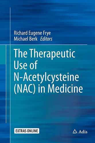
Support this website:
Order from Amazon
We will earn a commission.
Focusing on the practical use of N-Acetyl-Cysteine (NAC) in medicine, this book provides a comprehensive review of the basic biological and clinical studies documenting its benefits in treating medical disease. NAC is perhaps best known as an antidote for acetaminophen, but its therapeutic effect in a wide range of medical diseases has recently been realized. In addition to its well recognized use in radiological contrast prophylaxis for renal disease and pulmonary disorders, studies have suggested significant promise in psychiatric and neurological disorders such as addiction, Alzheimer’s disease, ataxia, autism, bipolar disorder, depression, epilepsy, neuropathy, obsessive-compulsive disorder, schizophrenia, traumatic brain injury and trichotillomania in addition to promising studies in audiology, cardiology, exercise physiology, gastroenterology, hematology, infectious disease, infertility and ophthalmology. Given the promising studies for a wide range of medical conditions, coupled with a excellent safety profile, the potential for NAC in the treatment of human disease appears considerable. Amazon
RJR Picks from Around the Web (updated 11 MAY 2018 )
Old Science
Weird Science

Treating Disease with Fecal Transplantation
Fossils of miniature humans (hobbits) discovered in Indonesia
Paleontology
Dinosaur tail, complete with feathers, found preserved in amber.
Astronomy

Mysterious fast radio burst (FRB) detected in the distant universe.
Big Data & Informatics

Big Data: Buzzword or Big Deal?
Hacking the genome: Identifying anonymized human subjects using publicly available data.