Other Sites:

Bibliography Options Menu

Robert J. Robbins is a biologist, an educator, a science administrator, a publisher, an information technologist, and an IT leader and manager who specializes in advancing biomedical knowledge and supporting education through the application of information technology. More About: RJR | OUR TEAM | OUR SERVICES | THIS WEBSITE
RJR: Recommended Bibliography 27 Jul 2026 at 01:50 Created:
DNA Barcoding
Wikipedia: DNA Barcoding
is a method of species identification using a short section of DNA
from a specific gene or genes. The premise of DNA barcoding is that
by comparison with a reference library of such DNA sections (also
called "sequences"), an individual sequence can be used to uniquely
identify an organism to species, just as a supermarket scanner uses
the familiar black stripes of the UPC barcode to identify an item
in its stock against its reference database. These "barcodes" are
sometimes used in an effort to identify unknown species or parts of
an organism, simply to catalog as many taxa as possible, or to
compare with traditional taxonomy in an effort to determine species
boundaries.
Different gene regions are used to identify the different organismal
groups using barcoding. The most commonly used barcode region for
animals and some protists is a portion of the cytochrome c oxidase I
(COI or COX1) gene, found in mitochondrial DNA. Other genes suitable
for DNA barcoding are the internal transcribed spacer (ITS) rRNA often
used for fungi and RuBisCO used for plants. Microorganisms
are detected using different gene regions.
See also: What is DNA barcoding? or
DNA barcoding workflows
Created with PubMed® Query: DNA[TIAB] barcode[TIAB] OR barcodes[TIAB] OR barcoding[TIAB] NOT pmcbook NOT ispreviousversion
Citations The Papers (from PubMed®)
RevDate: 2026-07-25
CmpDate: 2026-07-25
Selection and evaluation of DNase I hypersensitive sites for prenatal screening of trisomy 21 in the fetus.
Vavilovskii zhurnal genetiki i selektsii, 30(4):669-675.
This study introduces a novel approach for noninvasive prenatal testing (NIPT) of chromosomal abnormalities, based on analysis of epigenetic features in circulating cell-free DNA (cfDNA). The core innovation of our method leverages fundamental differences in chromatin organization between maternal and fetal cells. Specifically, we focused on genomic regions that exhibit open chromatin configuration in maternal blood cells but remain tightly packed in fetal tissues (DNase I hypersensitive sites or DHSs). These epigenetic differences create distinct cfDNA fragmentation signatures that allow selective identification of fetal DNA within the maternal cfDNA pool. The study workflow comprised several key steps: performing genome-wide screening to identify differentially accessible chromatin regions, selecting the most informative markers using a machine learning algorithm, and targeted sequencing of the selected epigenetic markers using molecular barcodes. Subsequently, a LASSO regression model was constructed and validated. As a proof of concept, the study demonstrates the method's efficacy in identifying trisomy 21 (Down syndrome), though the underlying principles can be readily adapted to other abnormalities. Complementing its robust performance, the technique offers practical advantages in terms of platform compatibility - the same epigenetic markers can be assessed using either next-generation sequencing or simpler, more cost-efficient methods like digital PCR. With further refinement, the approach could be extended to screen for additional aneuploidies (trisomies 13 and 18) and microdeletion syndromes. Therefore, this approach offers new opportunities for developing cost-effective testing systems suitable for widespread routine clinical implementation, combining high diagnostic accuracy with reduced analysis costs.
Additional Links: PMID-42499385
Full Text:
Publisher:
PubMed:
Citation:
show bibtex listing
hide bibtex listing
@article {pmid42499385,
year = {2026},
author = {Mazur, AM and Starshin, AS and Bogush, NV and Prokhortchouk, EB},
title = {Selection and evaluation of DNase I hypersensitive sites for prenatal screening of trisomy 21 in the fetus.},
journal = {Vavilovskii zhurnal genetiki i selektsii},
volume = {30},
number = {4},
pages = {669-675},
doi = {10.18699/vjgb-26-67},
pmid = {42499385},
issn = {2500-0462},
abstract = {This study introduces a novel approach for noninvasive prenatal testing (NIPT) of chromosomal abnormalities, based on analysis of epigenetic features in circulating cell-free DNA (cfDNA). The core innovation of our method leverages fundamental differences in chromatin organization between maternal and fetal cells. Specifically, we focused on genomic regions that exhibit open chromatin configuration in maternal blood cells but remain tightly packed in fetal tissues (DNase I hypersensitive sites or DHSs). These epigenetic differences create distinct cfDNA fragmentation signatures that allow selective identification of fetal DNA within the maternal cfDNA pool. The study workflow comprised several key steps: performing genome-wide screening to identify differentially accessible chromatin regions, selecting the most informative markers using a machine learning algorithm, and targeted sequencing of the selected epigenetic markers using molecular barcodes. Subsequently, a LASSO regression model was constructed and validated. As a proof of concept, the study demonstrates the method's efficacy in identifying trisomy 21 (Down syndrome), though the underlying principles can be readily adapted to other abnormalities. Complementing its robust performance, the technique offers practical advantages in terms of platform compatibility - the same epigenetic markers can be assessed using either next-generation sequencing or simpler, more cost-efficient methods like digital PCR. With further refinement, the approach could be extended to screen for additional aneuploidies (trisomies 13 and 18) and microdeletion syndromes. Therefore, this approach offers new opportunities for developing cost-effective testing systems suitable for widespread routine clinical implementation, combining high diagnostic accuracy with reduced analysis costs.},
}
RevDate: 2026-07-25
CmpDate: 2026-07-25
A new earthworm species of the genus Eisenia Malm, 1877 (Oligochaeta, Lumbricidae) from the Ili River valley, northwestern China.
ZooKeys, 1285:21-40.
The present study focuses on the taxonomy of the earthworm genus Eisenia Ealm, 1877 in China. The taxonomic framework for this genus in China has long been ambiguous, with studies in the northwestern region being particularly scarce. Prior to this research, only four subspecies within the Eisenia nordenskioldi (Eisen, 1879) complex, distributed across northeastern and northern China, had been described. Based on detailed morphological characteristics and molecular data, this paper describes a new species, E. ilensis sp. nov., collected from the Ili River valley in northwestern China. The key diagnostic characteristics of this new species include a medium body length (37-103 mm); two pairs of small, spherical spermathecae without ducts, located at the inter-segmental furrows 9/10 and 10/11; a pair of small, circular tubercula pubertatis on the clitellum situated between the ab setae; the first of dorsal pore beginning at the 4/5 inter-segmental furrow; and a pair of slit-like male pores on segment XV. Phylogenetic analyses based on the mitogenomic protein coding genes indicate that E. ilensis sp. nov. forms a distinct clade with E. balatonica Pop, 1943 supporting its status as an independent species and a basal evolutionary status. The discovery of the new species of E. ilensis sp. nov. not only enriches the species diversity of the genus Eisenia in China but also provides a crucial taxonomic foundation for subsequent research into the evolution and dispersal history of this genus in Eurasia.
Additional Links: PMID-42500350
PubMed:
Citation:
show bibtex listing
hide bibtex listing
@article {pmid42500350,
year = {2026},
author = {Xu, P and Zhang, Y and Shu, X and Han, D and Zhao, H},
title = {A new earthworm species of the genus Eisenia Malm, 1877 (Oligochaeta, Lumbricidae) from the Ili River valley, northwestern China.},
journal = {ZooKeys},
volume = {1285},
number = {},
pages = {21-40},
pmid = {42500350},
issn = {1313-2989},
abstract = {The present study focuses on the taxonomy of the earthworm genus Eisenia Ealm, 1877 in China. The taxonomic framework for this genus in China has long been ambiguous, with studies in the northwestern region being particularly scarce. Prior to this research, only four subspecies within the Eisenia nordenskioldi (Eisen, 1879) complex, distributed across northeastern and northern China, had been described. Based on detailed morphological characteristics and molecular data, this paper describes a new species, E. ilensis sp. nov., collected from the Ili River valley in northwestern China. The key diagnostic characteristics of this new species include a medium body length (37-103 mm); two pairs of small, spherical spermathecae without ducts, located at the inter-segmental furrows 9/10 and 10/11; a pair of small, circular tubercula pubertatis on the clitellum situated between the ab setae; the first of dorsal pore beginning at the 4/5 inter-segmental furrow; and a pair of slit-like male pores on segment XV. Phylogenetic analyses based on the mitogenomic protein coding genes indicate that E. ilensis sp. nov. forms a distinct clade with E. balatonica Pop, 1943 supporting its status as an independent species and a basal evolutionary status. The discovery of the new species of E. ilensis sp. nov. not only enriches the species diversity of the genus Eisenia in China but also provides a crucial taxonomic foundation for subsequent research into the evolution and dispersal history of this genus in Eurasia.},
}
RevDate: 2026-07-25
CmpDate: 2026-07-25
A new genus of Gnorimoschemini (Lepidoptera, Gelechiidae, Gelechiinae) from the Atacama and Sechura deserts.
ZooKeys, 1285:41-59.
Sexually dimorphic Gnorimoschemini moths (Lepidoptera, Gelechiidae, Gelechiinae) were reared from leaf mines collected on Tessaria absinthioides (Hook. & Arn.) DC. (Asteraceae) in the coastal Atacama Desert of northern Chile. Detailed examination revealed a combination of morphological characters not assignable to any known genus of the tribe, including 1) a scale tuft on the male hindwing, 2) an uncus with a triangular postero-medial projection, and 3) a U-shaped gnathos. COI sequences obtained from a male and a female showed minimal intraspecific divergence (0.6%), whereas interspecific divergences with representatives of other Gnorimoschemini genera ranged from 8.1% to 11.8%. Phylogenetic analyses based on COI sequences further confirmed the Atacama moths as a strongly supported, isolated clade, corroborating its morphological distinctiveness and supporting its recognition as a separate evolutionary lineage. The combined evidence justifies the establishment of Azaptilia azapensis Vargas, gen. nov. et sp. nov. Furthermore, based on the remarkable similarity in genitalia morphology, the Peruvian moth Scrobipalpula trichinaspis (Meyrick, 1917) from the Sechura Desert, originally described in Phthorimaea Meyrick, 1902, is also included in the new genus as Azaptilia trichinaspis (Meyrick, 1917), comb. nov. This discovery expands the known diversity of Gnorimoschemini in arid South American ecosystems of the Atacama and Sechura deserts and highlights the importance of integrating rearing, morphology, and DNA barcoding for uncovering hidden diversity in Neotropical Gelechiidae.
Additional Links: PMID-42500352
PubMed:
Citation:
show bibtex listing
hide bibtex listing
@article {pmid42500352,
year = {2026},
author = {Ampuero-Vega, J and Campos-Soto, R and Vargas, HA},
title = {A new genus of Gnorimoschemini (Lepidoptera, Gelechiidae, Gelechiinae) from the Atacama and Sechura deserts.},
journal = {ZooKeys},
volume = {1285},
number = {},
pages = {41-59},
pmid = {42500352},
issn = {1313-2989},
abstract = {Sexually dimorphic Gnorimoschemini moths (Lepidoptera, Gelechiidae, Gelechiinae) were reared from leaf mines collected on Tessaria absinthioides (Hook. & Arn.) DC. (Asteraceae) in the coastal Atacama Desert of northern Chile. Detailed examination revealed a combination of morphological characters not assignable to any known genus of the tribe, including 1) a scale tuft on the male hindwing, 2) an uncus with a triangular postero-medial projection, and 3) a U-shaped gnathos. COI sequences obtained from a male and a female showed minimal intraspecific divergence (0.6%), whereas interspecific divergences with representatives of other Gnorimoschemini genera ranged from 8.1% to 11.8%. Phylogenetic analyses based on COI sequences further confirmed the Atacama moths as a strongly supported, isolated clade, corroborating its morphological distinctiveness and supporting its recognition as a separate evolutionary lineage. The combined evidence justifies the establishment of Azaptilia azapensis Vargas, gen. nov. et sp. nov. Furthermore, based on the remarkable similarity in genitalia morphology, the Peruvian moth Scrobipalpula trichinaspis (Meyrick, 1917) from the Sechura Desert, originally described in Phthorimaea Meyrick, 1902, is also included in the new genus as Azaptilia trichinaspis (Meyrick, 1917), comb. nov. This discovery expands the known diversity of Gnorimoschemini in arid South American ecosystems of the Atacama and Sechura deserts and highlights the importance of integrating rearing, morphology, and DNA barcoding for uncovering hidden diversity in Neotropical Gelechiidae.},
}
RevDate: 2026-07-24
CmpDate: 2026-07-24
Huntsman spiders of the genus Sinopoda (Araneae, Sparassidae, Heteropodinae) from the Honghe Hani and Yi Autonomous Prefecture, southwestern China, with descriptions of two new species.
ZooKeys, 1280:301-320.
Spiders of the genus Sinopoda Jäger, 1999 from Honghe Hani and Yi Autonomous Prefecture, Yunnan Province, China are studied. A total of four species are reported and illustrated of which two, Sinopoda honghe Yu & Zhong, sp. nov. and Sinopoda kuan Yu & Zhong, sp. nov., are described as new to science. The other two previously described species from this region: S. tengchongensis Fu & Zhu, 2008 and S. tumefacta Zhong, Jäger, Chen & Liu, 2019 are also illustrated. Detailed descriptions, diagnoses, illustrations and DNA barcodes of the two new species are given. A distribution map of these four species in Honghe is provided.
Additional Links: PMID-42494839
PubMed:
Citation:
show bibtex listing
hide bibtex listing
@article {pmid42494839,
year = {2026},
author = {Zhang, C and Xing, Y and Zhong, Y and Yu, H},
title = {Huntsman spiders of the genus Sinopoda (Araneae, Sparassidae, Heteropodinae) from the Honghe Hani and Yi Autonomous Prefecture, southwestern China, with descriptions of two new species.},
journal = {ZooKeys},
volume = {1280},
number = {},
pages = {301-320},
pmid = {42494839},
issn = {1313-2989},
abstract = {Spiders of the genus Sinopoda Jäger, 1999 from Honghe Hani and Yi Autonomous Prefecture, Yunnan Province, China are studied. A total of four species are reported and illustrated of which two, Sinopoda honghe Yu & Zhong, sp. nov. and Sinopoda kuan Yu & Zhong, sp. nov., are described as new to science. The other two previously described species from this region: S. tengchongensis Fu & Zhu, 2008 and S. tumefacta Zhong, Jäger, Chen & Liu, 2019 are also illustrated. Detailed descriptions, diagnoses, illustrations and DNA barcodes of the two new species are given. A distribution map of these four species in Honghe is provided.},
}
RevDate: 2026-07-24
CmpDate: 2026-07-24
The development of single-cell lineage tracing technology and its application in immunotherapy.
Molecular therapy. Advances, 34(3):201802.
Immunotherapy stands as one of the most promising approaches in cancer treatment, with engineered T cell therapies, particularly chimeric antigen receptor T cell (CAR-T), leading the charge. However, relapse in some patients post-treatment suggests that research in this field remains incomplete. Tumor heterogeneity and the complexities of the immune microenvironment hinder a comprehensive understanding of the changes engineered T cells undergo once introduced into the human body. Single-cell lineage tracing (SCLT) technology facilitates the investigation of cellular development by monitoring the fate and differentiation of individual cells and their descendants within an organism. Employing methodologies such as CRISPR-based labeling and mitochondrial DNA tracking, SCLT allows for dynamic analysis of T cell clonal evolution, exhaustion mechanisms, and memory cell generation. This approach offers single-cell resolution data that contribute to resolving pertinent clinical challenges. This article provides a comprehensive review of recent developments and characteristics of the SCLT multi-omics approach. It elucidates the manner in which SCLT addresses the conventional constraints associated with spatiotemporal resolution and introduces a novel methodology for generating DNA barcodes to monitor CAR-T cells via CRISPR technology. These contributions offer valuable perspectives for the enhancement of cell therapy strategies.
Additional Links: PMID-42494981
PubMed:
Citation:
show bibtex listing
hide bibtex listing
@article {pmid42494981,
year = {2026},
author = {Wang, J and Li, Z and Chen, J and Li, H and Chen, F and Tan, L and Chen, X and Liu, S and Zhang, W and Shao, H},
title = {The development of single-cell lineage tracing technology and its application in immunotherapy.},
journal = {Molecular therapy. Advances},
volume = {34},
number = {3},
pages = {201802},
pmid = {42494981},
issn = {3117-387X},
abstract = {Immunotherapy stands as one of the most promising approaches in cancer treatment, with engineered T cell therapies, particularly chimeric antigen receptor T cell (CAR-T), leading the charge. However, relapse in some patients post-treatment suggests that research in this field remains incomplete. Tumor heterogeneity and the complexities of the immune microenvironment hinder a comprehensive understanding of the changes engineered T cells undergo once introduced into the human body. Single-cell lineage tracing (SCLT) technology facilitates the investigation of cellular development by monitoring the fate and differentiation of individual cells and their descendants within an organism. Employing methodologies such as CRISPR-based labeling and mitochondrial DNA tracking, SCLT allows for dynamic analysis of T cell clonal evolution, exhaustion mechanisms, and memory cell generation. This approach offers single-cell resolution data that contribute to resolving pertinent clinical challenges. This article provides a comprehensive review of recent developments and characteristics of the SCLT multi-omics approach. It elucidates the manner in which SCLT addresses the conventional constraints associated with spatiotemporal resolution and introduces a novel methodology for generating DNA barcodes to monitor CAR-T cells via CRISPR technology. These contributions offer valuable perspectives for the enhancement of cell therapy strategies.},
}
RevDate: 2026-07-24
CmpDate: 2026-07-24
Cell line authentication: a commercial service provider perspective.
Frontiers in cell and developmental biology, 14:1843943.
Cell line misidentification and cross contamination continue to undermine research quality despite improved laboratory practices. Short Tandem Repeat (STR) profiling remains the primary method for human cell line authentication and is increasingly required by journals, funders, and regulatory bodies. Analysis of authentication data generated at NorthGene™ in 2024-2025 demonstrates that misidentification and contamination remain prevalent, with 4.7% of lines misidentified and 1.8% contaminated in 2024, and 2.4% misidentified with 1.6% contaminated in 2025. Primary cell lines present additional challenges, as none submitted during this period met ASN 0002 standards for full traceability, posing risks to reproducibility and downstream biological interpretation. Mixture analysis proved essential for resolving inconclusive STR results and identifying specific contaminants, although progress is hindered when reference STR profiles are not publicly available. While STR profiling is highly discriminatory for human cells, complementary methods-such as species specific PCR and CO1 barcoding-may be required to detect interspecies contamination or authenticate non human lines. Overall, these findings highlight the ongoing need for rigorous authentication workflows and complete traceability to ensure scientific integrity and prevent invalid research outcomes.
Additional Links: PMID-42495720
PubMed:
Citation:
show bibtex listing
hide bibtex listing
@article {pmid42495720,
year = {2026},
author = {Ralston, E and Haskayne, C},
title = {Cell line authentication: a commercial service provider perspective.},
journal = {Frontiers in cell and developmental biology},
volume = {14},
number = {},
pages = {1843943},
pmid = {42495720},
issn = {2296-634X},
abstract = {Cell line misidentification and cross contamination continue to undermine research quality despite improved laboratory practices. Short Tandem Repeat (STR) profiling remains the primary method for human cell line authentication and is increasingly required by journals, funders, and regulatory bodies. Analysis of authentication data generated at NorthGene™ in 2024-2025 demonstrates that misidentification and contamination remain prevalent, with 4.7% of lines misidentified and 1.8% contaminated in 2024, and 2.4% misidentified with 1.6% contaminated in 2025. Primary cell lines present additional challenges, as none submitted during this period met ASN 0002 standards for full traceability, posing risks to reproducibility and downstream biological interpretation. Mixture analysis proved essential for resolving inconclusive STR results and identifying specific contaminants, although progress is hindered when reference STR profiles are not publicly available. While STR profiling is highly discriminatory for human cells, complementary methods-such as species specific PCR and CO1 barcoding-may be required to detect interspecies contamination or authenticate non human lines. Overall, these findings highlight the ongoing need for rigorous authentication workflows and complete traceability to ensure scientific integrity and prevent invalid research outcomes.},
}
RevDate: 2026-07-24
Small RNA barcodes for sensitive labeling of AAV transcripts.
eNeuro pii:ENEURO.0034-26.2026 [Epub ahead of print].
Adeno-associated virus (AAV) vectors are widely used for neuroscience research. Labeling AAV mRNA by in situ hybridization can track AAV transduction with high sensitivity and without the need for reporter genes. However, it is challenging to detect similar mRNA transcripts when multiple AAVs are injected into the same animal, or when a single AAV produces multiple distinct transcripts. To address these challenges, we developed methods for sensitive labeling of small barcodes on AAV mRNA at single-transcript resolution. These small RNA barcodes (ranging from 40 to 44 bases) can be easily added to any AAV vector genome. We provide detailed step-by-step protocols for sensitive and flexible labeling of barcoded AAV mRNA by BaseScope in situ hybridization. We validated these labeling methods in male and female mice, and in a female rhesus macaque. RNA barcoding can improve the reliability of multiplexed in vivo screening: by adding one barcode to half of the AAVs in a pooled library and a second barcode to the other half, barcode labeling can identify cells that receive multiple AAVs without the need for reporter proteins. This approach is also useful for intersectional experiments that combine multiple AAV vectors: multiple unique barcodes can track the spatial distribution of each AAV and can separately label transcripts from different AAV genome conformations, such as before or after Cre-Lox recombination. These new methods for sensitive multiplexed labeling of AAV mRNA will support greater rigor and reliability in neuroscience research.Significance Statement AAV vectors are commonly used in neuroscience research. Modern experimental designs are increasingly complex, often injecting two or more AAVs into different regions, or screening a library of many unique AAVs injected together into a single animal. In such complex experiments, it is challenging to track spatial distribution and rates of co-infection among multiple AAV vectors, especially if the AAVs do not carry reporter genes. To address these challenges, we developed small RNA barcodes and methods for highly sensitive labeling of barcoded AAV mRNA. These small barcodes can be easily added to any AAV genome without altering the experimental design, supporting the assessment of injection targeting and co-infection rates among multiple AAVs without the need for reporter genes.
Additional Links: PMID-42498665
Publisher:
PubMed:
Citation:
show bibtex listing
hide bibtex listing
@article {pmid42498665,
year = {2026},
author = {Magaña, Y and Wang, S and Torreblanca-Zanca, A and Wei, L and Castle, MJ},
title = {Small RNA barcodes for sensitive labeling of AAV transcripts.},
journal = {eNeuro},
volume = {},
number = {},
pages = {},
doi = {10.1523/ENEURO.0034-26.2026},
pmid = {42498665},
issn = {2373-2822},
abstract = {Adeno-associated virus (AAV) vectors are widely used for neuroscience research. Labeling AAV mRNA by in situ hybridization can track AAV transduction with high sensitivity and without the need for reporter genes. However, it is challenging to detect similar mRNA transcripts when multiple AAVs are injected into the same animal, or when a single AAV produces multiple distinct transcripts. To address these challenges, we developed methods for sensitive labeling of small barcodes on AAV mRNA at single-transcript resolution. These small RNA barcodes (ranging from 40 to 44 bases) can be easily added to any AAV vector genome. We provide detailed step-by-step protocols for sensitive and flexible labeling of barcoded AAV mRNA by BaseScope in situ hybridization. We validated these labeling methods in male and female mice, and in a female rhesus macaque. RNA barcoding can improve the reliability of multiplexed in vivo screening: by adding one barcode to half of the AAVs in a pooled library and a second barcode to the other half, barcode labeling can identify cells that receive multiple AAVs without the need for reporter proteins. This approach is also useful for intersectional experiments that combine multiple AAV vectors: multiple unique barcodes can track the spatial distribution of each AAV and can separately label transcripts from different AAV genome conformations, such as before or after Cre-Lox recombination. These new methods for sensitive multiplexed labeling of AAV mRNA will support greater rigor and reliability in neuroscience research.Significance Statement AAV vectors are commonly used in neuroscience research. Modern experimental designs are increasingly complex, often injecting two or more AAVs into different regions, or screening a library of many unique AAVs injected together into a single animal. In such complex experiments, it is challenging to track spatial distribution and rates of co-infection among multiple AAV vectors, especially if the AAVs do not carry reporter genes. To address these challenges, we developed small RNA barcodes and methods for highly sensitive labeling of barcoded AAV mRNA. These small barcodes can be easily added to any AAV genome without altering the experimental design, supporting the assessment of injection targeting and co-infection rates among multiple AAVs without the need for reporter genes.},
}
RevDate: 2026-07-24
CmpDate: 2026-07-24
Integrated ecological, anatomical, phytochemical and genetic characterization of Peganum harmala L. along a soil salinity gradient in the Taif Region, Saudi Arabia.
BMC plant biology, 26(1):.
BACKGROUND: Peganum harmala L. is a medicinal shrub that tolerates harsh, dry conditions, but the links between its habitat, structure, chemistry, and genetics are still not well understood. In this study, we investigated five geo‑referenced natural populations from the Taif region in Saudi Arabia, using an integrated ecological, anatomical, phytochemical, and molecular approach.
RESULTS: All soils samples were slightly alkaline (pH 7.61-7.79), and fell within the non-saline to slightly saline range based on electrical conductivity (EC) classifications, but they differed in salinity. Sites P1 (Al Ahlam Park) and P2 (C36) had lower EC values, while P3 (Ashayra), P4 (As Sayl al Kabir), and P5 (Al Sir) showed relatively higher EC values within this overall low salinity range. In all locations, P. harmala L. formed well-established, low, spreading to dense dome-shaped shrubs on sandy-stony substrates. Multivariate analysis of soil variables (pH, EC, CaCO₃, major anions and cations) set P3 apart from the other sites, and both clustering and principal component analysis (PCA) grouped P1, P2, P4, and P5 together, with P3 forming its own distinct cluster. Anatomically, most populations had stem cross sections with well-organized vascular cylinders. By contrast, stems from P3 displayed an irregular outline, a compact multilayered cortex, and scattered vascular strands with visibly reduced secondary xylem. Total alkaloid content ranged from 3.208 to 4.106%, with the highest values at P1 and P2 and the lowest at P3. Molecular analyses using SCoT markers (10 primers; 121 bands, 54 polymorphic) and IRAP markers (10 primers; 130 bands, 76 polymorphic) revealed moderate to high nuclear genetic diversity (expected heterozygosity up to 0.50; Shannon's index up to 0.69). Overall genetic similarity among populations was high, but their structure was non-random: populations 2, 4, and 5 consistently clustered together, whereas populations 1 and especially 3 were more divergent. Chloroplast barcodes (matK: PX089557; rpoC1: PV942406) from a representative voucher from Population 1 were consistent with its identification as P. harmala L. and clearly separated this sequence from those of related species in phylogenetic trees. In combination with morphological identification, this supports assignment of all sampled populations to P. harmala L.
CONCLUSIONS: Among the studied populations, P3 emerges as an ecologically and genetically distinct unit within P. harmala L. in the Taif region, characterized by its unique nuclear genetic profile, modified stem anatomy, and reduced total alkaloid content associated with higher soil salinity and altered major-ion composition. This integrative framework-combining soil characterization, quantitative anatomy, total alkaloids, SCoT/IRAP markers, and plastid barcodes illustrate how fine-scale edaphic heterogeneity is associated with anatomical, chemical, and genetic variation in this key desert medicinal shrub.
Additional Links: PMID-42498932
PubMed:
Citation:
show bibtex listing
hide bibtex listing
@article {pmid42498932,
year = {2026},
author = {Almutairi, ZM and Abd-Elgawad, ME},
title = {Integrated ecological, anatomical, phytochemical and genetic characterization of Peganum harmala L. along a soil salinity gradient in the Taif Region, Saudi Arabia.},
journal = {BMC plant biology},
volume = {26},
number = {1},
pages = {},
pmid = {42498932},
issn = {1471-2229},
support = {PSAU/2024/01/31574//Prince Sattam bin Abdulaziz University/ ; },
mesh = {Saudi Arabia ; *Peganum/genetics/anatomy & histology/chemistry ; *Salinity ; *Soil/chemistry ; Ecosystem ; Genetic Variation ; },
abstract = {BACKGROUND: Peganum harmala L. is a medicinal shrub that tolerates harsh, dry conditions, but the links between its habitat, structure, chemistry, and genetics are still not well understood. In this study, we investigated five geo‑referenced natural populations from the Taif region in Saudi Arabia, using an integrated ecological, anatomical, phytochemical, and molecular approach.
RESULTS: All soils samples were slightly alkaline (pH 7.61-7.79), and fell within the non-saline to slightly saline range based on electrical conductivity (EC) classifications, but they differed in salinity. Sites P1 (Al Ahlam Park) and P2 (C36) had lower EC values, while P3 (Ashayra), P4 (As Sayl al Kabir), and P5 (Al Sir) showed relatively higher EC values within this overall low salinity range. In all locations, P. harmala L. formed well-established, low, spreading to dense dome-shaped shrubs on sandy-stony substrates. Multivariate analysis of soil variables (pH, EC, CaCO₃, major anions and cations) set P3 apart from the other sites, and both clustering and principal component analysis (PCA) grouped P1, P2, P4, and P5 together, with P3 forming its own distinct cluster. Anatomically, most populations had stem cross sections with well-organized vascular cylinders. By contrast, stems from P3 displayed an irregular outline, a compact multilayered cortex, and scattered vascular strands with visibly reduced secondary xylem. Total alkaloid content ranged from 3.208 to 4.106%, with the highest values at P1 and P2 and the lowest at P3. Molecular analyses using SCoT markers (10 primers; 121 bands, 54 polymorphic) and IRAP markers (10 primers; 130 bands, 76 polymorphic) revealed moderate to high nuclear genetic diversity (expected heterozygosity up to 0.50; Shannon's index up to 0.69). Overall genetic similarity among populations was high, but their structure was non-random: populations 2, 4, and 5 consistently clustered together, whereas populations 1 and especially 3 were more divergent. Chloroplast barcodes (matK: PX089557; rpoC1: PV942406) from a representative voucher from Population 1 were consistent with its identification as P. harmala L. and clearly separated this sequence from those of related species in phylogenetic trees. In combination with morphological identification, this supports assignment of all sampled populations to P. harmala L.
CONCLUSIONS: Among the studied populations, P3 emerges as an ecologically and genetically distinct unit within P. harmala L. in the Taif region, characterized by its unique nuclear genetic profile, modified stem anatomy, and reduced total alkaloid content associated with higher soil salinity and altered major-ion composition. This integrative framework-combining soil characterization, quantitative anatomy, total alkaloids, SCoT/IRAP markers, and plastid barcodes illustrate how fine-scale edaphic heterogeneity is associated with anatomical, chemical, and genetic variation in this key desert medicinal shrub.},
}
MeSH Terms:
show MeSH Terms
hide MeSH Terms
Saudi Arabia
*Peganum/genetics/anatomy & histology/chemistry
*Salinity
*Soil/chemistry
Ecosystem
Genetic Variation
RevDate: 2026-07-23
CmpDate: 2026-07-23
The Lycianthes plastome and its comparison with Capsicum (Capsiceae, Solanaceae).
Genetica, 154(1):.
Solanaceae is an important crop plant family and its plastomes have been extensively studied to understand evolution and breeding. Capsicum (chili peppers) includes five domesticated species and worldwide are important economic resources. Lycianthes is the third most species rich genus within the family. Capsicum and Lycianthes are closely related genera and form the Capsiceae. While plastomes are available in public databases for some species of Capsicum, no plastomes for Lycianthes have been published. We anticipate that the plastomes of Lycianthes and Capsicum will be similar. In addition, point sequence variation will generate robust phylogenetic hypotheses. We newly sequenced, assembled and annotated the plastomes of six species of Lycianthes to investigate their genomic characteristics. Also, we carried out comparative and phylogenetic analysis within Capsiceae. Lycianthes presented the typical circular quadripartite structure and organization. Plastomes range from 156,108 to 156,721 bp. All contained 133 genes, divided into 84 Coding DNA Sequence, four pseudogenes, 37 tRNAs and eight rRNAs. Plastomes of Capsiceae were highly conserved, were similar in size and showed the same structure and organization. Comparative analyses revealed that 17 genes made the Capsicum plastome longer than the plastome of Lycianthes. However, the variable regions did not match with the length of the genes. Based on plastome sequences, phylogeny recovered the monophyly of Capsiceae. Also, Capsicum and Lycianthes formed monophyletic genera and had a sister group relationship with each other. The plastome of Lycianthes and Capsicum were similar. Phylogenomics supported the monophyly of both genera and a sister group relationship.
Additional Links: PMID-42489757
PubMed:
Citation:
show bibtex listing
hide bibtex listing
@article {pmid42489757,
year = {2026},
author = {Anguiano Constante, MA and Rodríguez, A},
title = {The Lycianthes plastome and its comparison with Capsicum (Capsiceae, Solanaceae).},
journal = {Genetica},
volume = {154},
number = {1},
pages = {},
pmid = {42489757},
issn = {1573-6857},
support = {855486/2020-000026-02NACF-19054//Secretaria de Ciencias, Humanidades, Tecnología e Inovación (SECIHTI)/ ; },
mesh = {Phylogeny ; *Capsicum/genetics/classification ; Evolution, Molecular ; *Solanaceae/genetics ; *Genome, Plastid ; *Genome, Chloroplast ; Sequence Analysis, DNA ; },
abstract = {Solanaceae is an important crop plant family and its plastomes have been extensively studied to understand evolution and breeding. Capsicum (chili peppers) includes five domesticated species and worldwide are important economic resources. Lycianthes is the third most species rich genus within the family. Capsicum and Lycianthes are closely related genera and form the Capsiceae. While plastomes are available in public databases for some species of Capsicum, no plastomes for Lycianthes have been published. We anticipate that the plastomes of Lycianthes and Capsicum will be similar. In addition, point sequence variation will generate robust phylogenetic hypotheses. We newly sequenced, assembled and annotated the plastomes of six species of Lycianthes to investigate their genomic characteristics. Also, we carried out comparative and phylogenetic analysis within Capsiceae. Lycianthes presented the typical circular quadripartite structure and organization. Plastomes range from 156,108 to 156,721 bp. All contained 133 genes, divided into 84 Coding DNA Sequence, four pseudogenes, 37 tRNAs and eight rRNAs. Plastomes of Capsiceae were highly conserved, were similar in size and showed the same structure and organization. Comparative analyses revealed that 17 genes made the Capsicum plastome longer than the plastome of Lycianthes. However, the variable regions did not match with the length of the genes. Based on plastome sequences, phylogeny recovered the monophyly of Capsiceae. Also, Capsicum and Lycianthes formed monophyletic genera and had a sister group relationship with each other. The plastome of Lycianthes and Capsicum were similar. Phylogenomics supported the monophyly of both genera and a sister group relationship.},
}
MeSH Terms:
show MeSH Terms
hide MeSH Terms
Phylogeny
*Capsicum/genetics/classification
Evolution, Molecular
*Solanaceae/genetics
*Genome, Plastid
*Genome, Chloroplast
Sequence Analysis, DNA
RevDate: 2026-07-23
Diverse Onchocercidae from Malaysian cats and Indonesian macaques: Morphological and molecular analysis of individual microfilariae using mitochondrial genomes, 28S rRNA, and Wolbachia endosymbiont sequences.
PLoS neglected tropical diseases, 20(7):e0014015 pii:PNTD-D-26-00252 [Epub ahead of print].
During an investigation of animals as reservoirs for the filarial parasite Brugia malayi, three molecularly undescribed filarial species were co-detected. Individual microfilariae (Mf) were isolated and analyzed from blood samples of crab-eating macaques (Macaca fascicularis) from Belitung, Indonesia, and from pet dogs and cats in Sabah, Malaysia. Among 163 macaques, 33 (20.2%) were positive for large Mf (mean length 498.9 µm) similar to Dirofilaria ('Belitung I'). One macaque was infected with small Mf (mean length 150.4 µm) ('Belitung II'), with a high density of 17,150 Mf/mL. In two cats co-infected with B. malayi, Mf of a Dirofilaria species ('Sabah') with an average length of 299.1 µm were detected. Morphometric analysis of Mf showed distinct differences between these three species and other Mf described in the area. Whole genome amplification and genome sequencing of 24 individual Mf enabled phylogenetic analysis of mitochondrial genomes, and analysis of specific mitochondrial and nuclear barcode regions. The three Mf groups formed distinct clusters and could not be identified by comparison with available reference sequence. Cluster 'Belitung I' from macaques formed a sister group to other characterized Dirofilaria. Cluster 'Belitung II' included bird filariae and primate filariae of the genus Mansonella as close relatives. The cluster 'Sabah' formed a monophyletic group with the zoonotic species Dirofilaria asiatica and Dirofilaria sp. 'Thailand'. DNA of Wolbachia endobacteria was detected in Mf of 'Belitung I' and 'Sabah', but not in 'Belitung II'. These findings highlight the limited understanding of filarial diversity in macaques and cats in Asia and underscore the need for a more comprehensive approach that combines morphological and molecular data to identify and assess the pathogenicity and zoonotic potential of these parasites.
Additional Links: PMID-42490671
Publisher:
PubMed:
Citation:
show bibtex listing
hide bibtex listing
@article {pmid42490671,
year = {2026},
author = {Diekmann, I and Choi, YJ and Supali, T and Alfian, R and Destani, Y and Iskandar, E and Sugianto, N and Mutalip, MHA and Aziz, NAA and Ibrahim, K and Fischer, K and Mitreva, M and Fischer, PU},
title = {Diverse Onchocercidae from Malaysian cats and Indonesian macaques: Morphological and molecular analysis of individual microfilariae using mitochondrial genomes, 28S rRNA, and Wolbachia endosymbiont sequences.},
journal = {PLoS neglected tropical diseases},
volume = {20},
number = {7},
pages = {e0014015},
doi = {10.1371/journal.pntd.0014015},
pmid = {42490671},
issn = {1935-2735},
abstract = {During an investigation of animals as reservoirs for the filarial parasite Brugia malayi, three molecularly undescribed filarial species were co-detected. Individual microfilariae (Mf) were isolated and analyzed from blood samples of crab-eating macaques (Macaca fascicularis) from Belitung, Indonesia, and from pet dogs and cats in Sabah, Malaysia. Among 163 macaques, 33 (20.2%) were positive for large Mf (mean length 498.9 µm) similar to Dirofilaria ('Belitung I'). One macaque was infected with small Mf (mean length 150.4 µm) ('Belitung II'), with a high density of 17,150 Mf/mL. In two cats co-infected with B. malayi, Mf of a Dirofilaria species ('Sabah') with an average length of 299.1 µm were detected. Morphometric analysis of Mf showed distinct differences between these three species and other Mf described in the area. Whole genome amplification and genome sequencing of 24 individual Mf enabled phylogenetic analysis of mitochondrial genomes, and analysis of specific mitochondrial and nuclear barcode regions. The three Mf groups formed distinct clusters and could not be identified by comparison with available reference sequence. Cluster 'Belitung I' from macaques formed a sister group to other characterized Dirofilaria. Cluster 'Belitung II' included bird filariae and primate filariae of the genus Mansonella as close relatives. The cluster 'Sabah' formed a monophyletic group with the zoonotic species Dirofilaria asiatica and Dirofilaria sp. 'Thailand'. DNA of Wolbachia endobacteria was detected in Mf of 'Belitung I' and 'Sabah', but not in 'Belitung II'. These findings highlight the limited understanding of filarial diversity in macaques and cats in Asia and underscore the need for a more comprehensive approach that combines morphological and molecular data to identify and assess the pathogenicity and zoonotic potential of these parasites.},
}
RevDate: 2026-07-23
CmpDate: 2026-07-23
Two new species of Symphylella (Symphyla, Scolopendrellidae) from North China, with an analysis of genetic divergence in Chinese species.
ZooKeys, 1284:251-270.
Symphylans from Liaoning Province, the Inner Mongolia Autonomous Region, and Shanxi Province, North China, were investigated and studied for the first time. Two new species of the Symphylella isabellae group, S. hylophila sp. nov. and S. rotundata sp. nov., are identified and described. Symphylella hylophila sp. nov. is characterized by having blunt ends of the processes, long, subconical styli each with a blunt apex, and the absence of long, erect setae on the cerci. Symphylella rotundata sp. nov. is characterized by having noticeably roundish ends of the tergal processes, short, subconical styli each with a pointed apex, and long, erect setae on the outer and ventral sides of the cerci. These new species are carefully delimited from similar species. Morphological analyses were supported by DNA barcodes, which were used to analyse the genetic divergence of Chinese species of the genus Symphylella.
Additional Links: PMID-42491421
PubMed:
Citation:
show bibtex listing
hide bibtex listing
@article {pmid42491421,
year = {2026},
author = {Jin, YL and Bu, Y},
title = {Two new species of Symphylella (Symphyla, Scolopendrellidae) from North China, with an analysis of genetic divergence in Chinese species.},
journal = {ZooKeys},
volume = {1284},
number = {},
pages = {251-270},
pmid = {42491421},
issn = {1313-2989},
abstract = {Symphylans from Liaoning Province, the Inner Mongolia Autonomous Region, and Shanxi Province, North China, were investigated and studied for the first time. Two new species of the Symphylella isabellae group, S. hylophila sp. nov. and S. rotundata sp. nov., are identified and described. Symphylella hylophila sp. nov. is characterized by having blunt ends of the processes, long, subconical styli each with a blunt apex, and the absence of long, erect setae on the cerci. Symphylella rotundata sp. nov. is characterized by having noticeably roundish ends of the tergal processes, short, subconical styli each with a pointed apex, and long, erect setae on the outer and ventral sides of the cerci. These new species are carefully delimited from similar species. Morphological analyses were supported by DNA barcodes, which were used to analyse the genetic divergence of Chinese species of the genus Symphylella.},
}
RevDate: 2026-07-23
Molecular detection of Coxiella spp. and characterization of blood-feeding sources in tick species parasitizing domestic animals in cattle farms from Magdalena Medio, Colombia.
Veterinary microbiology, 320:111156 pii:S0378-1135(26)00293-2 [Epub ahead of print].
Coxiella burnetii is a globally distributed zoonotic bacterium responsible for Q fever in humans, with domestic ruminants serving as primary reservoirs. In Colombia, while livestock production is a vital economic sector, the dynamics of C. burnetii circulation among ticks and domestic animal hosts remain poorly understood. This study aimed to identify C. burnetii in hosts and ticks from two cattle farms in the Magdalena Medio region. Ticks were collected directly from domestic animal hosts through physical inspections and the use of the dragging technique on pasture vegetation and identified via morphological keys and COI barcoding as Rhipicephalus microplus s.l., predominating on cattle, Dermacentor nitens on equines, and Rhipicephalus sanguineus s.l. on dogs. Blood-meal analysis using PCR-HRM and sequencing identified feeding sources in equines, bovines, and humans, with human DNA detected in all larval pools. Following tick characterization, Coxiella spp. were screened by qPCR targeting the IS1111 insertion sequence, revealing a frequency of 10.5% in domestic animal hosts and 1.36% in ticks. Finally, phylogenetic analysis of 16S rRNA and rpoB genes confirmed the coexistence of C. burnetii (Clade A) in hosts and ticks, alongside Coxiella-like endosymbionts (Clade C), primarily in R. microplus s.l. These findings reveal complex tick-host interactions and confirm the molecular circulation of Coxiella spp. in tropical livestock systems. The results underscore the need to use multigene markers to differentiate pathogens from endosymbionts and suggest a potential human exposure risk, highlighting the importance of One Health surveillance in these agroecosystems.
Additional Links: PMID-42492119
Publisher:
PubMed:
Citation:
show bibtex listing
hide bibtex listing
@article {pmid42492119,
year = {2026},
author = {Cabrera, R and Zapata, S and Durango-Manrique, Y and López, L and Gómez, GF and Gutiérrez, LA},
title = {Molecular detection of Coxiella spp. and characterization of blood-feeding sources in tick species parasitizing domestic animals in cattle farms from Magdalena Medio, Colombia.},
journal = {Veterinary microbiology},
volume = {320},
number = {},
pages = {111156},
doi = {10.1016/j.vetmic.2026.111156},
pmid = {42492119},
issn = {1873-2542},
abstract = {Coxiella burnetii is a globally distributed zoonotic bacterium responsible for Q fever in humans, with domestic ruminants serving as primary reservoirs. In Colombia, while livestock production is a vital economic sector, the dynamics of C. burnetii circulation among ticks and domestic animal hosts remain poorly understood. This study aimed to identify C. burnetii in hosts and ticks from two cattle farms in the Magdalena Medio region. Ticks were collected directly from domestic animal hosts through physical inspections and the use of the dragging technique on pasture vegetation and identified via morphological keys and COI barcoding as Rhipicephalus microplus s.l., predominating on cattle, Dermacentor nitens on equines, and Rhipicephalus sanguineus s.l. on dogs. Blood-meal analysis using PCR-HRM and sequencing identified feeding sources in equines, bovines, and humans, with human DNA detected in all larval pools. Following tick characterization, Coxiella spp. were screened by qPCR targeting the IS1111 insertion sequence, revealing a frequency of 10.5% in domestic animal hosts and 1.36% in ticks. Finally, phylogenetic analysis of 16S rRNA and rpoB genes confirmed the coexistence of C. burnetii (Clade A) in hosts and ticks, alongside Coxiella-like endosymbionts (Clade C), primarily in R. microplus s.l. These findings reveal complex tick-host interactions and confirm the molecular circulation of Coxiella spp. in tropical livestock systems. The results underscore the need to use multigene markers to differentiate pathogens from endosymbionts and suggest a potential human exposure risk, highlighting the importance of One Health surveillance in these agroecosystems.},
}
RevDate: 2026-07-23
Cryptic taxa in the morphospecies Bichromomyia flaviscutellata uncovered by expanded geographic sampling and COI DNA barcodes.
Acta tropica pii:S0001-706X(26)00283-4 [Epub ahead of print].
The Neotropical sand fly species Bichromomyia flaviscutellata is widely distributed and the main vector of Leishmania amazonensis. Previous molecular studies suggested that it comprises at least two morphologically cryptic taxa, but sampling has remained geographically limited. In this study, we expanded specimen collection across 12 Brazilian municipalities spanning the Amazon, Cerrado, and Atlantic Forest biomes, generating 62 new sequences of the mitochondrial cytochrome c oxidase subunit I (COI) gene and combining them with publicly available data. A phylogenetic gene tree, and distance- and coalescent-based delimitation methods, were used to evaluate lineage structure and assess potential species boundaries. Our results consistently revealed two deeply divergent and well‑supported mitochondrial lineages within B. flaviscutellata. One lineage (PS1) occurs across the Amazon basin, Cerrado, and an inland fragment of the western region of the Atlantic Forest, while the other (PS2) is restricted to the coastal Atlantic Forest. Additional internal structure was recovered within both lineages. Pairwise genetic distances were high between PS1 and PS2 (14%-15.7%) but remained comparatively low (< 5%) within each lineage. Other species included in the analysis, such as B. bicolor and B. olmeca, each formed a single, cohesive molecular cluster. The pronounced COI divergence observed here indicates long‑term isolation, but confirmation of these candidate species will require multilocus nuclear data. This study provides the most comprehensive molecular assessment of the B. flaviscutellata complex to date and highlights the need for integrative taxonomic approaches to formally evaluate species boundaries.
Additional Links: PMID-42492875
Publisher:
PubMed:
Citation:
show bibtex listing
hide bibtex listing
@article {pmid42492875,
year = {2026},
author = {Rodrigues, BL and Pinto, IS and de Oliveira, AG and Moreno, ES and Baton, LA and Lopes, TA and Shimabukuro, PHF and Galati, EAB},
title = {Cryptic taxa in the morphospecies Bichromomyia flaviscutellata uncovered by expanded geographic sampling and COI DNA barcodes.},
journal = {Acta tropica},
volume = {},
number = {},
pages = {108250},
doi = {10.1016/j.actatropica.2026.108250},
pmid = {42492875},
issn = {1873-6254},
abstract = {The Neotropical sand fly species Bichromomyia flaviscutellata is widely distributed and the main vector of Leishmania amazonensis. Previous molecular studies suggested that it comprises at least two morphologically cryptic taxa, but sampling has remained geographically limited. In this study, we expanded specimen collection across 12 Brazilian municipalities spanning the Amazon, Cerrado, and Atlantic Forest biomes, generating 62 new sequences of the mitochondrial cytochrome c oxidase subunit I (COI) gene and combining them with publicly available data. A phylogenetic gene tree, and distance- and coalescent-based delimitation methods, were used to evaluate lineage structure and assess potential species boundaries. Our results consistently revealed two deeply divergent and well‑supported mitochondrial lineages within B. flaviscutellata. One lineage (PS1) occurs across the Amazon basin, Cerrado, and an inland fragment of the western region of the Atlantic Forest, while the other (PS2) is restricted to the coastal Atlantic Forest. Additional internal structure was recovered within both lineages. Pairwise genetic distances were high between PS1 and PS2 (14%-15.7%) but remained comparatively low (< 5%) within each lineage. Other species included in the analysis, such as B. bicolor and B. olmeca, each formed a single, cohesive molecular cluster. The pronounced COI divergence observed here indicates long‑term isolation, but confirmation of these candidate species will require multilocus nuclear data. This study provides the most comprehensive molecular assessment of the B. flaviscutellata complex to date and highlights the need for integrative taxonomic approaches to formally evaluate species boundaries.},
}
RevDate: 2026-07-24
CmpDate: 2026-07-24
The different routes of parallel evolution in epiarenic growth in a hyperarid desert environment.
Frontiers in plant science, 17:1822909.
The Atacama Desert, one of the driest and oldest regions on Earth, represents an extreme environment that has driven remarkable adaptive evolution over millions of years. Within this setting, the genus Tillandsia comprises specialists capable of surviving at the dry limit of plant life. These species exhibit crassulacean acid metabolism (CAM), lack functional roots, and possess trichomes adapted for water and nutrient absorption. Of the more c. 750 known Tillandsia species, nine occur in the Chilean-Peruvian Atacama Desert, where they colonize bare sand surfaces (epiarenic growth) under hyperarid conditions without significant rainfall, relying solely on nocturnal fog for moisture. Despite their striking adaptations, the evolutionary mechanisms and timing underlying the emergence of epiarenic growth remain poorly understood. Here, we reconstructed a maximum-likelihood phylogeny based on 278 plastome sequences of Tillandsioideae and other Bromeliaceae subfamilies to study respective sister species relationships. Further, divergence time estimates for the origin of epiarenic Tillandsia were estimated using Bayesian inference in BEAST2. In addition, we analyzed variation in orthologous copies of the nuclear-encoded Agt1 gene to test for interspecific and interploidal gene flow among epiarenic taxa and their closest relatives. This gene has been previously established as a barcoding marker in bromeliads. The results indicate that epiarenic Tillandsia evolved multiple times independently from the Late Pliocene through the Pleistocene, consistent with a long-term hyperarid evolutionary arena promoting extreme adaptations. Moreover, evidence for frequent interspecific hybridization and gene flow suggests that hybridization may have also contributed to the long-term success of epiarenic Tillandsia species and is reflecting also the spatio-temporal dynamics of the Atacama's hyperarid landscapes during the Pleistocene.
Additional Links: PMID-42494632
PubMed:
Citation:
show bibtex listing
hide bibtex listing
@article {pmid42494632,
year = {2026},
author = {Koch, MA and Kiefer, C and Bergmann, E and Stein, RE and Barfuss, MHJ},
title = {The different routes of parallel evolution in epiarenic growth in a hyperarid desert environment.},
journal = {Frontiers in plant science},
volume = {17},
number = {},
pages = {1822909},
pmid = {42494632},
issn = {1664-462X},
abstract = {The Atacama Desert, one of the driest and oldest regions on Earth, represents an extreme environment that has driven remarkable adaptive evolution over millions of years. Within this setting, the genus Tillandsia comprises specialists capable of surviving at the dry limit of plant life. These species exhibit crassulacean acid metabolism (CAM), lack functional roots, and possess trichomes adapted for water and nutrient absorption. Of the more c. 750 known Tillandsia species, nine occur in the Chilean-Peruvian Atacama Desert, where they colonize bare sand surfaces (epiarenic growth) under hyperarid conditions without significant rainfall, relying solely on nocturnal fog for moisture. Despite their striking adaptations, the evolutionary mechanisms and timing underlying the emergence of epiarenic growth remain poorly understood. Here, we reconstructed a maximum-likelihood phylogeny based on 278 plastome sequences of Tillandsioideae and other Bromeliaceae subfamilies to study respective sister species relationships. Further, divergence time estimates for the origin of epiarenic Tillandsia were estimated using Bayesian inference in BEAST2. In addition, we analyzed variation in orthologous copies of the nuclear-encoded Agt1 gene to test for interspecific and interploidal gene flow among epiarenic taxa and their closest relatives. This gene has been previously established as a barcoding marker in bromeliads. The results indicate that epiarenic Tillandsia evolved multiple times independently from the Late Pliocene through the Pleistocene, consistent with a long-term hyperarid evolutionary arena promoting extreme adaptations. Moreover, evidence for frequent interspecific hybridization and gene flow suggests that hybridization may have also contributed to the long-term success of epiarenic Tillandsia species and is reflecting also the spatio-temporal dynamics of the Atacama's hyperarid landscapes during the Pleistocene.},
}
RevDate: 2026-07-21
CmpDate: 2026-07-21
First Records and Expanding Distribution of a Small Big-Headed Ant, Pheidole parva, in Florida, USA.
Neotropical entomology, 55(1):.
Pheidole parva Mayr (1865) is a small ubiquitous ant native to the Indomalayan region where it is considered a pest in healthcare facilities. This tramp ant was first reported outside its native range more than 100 years ago in the Seychelles Islands. Since then, it has been found in other areas of the Old World, where it was likely accidentally introduced via trading ships. In the 2000s, this ant was detected in Japan (2001), the Arabian Peninsula (2009), and recently in the Mediterranean island of Cyprus (2023) and Lebanon (2025). Here, we describe its first occurrence in North America, in the state of Florida where both Nearctic and Neotropical realms meet. Our findings are based on recent Pheidole parva specimens collected throughout the state, curated iNaturalist observations, Antweb data, and DNA barcoding based on the mitochondrial cytochrome oxidase subunit I (COI) gene. Among the 26 P. parva specimens analyzed from Florida, we found three different COI haplotypes. Two of them were also found in other introduced areas; however, one of the haplotypes has only been detected in a native population. The three haplotypes were distributed throughout the state with no clear geographical structure, consistent with multiple introductions and broad establishment of this non-native ant in Florida.
Additional Links: PMID-42479329
PubMed:
Citation:
show bibtex listing
hide bibtex listing
@article {pmid42479329,
year = {2026},
author = {Ascunce, MS and Booher, DB and Stoll, AC and San Juan, A and Lee, A and Porter, SD},
title = {First Records and Expanding Distribution of a Small Big-Headed Ant, Pheidole parva, in Florida, USA.},
journal = {Neotropical entomology},
volume = {55},
number = {1},
pages = {},
pmid = {42479329},
issn = {1678-8052},
support = {Imported Fire Ant//Agricultural Research Service/ ; Household Insects (IFAHI) Unit//Agricultural Research Service/ ; },
mesh = {Animals ; Florida ; *Ants/genetics ; *Animal Distribution ; },
abstract = {Pheidole parva Mayr (1865) is a small ubiquitous ant native to the Indomalayan region where it is considered a pest in healthcare facilities. This tramp ant was first reported outside its native range more than 100 years ago in the Seychelles Islands. Since then, it has been found in other areas of the Old World, where it was likely accidentally introduced via trading ships. In the 2000s, this ant was detected in Japan (2001), the Arabian Peninsula (2009), and recently in the Mediterranean island of Cyprus (2023) and Lebanon (2025). Here, we describe its first occurrence in North America, in the state of Florida where both Nearctic and Neotropical realms meet. Our findings are based on recent Pheidole parva specimens collected throughout the state, curated iNaturalist observations, Antweb data, and DNA barcoding based on the mitochondrial cytochrome oxidase subunit I (COI) gene. Among the 26 P. parva specimens analyzed from Florida, we found three different COI haplotypes. Two of them were also found in other introduced areas; however, one of the haplotypes has only been detected in a native population. The three haplotypes were distributed throughout the state with no clear geographical structure, consistent with multiple introductions and broad establishment of this non-native ant in Florida.},
}
MeSH Terms:
show MeSH Terms
hide MeSH Terms
Animals
Florida
*Ants/genetics
*Animal Distribution
RevDate: 2026-07-21
Authentication of Alpinia galanga extracts using DNA barcoding, HPTLC and HPLC analysis.
Natural product research [Epub ahead of print].
Alpinia galanga (Linn.) Willd, commonly known as greater galangal, is a perennial rhizomatous plant of the Zingiberaceae family valued in Ayurveda and Traditional Chinese Medicine for its therapeutic properties. Rising demand in the nutraceutical sector requires reliable authentication methods. This study integrates DNA barcoding, HPTLC, and HPLC to authenticate A. galanga rhizomes and extracts. DNA was successfully extracted from all samples, and the rbcL and trnL-trnF regions amplified effectively, proving highly reliable for species-level identification. Complementary chemical analyses supported molecular findings. HPTLC fingerprinting showed consistent banding patterns across botanical reference material, rhizome, and extract samples, with a prominent RF 0.90 band serving as a major marker compound. HPLC profiling confirmed the presence of galangin in all samples, with retention time matching the reference standard. Together, molecular and chemical profiling provide a robust approach to authenticate A. galanga and ensure quality standards while reducing risks of adulteration in plant-based products.
Additional Links: PMID-42480010
Publisher:
PubMed:
Citation:
show bibtex listing
hide bibtex listing
@article {pmid42480010,
year = {2026},
author = {Urumarudappa, SKJ and Bommuluri, V and J, S and Mittal, AK and Faller, A and Lu, Z and Chang, P and Swanson, G},
title = {Authentication of Alpinia galanga extracts using DNA barcoding, HPTLC and HPLC analysis.},
journal = {Natural product research},
volume = {},
number = {},
pages = {1-8},
doi = {10.1080/14786419.2026.2704039},
pmid = {42480010},
issn = {1478-6427},
abstract = {Alpinia galanga (Linn.) Willd, commonly known as greater galangal, is a perennial rhizomatous plant of the Zingiberaceae family valued in Ayurveda and Traditional Chinese Medicine for its therapeutic properties. Rising demand in the nutraceutical sector requires reliable authentication methods. This study integrates DNA barcoding, HPTLC, and HPLC to authenticate A. galanga rhizomes and extracts. DNA was successfully extracted from all samples, and the rbcL and trnL-trnF regions amplified effectively, proving highly reliable for species-level identification. Complementary chemical analyses supported molecular findings. HPTLC fingerprinting showed consistent banding patterns across botanical reference material, rhizome, and extract samples, with a prominent RF 0.90 band serving as a major marker compound. HPLC profiling confirmed the presence of galangin in all samples, with retention time matching the reference standard. Together, molecular and chemical profiling provide a robust approach to authenticate A. galanga and ensure quality standards while reducing risks of adulteration in plant-based products.},
}
RevDate: 2026-07-21
pepitope facilitates TCR-neoantigen screen analysis in the R language.
Bioinformatics (Oxford, England) pii:8739520 [Epub ahead of print].
MOTIVATION: Functional screening of patient-derived T cell receptor (TCR)-neoantigen pairs via co-culture experiments is a way to design personalised immunotherapy or to investigate its mechanism of action. Current computational toolkits can either generate and prioritise candidate epitopes from tumour variants or count barcodes in sequencing data. However, they lack modules to support experimental screening, such as sample demultiplexing, construct quality control, and downstream analysis. To bridge these gaps, we present pepitope, an R package that integrates minigene library generation, sequencing-based quality control (QC), and differential abundance analysis of co-culture screens into a single software package within the accessible R/Bioconductor ecosystem.
RESULTS: pepitope workflows include the extraction of mutant and reference peptides with customisable flanking regions from tumour variant calls using Bioconductor annotation resources; demultiplexing and barcode counting for construct QC; and negative-binomial-based differential testing built on DESeq2 to identify immunogenic epitopes in TCR co-culture assays. By remaining within R, pepitope lowers the barrier for lab-based biologists familiar with R and Bioconductor to perform end-to-end co-culture screen analyses without needing dedicated computational support.
AVAILABILITY: pepitope (R ≥ 4.5.0) is freely available on GitHub under the GPL-3.0 license, with detailed vignettes hosted at https://mschubert.github.io/pepitope/. Installation is facilitated via the remotes package in R.
SUPPLEMENTARY INFORMATION: Supplementary data are available at Bioinformatics online.
Additional Links: PMID-42482167
Publisher:
PubMed:
Citation:
show bibtex listing
hide bibtex listing
@article {pmid42482167,
year = {2026},
author = {Broft, M and Scheper, W and Schubert, M},
title = {pepitope facilitates TCR-neoantigen screen analysis in the R language.},
journal = {Bioinformatics (Oxford, England)},
volume = {},
number = {},
pages = {},
doi = {10.1093/bioinformatics/btag542},
pmid = {42482167},
issn = {1367-4811},
abstract = {MOTIVATION: Functional screening of patient-derived T cell receptor (TCR)-neoantigen pairs via co-culture experiments is a way to design personalised immunotherapy or to investigate its mechanism of action. Current computational toolkits can either generate and prioritise candidate epitopes from tumour variants or count barcodes in sequencing data. However, they lack modules to support experimental screening, such as sample demultiplexing, construct quality control, and downstream analysis. To bridge these gaps, we present pepitope, an R package that integrates minigene library generation, sequencing-based quality control (QC), and differential abundance analysis of co-culture screens into a single software package within the accessible R/Bioconductor ecosystem.
RESULTS: pepitope workflows include the extraction of mutant and reference peptides with customisable flanking regions from tumour variant calls using Bioconductor annotation resources; demultiplexing and barcode counting for construct QC; and negative-binomial-based differential testing built on DESeq2 to identify immunogenic epitopes in TCR co-culture assays. By remaining within R, pepitope lowers the barrier for lab-based biologists familiar with R and Bioconductor to perform end-to-end co-culture screen analyses without needing dedicated computational support.
AVAILABILITY: pepitope (R ≥ 4.5.0) is freely available on GitHub under the GPL-3.0 license, with detailed vignettes hosted at https://mschubert.github.io/pepitope/. Installation is facilitated via the remotes package in R.
SUPPLEMENTARY INFORMATION: Supplementary data are available at Bioinformatics online.},
}
RevDate: 2026-07-22
CmpDate: 2026-07-22
Proteomic profiling of single extracellular vesicles reveals association of CD31[+] EV subpopulation with immune dysregulation in people living with HIV.
Frontiers in immunology, 17:1871641.
BACKGROUND: HIV remains globally prevalent, and the long-term complications experienced by people living with HIV (PLWH) have increased interest in the functional heterogeneity of extracellular vesicle (EV) subpopulations. Defining these EV subsets may help clarify HIV-associated immune dysregulation and inform future diagnostic and therapeutic strategies.
METHODS: Plasma samples were obtained from three PLWH (HIV[+] group) and three healthy controls (HC group). Single-EV surface membrane proteins were profiled using the proximity barcoding assay (PBA), and EV subpopulations were identified by FlowSOM clustering with t-SNE visualization. Functional enrichment analysis, protein-protein interaction (PPI) network analysis, and database-based annotation were used to infer putative subpopulation functions and cellular origins. Differentially expressed EV proteins (DEPs) were screened using limma, and ligand-receptor interaction networks were constructed with CellPhoneDB and cross-referenced with publicly available single-cell RNA sequencing data.
RESULTS: HIV infection did not appear to alter overall EV abundance but was associated with selective remodeling of EV heterogeneity and expansion of distinct EV subpopulations. CD31[+] EVs (cluster 2), inferred to be endothelial-derived, were markedly enriched in PLWH. Ligand-receptor analysis suggested that CD31[+] EVs may communicate with CD16[+] monocytes through F11R-ITGAL/ITGB2 and with plasmablasts through CD31-CD38 interactions; these interactions were associated with inflammatory, leukocyte transendothelial migration, and metabolic pathways. In addition, HIV-enriched B2M[+] EVs (clusters 3) and MUC16[+] EVs (clusters 9) showed predicted interactions with CD4[+] T cells and with CD8[+] effector memory T and NK cells, respectively, suggesting potential effects on reservoir-related and cytotoxicity-related programs.
CONCLUSIONS: These exploratory findings suggest that EV heterogeneity may encode cell-type-specific immune regulatory information in HIV infection and highlight CD31[+] EVs as a candidate EV subset associated with HIV-related immune dysregulation. Larger cohorts and functional validation are required before these EV populations can be considered therapeutic targets for HIV cure strategies.
Additional Links: PMID-42483197
PubMed:
Citation:
show bibtex listing
hide bibtex listing
@article {pmid42483197,
year = {2026},
author = {Ding, N and Sun, J and Zhu, A and Li, J and Sun, J and Yan, H and Ma, Y and He, X and Zhang, T and Yao, Q and Su, B},
title = {Proteomic profiling of single extracellular vesicles reveals association of CD31[+] EV subpopulation with immune dysregulation in people living with HIV.},
journal = {Frontiers in immunology},
volume = {17},
number = {},
pages = {1871641},
pmid = {42483197},
issn = {1664-3224},
mesh = {Humans ; *HIV Infections/immunology/metabolism ; *Extracellular Vesicles/metabolism/immunology ; *Proteomics/methods ; Female ; Male ; Adult ; Protein Interaction Maps ; Middle Aged ; },
abstract = {BACKGROUND: HIV remains globally prevalent, and the long-term complications experienced by people living with HIV (PLWH) have increased interest in the functional heterogeneity of extracellular vesicle (EV) subpopulations. Defining these EV subsets may help clarify HIV-associated immune dysregulation and inform future diagnostic and therapeutic strategies.
METHODS: Plasma samples were obtained from three PLWH (HIV[+] group) and three healthy controls (HC group). Single-EV surface membrane proteins were profiled using the proximity barcoding assay (PBA), and EV subpopulations were identified by FlowSOM clustering with t-SNE visualization. Functional enrichment analysis, protein-protein interaction (PPI) network analysis, and database-based annotation were used to infer putative subpopulation functions and cellular origins. Differentially expressed EV proteins (DEPs) were screened using limma, and ligand-receptor interaction networks were constructed with CellPhoneDB and cross-referenced with publicly available single-cell RNA sequencing data.
RESULTS: HIV infection did not appear to alter overall EV abundance but was associated with selective remodeling of EV heterogeneity and expansion of distinct EV subpopulations. CD31[+] EVs (cluster 2), inferred to be endothelial-derived, were markedly enriched in PLWH. Ligand-receptor analysis suggested that CD31[+] EVs may communicate with CD16[+] monocytes through F11R-ITGAL/ITGB2 and with plasmablasts through CD31-CD38 interactions; these interactions were associated with inflammatory, leukocyte transendothelial migration, and metabolic pathways. In addition, HIV-enriched B2M[+] EVs (clusters 3) and MUC16[+] EVs (clusters 9) showed predicted interactions with CD4[+] T cells and with CD8[+] effector memory T and NK cells, respectively, suggesting potential effects on reservoir-related and cytotoxicity-related programs.
CONCLUSIONS: These exploratory findings suggest that EV heterogeneity may encode cell-type-specific immune regulatory information in HIV infection and highlight CD31[+] EVs as a candidate EV subset associated with HIV-related immune dysregulation. Larger cohorts and functional validation are required before these EV populations can be considered therapeutic targets for HIV cure strategies.},
}
MeSH Terms:
show MeSH Terms
hide MeSH Terms
Humans
*HIV Infections/immunology/metabolism
*Extracellular Vesicles/metabolism/immunology
*Proteomics/methods
Female
Male
Adult
Protein Interaction Maps
Middle Aged
RevDate: 2026-07-22
CmpDate: 2026-07-22
Data Resource Profile: The Enhanced Prescribing Database (EPD) in Northern Ireland.
International journal of population data science, 6(1):3354.
INTRODUCTION: The Enhanced Prescribing Database (EPD) is a comprehensive electronic database capturing detailed information on all Health and Social Care Northern Ireland (HSCNI) prescriptions dispensed in community pharmacies across Northern Ireland (the publicly funded healthcare system in Northern Ireland, equivalent to the NHS elsewhere in the United Kingdom). Established in 2010 by the Health and Social Care Business Services Organisation (HSC BSO), the EPD provides a valuable resource for monitoring prescribing practices and supporting population-based research.
METHODS: When pharmacies in Northern Ireland submit dispensed prescriptions to the HSC BSO for reimbursement, these forms are scanned, and information is extracted from 2D barcodes. This data is initially stored in the Electronic Prescription and Eligibility System (EPES) for payment processing. The EPD is then created as a separate research database, derived from the EPES but tailored for research purposes. The inclusion of the unique Health and Care Number (HCN) facilitates linkage to other health and social care datasets within approved projects. Direct identifiers (including the HCN) are retained only within the secure environment for linkage, and are replaced with project-specific anonymous identifiers prior to researcher access.
RESULTS: The EPD is held within the Health and Social Care Honest Broker Service (HSC HBS), the Secure Data Environment for Health and Social Care research in Northern Ireland. The database captures prescribing data from January 2010 onwards and is updated monthly, with data available up to December 2025 at the time of manuscript preparation. The EPD contains information on over 44 million prescribed items annually, with detailed information on dispensed items, prescriber information, patient demographics and classification data. Data quality is high, with product codes populated for over 99% of records, HCNs populated for approximately 70-90% of records, and scan rates (the proportion of prescriptions for which patient identifiers are successfully captured) improving from 74.9% in 2018/19 to 91.1% in 2024/25.
CONCLUSION: Researchers can access de-identified data via the HSC HBS following approval from the Health and Social Care Data Access Committee (HSCDAC). The EPD supports a wide range of research, including drug utilisation studies, prescribing practice evaluations, and analyses that use prescribing data as proxy indicators of treated disease. Ongoing system enhancements, including Optical Character Recognition, support incremental improvements in patient information capture and linkage completeness.
Additional Links: PMID-42483311
PubMed:
Citation:
show bibtex listing
hide bibtex listing
@article {pmid42483311,
year = {2023},
author = {Ross, E and Cooper, J and O'Reilly, D and Maguire, A},
title = {Data Resource Profile: The Enhanced Prescribing Database (EPD) in Northern Ireland.},
journal = {International journal of population data science},
volume = {6},
number = {1},
pages = {3354},
pmid = {42483311},
issn = {2399-4908},
mesh = {Northern Ireland ; *Databases, Factual ; Humans ; *Drug Prescriptions/statistics & numerical data ; },
abstract = {INTRODUCTION: The Enhanced Prescribing Database (EPD) is a comprehensive electronic database capturing detailed information on all Health and Social Care Northern Ireland (HSCNI) prescriptions dispensed in community pharmacies across Northern Ireland (the publicly funded healthcare system in Northern Ireland, equivalent to the NHS elsewhere in the United Kingdom). Established in 2010 by the Health and Social Care Business Services Organisation (HSC BSO), the EPD provides a valuable resource for monitoring prescribing practices and supporting population-based research.
METHODS: When pharmacies in Northern Ireland submit dispensed prescriptions to the HSC BSO for reimbursement, these forms are scanned, and information is extracted from 2D barcodes. This data is initially stored in the Electronic Prescription and Eligibility System (EPES) for payment processing. The EPD is then created as a separate research database, derived from the EPES but tailored for research purposes. The inclusion of the unique Health and Care Number (HCN) facilitates linkage to other health and social care datasets within approved projects. Direct identifiers (including the HCN) are retained only within the secure environment for linkage, and are replaced with project-specific anonymous identifiers prior to researcher access.
RESULTS: The EPD is held within the Health and Social Care Honest Broker Service (HSC HBS), the Secure Data Environment for Health and Social Care research in Northern Ireland. The database captures prescribing data from January 2010 onwards and is updated monthly, with data available up to December 2025 at the time of manuscript preparation. The EPD contains information on over 44 million prescribed items annually, with detailed information on dispensed items, prescriber information, patient demographics and classification data. Data quality is high, with product codes populated for over 99% of records, HCNs populated for approximately 70-90% of records, and scan rates (the proportion of prescriptions for which patient identifiers are successfully captured) improving from 74.9% in 2018/19 to 91.1% in 2024/25.
CONCLUSION: Researchers can access de-identified data via the HSC HBS following approval from the Health and Social Care Data Access Committee (HSCDAC). The EPD supports a wide range of research, including drug utilisation studies, prescribing practice evaluations, and analyses that use prescribing data as proxy indicators of treated disease. Ongoing system enhancements, including Optical Character Recognition, support incremental improvements in patient information capture and linkage completeness.},
}
MeSH Terms:
show MeSH Terms
hide MeSH Terms
Northern Ireland
*Databases, Factual
Humans
*Drug Prescriptions/statistics & numerical data
RevDate: 2026-07-22
Comparative and Pan-Chloroplast Genome Reveals Molecular Markers, High Haplotype Diversity, and Geographic Structure in Medicinal Leymus Species (Triticeae, Poaceae).
Phytomedicine : international journal of phytotherapy and phytopharmacology, 159:158595 pii:S0944-7113(26)00826-3 [Epub ahead of print].
BACKGROUND: The genus Leymus includes traditional medicinal species. A lack of comprehensive chloroplast genome analyses, coupled with morphological similarities, has impeded advances in Leymus species identification, molecular evolution, and phylogeny. Qinghai Province, the primary habitat of L. secalinus, requires characterization of its chloroplast genome variation for elucidating adaptive mechanisms.
PURPOSE: To elucidate the phylogenetic relationships and genomic features of Leymus chloroplasts, as well as the adaptive variation of Leymus secalinus populations in Qinghai Province.
METHODS: Chloroplast genomes of 28 Leymus secalinus and 2 Leymus flexus individuals from Qinghai were de novo sequenced and analyzed alongside 36 public accessions. Comparative genomic analyses revealed features and evolutionary patterns. We also constructed a L. secalinus pan-plastome atlas for genomic diversity analysis.
RESULT: Leymus chloroplast genomes exhibit a typical Poaceae quadripartite structure and lack ycf1, ycf2, and accD genes. Sequence variation is mainly concentrated in intergenic spacer regions, while coding regions are highly conserved. Phylogenetic analyses indicate that L. secalinus is most closely related to L. flexus. Highly variable regions (petA and trnL-UAA intron) partially resolve phylogenetic relationships, suggesting their potential utility as supplementary DNA barcodes for species identification. Pan-plastome analyses reveal weak phylogeographic structure across saline-alkaline habitats, suggesting that historical dispersal and lineage admixture have played a dominant role in shaping current chloroplast haplotype distributions.
CONCLUSION: Comparative analyses of Leymus chloroplast genomes provide insights into genome evolution and phylogenetic relationships. Pan-plastome analysis further indicates a highly conserved chloroplast genomic background across environmentally heterogeneous populations of L. secalinus.
Additional Links: PMID-42486041
Publisher:
PubMed:
Citation:
show bibtex listing
hide bibtex listing
@article {pmid42486041,
year = {2026},
author = {Zhang, Y and Yuan, Z and Lyu, L and Xu, Z and Feng, B and Zhou, Q and Fan, X and Wang, T},
title = {Comparative and Pan-Chloroplast Genome Reveals Molecular Markers, High Haplotype Diversity, and Geographic Structure in Medicinal Leymus Species (Triticeae, Poaceae).},
journal = {Phytomedicine : international journal of phytotherapy and phytopharmacology},
volume = {159},
number = {},
pages = {158595},
doi = {10.1016/j.phymed.2026.158595},
pmid = {42486041},
issn = {1618-095X},
abstract = {BACKGROUND: The genus Leymus includes traditional medicinal species. A lack of comprehensive chloroplast genome analyses, coupled with morphological similarities, has impeded advances in Leymus species identification, molecular evolution, and phylogeny. Qinghai Province, the primary habitat of L. secalinus, requires characterization of its chloroplast genome variation for elucidating adaptive mechanisms.
PURPOSE: To elucidate the phylogenetic relationships and genomic features of Leymus chloroplasts, as well as the adaptive variation of Leymus secalinus populations in Qinghai Province.
METHODS: Chloroplast genomes of 28 Leymus secalinus and 2 Leymus flexus individuals from Qinghai were de novo sequenced and analyzed alongside 36 public accessions. Comparative genomic analyses revealed features and evolutionary patterns. We also constructed a L. secalinus pan-plastome atlas for genomic diversity analysis.
RESULT: Leymus chloroplast genomes exhibit a typical Poaceae quadripartite structure and lack ycf1, ycf2, and accD genes. Sequence variation is mainly concentrated in intergenic spacer regions, while coding regions are highly conserved. Phylogenetic analyses indicate that L. secalinus is most closely related to L. flexus. Highly variable regions (petA and trnL-UAA intron) partially resolve phylogenetic relationships, suggesting their potential utility as supplementary DNA barcodes for species identification. Pan-plastome analyses reveal weak phylogeographic structure across saline-alkaline habitats, suggesting that historical dispersal and lineage admixture have played a dominant role in shaping current chloroplast haplotype distributions.
CONCLUSION: Comparative analyses of Leymus chloroplast genomes provide insights into genome evolution and phylogenetic relationships. Pan-plastome analysis further indicates a highly conserved chloroplast genomic background across environmentally heterogeneous populations of L. secalinus.},
}
RevDate: 2026-07-22
DNA barcoding reveals taxonomic gaps and cryptic diversity in mosquito vectors from arbovirus-active coastal ecosystems of Colombia.
Acta tropica pii:S0001-706X(26)00282-2 [Epub ahead of print].
BACKGROUND: Mangrove-coastal ecosystems in the Colombian Caribbean combine high mosquito diversity, migratory birds, vertebrate reservoirs, and documented circulation of medically important arboviruses. Reliable vector identification in these systems is limited by morphological complexity and incomplete public barcode reference libraries.
METHODS: Adult mosquitoes collected in coastal rural localities of Córdoba department, Colombia, were morphologically identified and analyzed using COI DNA barcoding. A curated dataset of 181 quality-controlled COI sequences standardized to 696 alignment positions was generated after removal of exact duplicate records and sequences with internal stop codons. K2P distances and Neighbor-Joining trees were estimated in MEGA12. To address the limitations of distance-based inference, maximum-likelihood analysis was performed in IQ-TREE with ModelFinder, SH-aLRT, and ultrafast bootstrap support; Bayesian inference was performed in MrBayes; and maximum parsimony was used as a complementary topology check. Public BOLD searches were used as an independent reference-library check.
RESULTS: The dataset represented 15 mosquito taxa across medically and ecologically relevant genera. Mean intraspecific K2P distances ranged from 0.0000 to 0.0143, with the highest values in An. aquasalis and Ps. confinnis. Mean between-taxon K2P distances ranged from 0.0638 to 0.2542. IQ-TREE selected GTR+F+G4 as the best-fit model and recovered the principal barcode clusters observed in the NJ/K2P tree; Bayesian inference produced a broadly congruent topology with acceptable convergence diagnostics. Public BOLD searches supported several morphology-based identifications but returned genus-level, ambiguous, or discordant placements for difficult groups, especially Culex, Psorophora, Haemagogus, and Culex grupo Salinarius.
CONCLUSIONS: This expanded COI barcode dataset provides a molecular baseline for mosquito surveillance in arbovirus-active coastal ecosystems of Colombia. The results support the operational value of COI while showing that single-marker identifications must be interpreted cautiously in groups affected by incomplete reference libraries, species complexes, and unresolved mitochondrial structure.
Additional Links: PMID-42486426
Publisher:
PubMed:
Citation:
show bibtex listing
hide bibtex listing
@article {pmid42486426,
year = {2026},
author = {Hoyos-López, RO and Toro-Cantillo, A and Fernández, N and Gallego-Gómez, JC},
title = {DNA barcoding reveals taxonomic gaps and cryptic diversity in mosquito vectors from arbovirus-active coastal ecosystems of Colombia.},
journal = {Acta tropica},
volume = {},
number = {},
pages = {108249},
doi = {10.1016/j.actatropica.2026.108249},
pmid = {42486426},
issn = {1873-6254},
abstract = {BACKGROUND: Mangrove-coastal ecosystems in the Colombian Caribbean combine high mosquito diversity, migratory birds, vertebrate reservoirs, and documented circulation of medically important arboviruses. Reliable vector identification in these systems is limited by morphological complexity and incomplete public barcode reference libraries.
METHODS: Adult mosquitoes collected in coastal rural localities of Córdoba department, Colombia, were morphologically identified and analyzed using COI DNA barcoding. A curated dataset of 181 quality-controlled COI sequences standardized to 696 alignment positions was generated after removal of exact duplicate records and sequences with internal stop codons. K2P distances and Neighbor-Joining trees were estimated in MEGA12. To address the limitations of distance-based inference, maximum-likelihood analysis was performed in IQ-TREE with ModelFinder, SH-aLRT, and ultrafast bootstrap support; Bayesian inference was performed in MrBayes; and maximum parsimony was used as a complementary topology check. Public BOLD searches were used as an independent reference-library check.
RESULTS: The dataset represented 15 mosquito taxa across medically and ecologically relevant genera. Mean intraspecific K2P distances ranged from 0.0000 to 0.0143, with the highest values in An. aquasalis and Ps. confinnis. Mean between-taxon K2P distances ranged from 0.0638 to 0.2542. IQ-TREE selected GTR+F+G4 as the best-fit model and recovered the principal barcode clusters observed in the NJ/K2P tree; Bayesian inference produced a broadly congruent topology with acceptable convergence diagnostics. Public BOLD searches supported several morphology-based identifications but returned genus-level, ambiguous, or discordant placements for difficult groups, especially Culex, Psorophora, Haemagogus, and Culex grupo Salinarius.
CONCLUSIONS: This expanded COI barcode dataset provides a molecular baseline for mosquito surveillance in arbovirus-active coastal ecosystems of Colombia. The results support the operational value of COI while showing that single-marker identifications must be interpreted cautiously in groups affected by incomplete reference libraries, species complexes, and unresolved mitochondrial structure.},
}
RevDate: 2026-07-23
DOM: Dual Optical Mapping Integrating Sequence-Specific Barcodes and A/T Density Profiles.
Small methods [Epub ahead of print].
Accurate single-molecule optical genome mapping remains challenging due to the limited information content of barcode-only or profile-only strategies. Here, we present Dual Optical Mapping (DOM), which integrates sequence-specific barcode markers with AT frequency-dependent intensity profiles on the same DNA molecules to enhance mapping accuracy. Conventional optical mapping approaches rely either on sequence-specific barcodes generated by restriction endonucleases, nicking enzymes, or methyltransferases, or on dense profile mapping using DNA-binding molecules. By combining these complementary sources of information within a unified dual-channel framework, DOM increases positional specificity and alignment confidence. As a model system, we applied DOM to the E. coli genome (4.6 Mbp), where the combined dual-channel scoring metric (cc_rg2) ranked 172 of 182 molecules (95%) at the correct genomic locus as the top match, while 179 of 182 molecules (98%) could be validated after quality-control review. We further extended DOM to the human genome through genome-wide cross-correlation scanning coupled with placement score ranking. This quantitative framework enables objective evaluation of alignment distinctiveness across the entire genome, demonstrating scalability to large and complex genomic contexts. These results establish DOM as a scalable dual-channel framework for high-accuracy single-molecule genome mapping across both bacterial and human genomes.
Additional Links: PMID-42487348
Publisher:
PubMed:
Citation:
show bibtex listing
hide bibtex listing
@article {pmid42487348,
year = {2026},
author = {Bae, J and Kim, Y and Choe, J and Hong, J and Lee, B and Hashiya, K and Bando, T and Sugiyama, H and Lee, S and Lee, JH and Jo, K},
title = {DOM: Dual Optical Mapping Integrating Sequence-Specific Barcodes and A/T Density Profiles.},
journal = {Small methods},
volume = {},
number = {},
pages = {e70883},
doi = {10.1002/smtd.70883},
pmid = {42487348},
issn = {2366-9608},
support = {SRFC-MA2101-07//Samsung Research Funding Center/ ; RS-2024-00441954//National Research Foundation of Korea/ ; RS-2025-00515166//National Research Foundation of Korea/ ; },
abstract = {Accurate single-molecule optical genome mapping remains challenging due to the limited information content of barcode-only or profile-only strategies. Here, we present Dual Optical Mapping (DOM), which integrates sequence-specific barcode markers with AT frequency-dependent intensity profiles on the same DNA molecules to enhance mapping accuracy. Conventional optical mapping approaches rely either on sequence-specific barcodes generated by restriction endonucleases, nicking enzymes, or methyltransferases, or on dense profile mapping using DNA-binding molecules. By combining these complementary sources of information within a unified dual-channel framework, DOM increases positional specificity and alignment confidence. As a model system, we applied DOM to the E. coli genome (4.6 Mbp), where the combined dual-channel scoring metric (cc_rg2) ranked 172 of 182 molecules (95%) at the correct genomic locus as the top match, while 179 of 182 molecules (98%) could be validated after quality-control review. We further extended DOM to the human genome through genome-wide cross-correlation scanning coupled with placement score ranking. This quantitative framework enables objective evaluation of alignment distinctiveness across the entire genome, demonstrating scalability to large and complex genomic contexts. These results establish DOM as a scalable dual-channel framework for high-accuracy single-molecule genome mapping across both bacterial and human genomes.},
}
RevDate: 2026-07-23
CmpDate: 2026-07-23
Cleptoparasitic bees (Apoidea, Anthophila) in rural protected areas in Latium (Italy): a faunistic survey through an integrative taxonomic approach.
Biodiversity data journal, 14:e193040.
BACKGROUND: Cleptoparasitic bees represent a poorly studied yet ecologically important component of wild bee communities, responding sensitively to environmental heterogeneity and reflecting local wild bee diversity. Despite representing approximately 13% of bee species globally, significant knowledge gaps remain regarding their species distribution, taxonomy, and conservation status, particularly within the Mediterranean region, which is recognized as a global hotspot for bee diversity.
NEW INFORMATION: This work provides a faunistic survey of cleptoparasitic bees collected in rural protected areas within the province of Rome (Latium, central Italy), employing an integrative taxonomic approach combining morphological identification and DNA barcoding (COI-5P gene). A total of 417 individuals belonging to 44 species from 10 genera and three families were collected. Five species are newly recorded for Latium. A total of 98 COI-5P sequences were generated and deposited in BOLD and GenBank, including the first COI-5P record for Nomada nausicaa Schmiedeknecht, 1882. The integrative approach proved particularly valuable for resolving challenging identifications and for highlighting some taxonomic issues - particularly within the genus Melecta - that clearly require further molecular and morphological studies. Our findings provide new distributional data on cleptoparasitic bees in central Italy and expand the availability of COI-5P sequences in public molecular databases.
Additional Links: PMID-42488589
PubMed:
Citation:
show bibtex listing
hide bibtex listing
@article {pmid42488589,
year = {2026},
author = {Annessi, M and Riccieri, A and Polidori, C and Mei, M and Di Giulio, A},
title = {Cleptoparasitic bees (Apoidea, Anthophila) in rural protected areas in Latium (Italy): a faunistic survey through an integrative taxonomic approach.},
journal = {Biodiversity data journal},
volume = {14},
number = {},
pages = {e193040},
pmid = {42488589},
issn = {1314-2828},
abstract = {BACKGROUND: Cleptoparasitic bees represent a poorly studied yet ecologically important component of wild bee communities, responding sensitively to environmental heterogeneity and reflecting local wild bee diversity. Despite representing approximately 13% of bee species globally, significant knowledge gaps remain regarding their species distribution, taxonomy, and conservation status, particularly within the Mediterranean region, which is recognized as a global hotspot for bee diversity.
NEW INFORMATION: This work provides a faunistic survey of cleptoparasitic bees collected in rural protected areas within the province of Rome (Latium, central Italy), employing an integrative taxonomic approach combining morphological identification and DNA barcoding (COI-5P gene). A total of 417 individuals belonging to 44 species from 10 genera and three families were collected. Five species are newly recorded for Latium. A total of 98 COI-5P sequences were generated and deposited in BOLD and GenBank, including the first COI-5P record for Nomada nausicaa Schmiedeknecht, 1882. The integrative approach proved particularly valuable for resolving challenging identifications and for highlighting some taxonomic issues - particularly within the genus Melecta - that clearly require further molecular and morphological studies. Our findings provide new distributional data on cleptoparasitic bees in central Italy and expand the availability of COI-5P sequences in public molecular databases.},
}
RevDate: 2026-07-23
Dive surveys and eDNA detect contrasting macroalgal community composition in Nova Scotia, Canada.
Journal of phycology [Epub ahead of print].
Morphological identification of macroalgae can be time-consuming and species may be difficult to observe, collect, or identify, preventing detailed study on the distribution and ecology of many species. The use of eDNA in species surveillance and biodiversity assessments has gained considerable traction in studying macroalgae, as it provides an opportunity for increased sampling efficiency and a reduced reliance on taxonomic expertise for morphological identification. However, it is crucial to understand how eDNA tools may bias results of species diversity and detection and how these biases differ from traditional morphological surveys. The present study compared species composition and detection rates of dive quadrat collections with a combination of morphological identification and DNA barcoding (i.e., traditional survey methods) to two types of eDNA tools: quantitative polymerase chain reaction (qPCR) using four species-specific qPCR assays and metabarcoding using two metabarcoding markers. Metabarcoding detected nearly two times more species than traditional surveys; however, species overlap was as low as 19.2%, and some large conspicuous and ecologically important macroalgae were not detected by metabarcoding. This result suggests that metabarcoding uncovers a host of taxa largely distinct from dive surveys. Metabarcoding and qPCR proved capable of detecting target species even at very low abundances, but both showed a high incidence of false negatives. Results presented here support a combined approach leveraging the considerable strengths of eDNA in concert with species validation and abundance metrics provided by tradtional surveys.
Additional Links: PMID-42489177
Publisher:
PubMed:
Citation:
show bibtex listing
hide bibtex listing
@article {pmid42489177,
year = {2026},
author = {Brooks, CM and DiBacco, C and McBride, MC and Krumhansl, KA},
title = {Dive surveys and eDNA detect contrasting macroalgal community composition in Nova Scotia, Canada.},
journal = {Journal of phycology},
volume = {},
number = {},
pages = {},
doi = {10.1111/jpy.70208},
pmid = {42489177},
issn = {1529-8817},
support = {//Fisheries and Oceans Canada/ ; },
abstract = {Morphological identification of macroalgae can be time-consuming and species may be difficult to observe, collect, or identify, preventing detailed study on the distribution and ecology of many species. The use of eDNA in species surveillance and biodiversity assessments has gained considerable traction in studying macroalgae, as it provides an opportunity for increased sampling efficiency and a reduced reliance on taxonomic expertise for morphological identification. However, it is crucial to understand how eDNA tools may bias results of species diversity and detection and how these biases differ from traditional morphological surveys. The present study compared species composition and detection rates of dive quadrat collections with a combination of morphological identification and DNA barcoding (i.e., traditional survey methods) to two types of eDNA tools: quantitative polymerase chain reaction (qPCR) using four species-specific qPCR assays and metabarcoding using two metabarcoding markers. Metabarcoding detected nearly two times more species than traditional surveys; however, species overlap was as low as 19.2%, and some large conspicuous and ecologically important macroalgae were not detected by metabarcoding. This result suggests that metabarcoding uncovers a host of taxa largely distinct from dive surveys. Metabarcoding and qPCR proved capable of detecting target species even at very low abundances, but both showed a high incidence of false negatives. Results presented here support a combined approach leveraging the considerable strengths of eDNA in concert with species validation and abundance metrics provided by tradtional surveys.},
}
RevDate: 2026-07-20
DNA Metabarcoding of Myotis lucifugus Guano Reveals Unexplored Arthropod Diversity and Diet Variability in Newfoundland.
Genome [Epub ahead of print].
All bats in Newfoundland, Canada are insectivores. While the dietary insects of several bat species have been investigated elsewhere in Canada and North America, little is known about the insect prey of Newfoundland bats. Here we use DNA metabarcode sequencing of the mitochondrial cytochrome oxidase subunit 1 (COI) gene to identify insect prey from Myotis lucifugus guano. Among 21 guano samples from six sites in Newfoundland, 346 amplicon sequence variants (ASVs) of arthropods were identified. Among these 346 ASVs, 343 and 330 have not been documented in the barcode of life database from Newfoundland and Canada, respectively. In addition, only 34 and 54 of the 346 ASVs were identified to 19 species and 24 genera respectively with high confidence. Overall, Trichoptera had the highest percentage of COI sequences while Coleoptera had the most ASVs. High arthropod variabilities were observed among guano samples from both within and among sites. In eastern Newfoundland, notable difference in prey arthropod compositions was observed between two sampling times. Together, our results show tremendous undiscovered arthropod diversity in Newfoundland and that bat guano DNA metabarcoding can be an effective tool to help reveal this biodiversity. The implications of this research to bat conservation were discussed.
Additional Links: PMID-42475732
Publisher:
PubMed:
Citation:
show bibtex listing
hide bibtex listing
@article {pmid42475732,
year = {2026},
author = {Insuk, C and MacDougall-Shackleton, H and Bertolo, S and Lightfoot, H and Humber, J and Xu, J},
title = {DNA Metabarcoding of Myotis lucifugus Guano Reveals Unexplored Arthropod Diversity and Diet Variability in Newfoundland.},
journal = {Genome},
volume = {},
number = {},
pages = {},
doi = {10.1139/gen-2025-0082},
pmid = {42475732},
issn = {1480-3321},
abstract = {All bats in Newfoundland, Canada are insectivores. While the dietary insects of several bat species have been investigated elsewhere in Canada and North America, little is known about the insect prey of Newfoundland bats. Here we use DNA metabarcode sequencing of the mitochondrial cytochrome oxidase subunit 1 (COI) gene to identify insect prey from Myotis lucifugus guano. Among 21 guano samples from six sites in Newfoundland, 346 amplicon sequence variants (ASVs) of arthropods were identified. Among these 346 ASVs, 343 and 330 have not been documented in the barcode of life database from Newfoundland and Canada, respectively. In addition, only 34 and 54 of the 346 ASVs were identified to 19 species and 24 genera respectively with high confidence. Overall, Trichoptera had the highest percentage of COI sequences while Coleoptera had the most ASVs. High arthropod variabilities were observed among guano samples from both within and among sites. In eastern Newfoundland, notable difference in prey arthropod compositions was observed between two sampling times. Together, our results show tremendous undiscovered arthropod diversity in Newfoundland and that bat guano DNA metabarcoding can be an effective tool to help reveal this biodiversity. The implications of this research to bat conservation were discussed.},
}
RevDate: 2026-07-20
DNA barcoding of the Amazon Continental Shelf ichthyoplankton: fish larvae assemblages inside and outside of the Amazon River plume.
Genome [Epub ahead of print].
The Amazon River plume (ARP) impacts the Amazon Continental Shelf's (ACS) marine ecosystem, but its effect on fish larvae remains uninvestigated. To investigate the influence of the ARP on the ichthyoplankton community, MIDI Multinet net integrative tows were carried in the ACS (September 2021) and samples were identified and separated into morphotypes for molecular analysis. Morphological assessment of 343 fish larvae initially suggested 30 families, but subsequent DNA barcoding revealed a 42% discrepancy, highlighting the limitations of traditional taxonomy for larvae identification. Eighty-three fish larvae, representatives of identified morphotypes were successfully assigned to 58 BINs, including sequences new to global databases and possibly five new occurrences in the region. The assemblage displayed a heterogeneous mix of neritic, oceanic, and estuarine taxa. However, no clear pattern emerged between species life-history traits and their distribution across the plume-generated hydrographic habitats. Although species composition varied across the plume's influence gradient, our sampling design cannot identify the underlying environmental drivers, underscoring the need for broader seasonal and spatial surveys in the region. These findings reveal the complexity of the ACS ecosystem and demonstrate the necessity of integrating molecular tools to accurately characterize biodiversity gaps and support conservation strategies in this dynamic region.
Additional Links: PMID-42475748
Publisher:
PubMed:
Citation:
show bibtex listing
hide bibtex listing
@article {pmid42475748,
year = {2026},
author = {Pantoja, JDSL and Corrêa, LDC and Díaz, XFG and Melo, NFAC and Hamoy, IG and Campos, PN and Durand, JD and Sá, A},
title = {DNA barcoding of the Amazon Continental Shelf ichthyoplankton: fish larvae assemblages inside and outside of the Amazon River plume.},
journal = {Genome},
volume = {},
number = {},
pages = {},
doi = {10.1139/gen-2026-0015},
pmid = {42475748},
issn = {1480-3321},
abstract = {The Amazon River plume (ARP) impacts the Amazon Continental Shelf's (ACS) marine ecosystem, but its effect on fish larvae remains uninvestigated. To investigate the influence of the ARP on the ichthyoplankton community, MIDI Multinet net integrative tows were carried in the ACS (September 2021) and samples were identified and separated into morphotypes for molecular analysis. Morphological assessment of 343 fish larvae initially suggested 30 families, but subsequent DNA barcoding revealed a 42% discrepancy, highlighting the limitations of traditional taxonomy for larvae identification. Eighty-three fish larvae, representatives of identified morphotypes were successfully assigned to 58 BINs, including sequences new to global databases and possibly five new occurrences in the region. The assemblage displayed a heterogeneous mix of neritic, oceanic, and estuarine taxa. However, no clear pattern emerged between species life-history traits and their distribution across the plume-generated hydrographic habitats. Although species composition varied across the plume's influence gradient, our sampling design cannot identify the underlying environmental drivers, underscoring the need for broader seasonal and spatial surveys in the region. These findings reveal the complexity of the ACS ecosystem and demonstrate the necessity of integrating molecular tools to accurately characterize biodiversity gaps and support conservation strategies in this dynamic region.},
}
RevDate: 2026-07-21
CmpDate: 2026-07-21
COI mtDNA barcodes of freshwater leeches (Hirudinea) from Azerbaijan reveal lineage structure within taxa.
Journal of genetics, 105:.
We present a COI-based assessment of freshwater leeches in Azerbaijan's Lankaran-Astara region. Between 2019 and 2021, we sampled 136 individuals from 33 wetlands across six districts and generated 15 new COI sequences, which were combined with curated GenBank records. Four species were confirmed in three families: Dina lineata (Erpobdellidae), Helobdella stagnalis (Glossiphoniidae), Hirudo orientalis (Hirudinidae), and Trocheta sp. (Erpobdellidae). Azerbaijani D. lineata populations occur as two distinct clades, one that includes an individual from Iraq (Lineage A) and one that includes an individual from Iran (Lineage B). These Asian lineages are separated from European D. lineata. An Azerbaijani Trocheta sp. specimen (FSM20) clusters with one from Iran on a distinct branch that is > 8% divergent from its closest congeners. We suggest this is likely a new species. Our results validate COI barcoding for resolving species boundaries in Caucasian leeches, and highlight the South Caucasus as a promising region for Trocheta diversity and discovery.
Additional Links: PMID-42478011
PubMed:
Citation:
show bibtex listing
hide bibtex listing
@article {pmid42478011,
year = {2026},
author = {Farzali, S and Saglam, N},
title = {COI mtDNA barcodes of freshwater leeches (Hirudinea) from Azerbaijan reveal lineage structure within taxa.},
journal = {Journal of genetics},
volume = {105},
number = {},
pages = {},
pmid = {42478011},
issn = {0973-7731},
mesh = {Animals ; *DNA Barcoding, Taxonomic/methods ; Phylogeny ; *DNA, Mitochondrial/genetics ; Azerbaijan ; *Electron Transport Complex IV/genetics ; Fresh Water ; Genetic Variation ; },
abstract = {We present a COI-based assessment of freshwater leeches in Azerbaijan's Lankaran-Astara region. Between 2019 and 2021, we sampled 136 individuals from 33 wetlands across six districts and generated 15 new COI sequences, which were combined with curated GenBank records. Four species were confirmed in three families: Dina lineata (Erpobdellidae), Helobdella stagnalis (Glossiphoniidae), Hirudo orientalis (Hirudinidae), and Trocheta sp. (Erpobdellidae). Azerbaijani D. lineata populations occur as two distinct clades, one that includes an individual from Iraq (Lineage A) and one that includes an individual from Iran (Lineage B). These Asian lineages are separated from European D. lineata. An Azerbaijani Trocheta sp. specimen (FSM20) clusters with one from Iran on a distinct branch that is > 8% divergent from its closest congeners. We suggest this is likely a new species. Our results validate COI barcoding for resolving species boundaries in Caucasian leeches, and highlight the South Caucasus as a promising region for Trocheta diversity and discovery.},
}
MeSH Terms:
show MeSH Terms
hide MeSH Terms
Animals
*DNA Barcoding, Taxonomic/methods
Phylogeny
*DNA, Mitochondrial/genetics
Azerbaijan
*Electron Transport Complex IV/genetics
Fresh Water
Genetic Variation
RevDate: 2026-07-19
CmpDate: 2026-07-19
DNA barcoding Diplostomum (Digenea: Diplostomidae) through the lens of taxonomy: Status and issues.
Current research in parasitology & vector-borne diseases, 10:100406.
Molecular barcoding is causing fundamental changes in our understanding of the species diversity in a large number of digenean families. Yet, after 16 years of barcoding, we are still far from reaching a global coverage and a clearer picture of the taxonomic diversity of Diplostomum, the most species-rich genus of the digenean family Diplostomidae, comprising important fish pathogens. Here, focusing on taxonomy, we provide an overview of the important developments during this period (2009-2025), an update of Diplostomum taxonomy and nomenclature for 49 species and species-level lineages barcoded to date, an assessment of the progress in several specific areas of current interest, such as species richness and taxonomic diversity, geographical distribution, life cycles, host specificity, and the status of barcode libraries, and identify problems with the use and interpretations of the new data gathered that may affect future work.
Additional Links: PMID-42472257
PubMed:
Citation:
show bibtex listing
hide bibtex listing
@article {pmid42472257,
year = {2026},
author = {Pérez-Del-Olmo, A and Kostadinova, A},
title = {DNA barcoding Diplostomum (Digenea: Diplostomidae) through the lens of taxonomy: Status and issues.},
journal = {Current research in parasitology & vector-borne diseases},
volume = {10},
number = {},
pages = {100406},
pmid = {42472257},
issn = {2667-114X},
abstract = {Molecular barcoding is causing fundamental changes in our understanding of the species diversity in a large number of digenean families. Yet, after 16 years of barcoding, we are still far from reaching a global coverage and a clearer picture of the taxonomic diversity of Diplostomum, the most species-rich genus of the digenean family Diplostomidae, comprising important fish pathogens. Here, focusing on taxonomy, we provide an overview of the important developments during this period (2009-2025), an update of Diplostomum taxonomy and nomenclature for 49 species and species-level lineages barcoded to date, an assessment of the progress in several specific areas of current interest, such as species richness and taxonomic diversity, geographical distribution, life cycles, host specificity, and the status of barcode libraries, and identify problems with the use and interpretations of the new data gathered that may affect future work.},
}
RevDate: 2026-07-20
Cavernicola pilosa Barber (Hemiptera, Reduviidae, Triatominae) infected by Trypanosoma cruzi marinkellei Baker et al. (Kinetoplastida, Trypanosomatidae): Household invasion and risk in the state of Acre, Western Brazilian Amazon.
Medical and veterinary entomology [Epub ahead of print].
The genus Cavernicola belongs to the subfamily Triatominae, which includes insect vectors of Chagas disease. This study reports the occurrence of Cavernicola pilosa Barber infected with Trypanosoma cruzi marinkellei Baker et al. (Kinetoplastida: Trypanosomatidae) in the state of Acre, Brazil. The triatomine bug was collected inside a house by a resident of Cruzeiro do Sul, Acre. Specimen identification was based on the morphological characters described by Galvão (2014). Trypanosomatid identification was performed through DNA extraction from the abdominal contents of the specimen, followed by fluorescent fragment length barcoding and phylogenetic analyses using the V7-V8 region of the SSU rRNA and the gGAPDH gene. The insect was identified as a male Cavernicola pilosa infected with Trypanosoma cruzi marinkellei. This record increases the known number of triatomine species in the state of Acre from 11 to 12 and the number of genera from four to five. Furthermore, it represents the first documented occurrence of a Triatominae species infected with T. c. marinkellei in Acre, highlighting the need for further studies to elucidate the distribution, ecology and vectorial potential of C. pilosa in the region.
Additional Links: PMID-42473110
Publisher:
PubMed:
Citation:
show bibtex listing
hide bibtex listing
@article {pmid42473110,
year = {2026},
author = {Madeira, FP and de Jesus, AC and Ribeiro, MAL and de Freitas, FHS and Lima, JD and Menezes, ALR and de Oliveira, J and da Rosa, JA and Santana, JKS and de Paiva, VF and Dos Reis, YV and Camargo, LMA and Alevi, KCC and Correia, JPSO and Galvão, C and de Oliveira Meneguetti, DU},
title = {Cavernicola pilosa Barber (Hemiptera, Reduviidae, Triatominae) infected by Trypanosoma cruzi marinkellei Baker et al. (Kinetoplastida, Trypanosomatidae): Household invasion and risk in the state of Acre, Western Brazilian Amazon.},
journal = {Medical and veterinary entomology},
volume = {},
number = {},
pages = {},
doi = {10.1111/mve.70094},
pmid = {42473110},
issn = {1365-2915},
support = {//Fundação de Amparo à Pesquisa do Estado de São Paulo/ ; //Fundação Carlos Chagas Filho de Amparo à Pesquisa do Estado do Rio de Janeiro/ ; //Fundação de Amparo à Pesquisa do Estado do Acre/ ; //Conselho Nacional de Desenvolvimento Científico e Tecnológico/ ; //Coordenação de Aperfeiçoamento de Pessoal de Nível Superior/ ; },
abstract = {The genus Cavernicola belongs to the subfamily Triatominae, which includes insect vectors of Chagas disease. This study reports the occurrence of Cavernicola pilosa Barber infected with Trypanosoma cruzi marinkellei Baker et al. (Kinetoplastida: Trypanosomatidae) in the state of Acre, Brazil. The triatomine bug was collected inside a house by a resident of Cruzeiro do Sul, Acre. Specimen identification was based on the morphological characters described by Galvão (2014). Trypanosomatid identification was performed through DNA extraction from the abdominal contents of the specimen, followed by fluorescent fragment length barcoding and phylogenetic analyses using the V7-V8 region of the SSU rRNA and the gGAPDH gene. The insect was identified as a male Cavernicola pilosa infected with Trypanosoma cruzi marinkellei. This record increases the known number of triatomine species in the state of Acre from 11 to 12 and the number of genera from four to five. Furthermore, it represents the first documented occurrence of a Triatominae species infected with T. c. marinkellei in Acre, highlighting the need for further studies to elucidate the distribution, ecology and vectorial potential of C. pilosa in the region.},
}
RevDate: 2026-07-20
Comparative plastome analyses and genomic resource development for five Neoporphyra haitanensis and two Neopyropia yezoensis cultivars based on genome skimming data.
Journal of phycology [Epub ahead of print].
Neoporphyra haitanensis and Neopyropia yezoensis are two economically important seaweeds in Asia, yet their germplasm identification is often hindered by high phenotypic plasticity and limited molecular resources. To address this, we utilized a genome skimming strategy on five No. haitanensis and two Ny. yezoensis cultivars to retrieve their plastome information (including whole plastome sequences, plastome-divergent hotspots, and plastome-derived SSRs) and to identify genome-wide polymorphic nuclear SSRs. The plastomes of No. haitanensis (201,110-201,310 bp) were markedly larger than those of Ny. yezoensis (191,974 bp), a difference largely driven by expansions in intergenic regions. Gene annotation identified 253 genes in No. haitanensis and 256 in Ny. yezoensis, with the latter uniquely containing two additional hypothetical ORFs (ORF33 and ORF35) and an extra rRNA gene (rrfB). Further comparative analysis revealed seven hypervariable regions (π > 0.1), five of which-apcE-tatC, psbW-trnR, psbX-accD, trnW-rpl11, and ycf32-rpl32-are intergenic spacers with strong potential as species-discriminatory barcodes. We also characterized 26-30 chloroplast-derived SSRs and identified notable interspecific variations in their repeat-type profiles. Importantly, we developed 230 and 505 high-quality polymorphic nuclear SSRs for No. haitanensis and Ny. yezoensis, respectively, predominantly trinucleotide repeats, offering a robust toolset for fine-scale genetic analyses. Phylogenetic reconstruction using 201 shared plastid protein-coding genes clearly separated the two species into distinct clades, supporting their independent evolutionary trajectories, and further revealed intraspecific genetic differentiation, particularly with the cultivar ZD-1 forming a distinct branch within No. haitanensis. This study provides comprehensive genomic resources-including complete plastomes, hypervariable regions, and polymorphic SSR markers-that will facilitate germplasm identification, phylogenetic studies, and molecular breeding in Porphyra sensu lato.
Additional Links: PMID-42474104
Publisher:
PubMed:
Citation:
show bibtex listing
hide bibtex listing
@article {pmid42474104,
year = {2026},
author = {Zhang, C and Gao, Z and Wang, H and Zhou, W and Deng, Y and Yang, R and Chen, H and Chen, J and Zhu, S},
title = {Comparative plastome analyses and genomic resource development for five Neoporphyra haitanensis and two Neopyropia yezoensis cultivars based on genome skimming data.},
journal = {Journal of phycology},
volume = {},
number = {},
pages = {},
doi = {10.1111/jpy.70205},
pmid = {42474104},
issn = {1529-8817},
support = {2024S130//Ningbo Public Welfare Science and Technology Program Project/ ; 32373099//NSFC/ ; //Ningbo Yongjiang Talent Program/ ; 2021Z004//Major Scientific and Technological Project of Ningbo/ ; 2021Z103//Major Scientific and Technological Project of Ningbo/ ; //China Agriculture Research System of MOF and MARA/ ; 2021C02069-9//Key Scientific and Technological Grant of Zhejiang for Breeding New Agricultural (Aquaculture) Varieties/ ; },
abstract = {Neoporphyra haitanensis and Neopyropia yezoensis are two economically important seaweeds in Asia, yet their germplasm identification is often hindered by high phenotypic plasticity and limited molecular resources. To address this, we utilized a genome skimming strategy on five No. haitanensis and two Ny. yezoensis cultivars to retrieve their plastome information (including whole plastome sequences, plastome-divergent hotspots, and plastome-derived SSRs) and to identify genome-wide polymorphic nuclear SSRs. The plastomes of No. haitanensis (201,110-201,310 bp) were markedly larger than those of Ny. yezoensis (191,974 bp), a difference largely driven by expansions in intergenic regions. Gene annotation identified 253 genes in No. haitanensis and 256 in Ny. yezoensis, with the latter uniquely containing two additional hypothetical ORFs (ORF33 and ORF35) and an extra rRNA gene (rrfB). Further comparative analysis revealed seven hypervariable regions (π > 0.1), five of which-apcE-tatC, psbW-trnR, psbX-accD, trnW-rpl11, and ycf32-rpl32-are intergenic spacers with strong potential as species-discriminatory barcodes. We also characterized 26-30 chloroplast-derived SSRs and identified notable interspecific variations in their repeat-type profiles. Importantly, we developed 230 and 505 high-quality polymorphic nuclear SSRs for No. haitanensis and Ny. yezoensis, respectively, predominantly trinucleotide repeats, offering a robust toolset for fine-scale genetic analyses. Phylogenetic reconstruction using 201 shared plastid protein-coding genes clearly separated the two species into distinct clades, supporting their independent evolutionary trajectories, and further revealed intraspecific genetic differentiation, particularly with the cultivar ZD-1 forming a distinct branch within No. haitanensis. This study provides comprehensive genomic resources-including complete plastomes, hypervariable regions, and polymorphic SSR markers-that will facilitate germplasm identification, phylogenetic studies, and molecular breeding in Porphyra sensu lato.},
}
RevDate: 2026-07-17
Potential of integrated national laboratory information management system in enhancing early HIV care for infants in Malawi: a report.
BMJ innovations [Epub ahead of print].
BACKGROUND: Initiating and retaining HIV-exposed infants (HEI) on antiretroviral therapy (ART) before 12 weeks of age remains a pressing public health concern in Malawi and other sub-Saharan African countries. Despite its known benefits in reducing mortality and morbidity, challenges persist, including long turnaround times (TAT) for DNA-PCR results, negative caregiver perspectives, and healthcare-worker barriers. This report discusses the potential of integrating different data collection methods in laboratory information systems to reduce TAT between testing and ART initiation.
METHODS: To address delays in the Malawi National Laboratory Information Management System (NLIMS), we integrated NLIMS into the electronic medical records(EMRs). We conducted this activity between November 2022 and June 2023 at the Elizabeth Glaser Paediatric Aids Foundation (EGPAF) in Malawi.
RESULTS: The integrated NLIMS featured offline capabilities, barcode standardization, and efficient order pulling from previously independent workflows. Electronic transmission to EMRs improved the efficiency of transmitting test statuses online. The average TAT decreased by 65 days (from 77 to 12 days) during months with both lower and higher testing volumes ranging from 2,595 to 5,349 samples.
CONCLUSIONS: Integrating data collection workflows significantly reduced TAT, demonstrating a non-random variation and overall improvement. Future studies should explore algorithms, workflows, and modules that alert healthcare providers and caregivers of the critical therapeutic point before 12 weeks.
Additional Links: PMID-42466118
PubMed:
Citation:
show bibtex listing
hide bibtex listing
@article {pmid42466118,
year = {2026},
author = {Chapuma, C and Bondwe, P and Singini, G and Chamanga, R and Nkosi, P and Ghobede, E and Maphosa, T},
title = {Potential of integrated national laboratory information management system in enhancing early HIV care for infants in Malawi: a report.},
journal = {BMJ innovations},
volume = {},
number = {},
pages = {},
pmid = {42466118},
issn = {2055-642X},
abstract = {BACKGROUND: Initiating and retaining HIV-exposed infants (HEI) on antiretroviral therapy (ART) before 12 weeks of age remains a pressing public health concern in Malawi and other sub-Saharan African countries. Despite its known benefits in reducing mortality and morbidity, challenges persist, including long turnaround times (TAT) for DNA-PCR results, negative caregiver perspectives, and healthcare-worker barriers. This report discusses the potential of integrating different data collection methods in laboratory information systems to reduce TAT between testing and ART initiation.
METHODS: To address delays in the Malawi National Laboratory Information Management System (NLIMS), we integrated NLIMS into the electronic medical records(EMRs). We conducted this activity between November 2022 and June 2023 at the Elizabeth Glaser Paediatric Aids Foundation (EGPAF) in Malawi.
RESULTS: The integrated NLIMS featured offline capabilities, barcode standardization, and efficient order pulling from previously independent workflows. Electronic transmission to EMRs improved the efficiency of transmitting test statuses online. The average TAT decreased by 65 days (from 77 to 12 days) during months with both lower and higher testing volumes ranging from 2,595 to 5,349 samples.
CONCLUSIONS: Integrating data collection workflows significantly reduced TAT, demonstrating a non-random variation and overall improvement. Future studies should explore algorithms, workflows, and modules that alert healthcare providers and caregivers of the critical therapeutic point before 12 weeks.},
}
RevDate: 2026-07-17
Molecular identification of some wild macrofungi from Southeastern Nigeria.
BMC genomic data pii:10.1186/s12863-026-01470-2 [Epub ahead of print].
Mushrooms are grossly under exploited and efforts to domesticate them are not yielding enough results as over 95% of mushrooms consumed in Africa and most parts of the world are still collected from the wild. However, the utility and commercialization of wild mushrooms has been hampered by incorrect morphological identifications. Molecular markers, including the internal transcribed spacer (ITS) region, have proven to be efficient in mushroom diversity studies. This research aimed to investigate the diversity of wild mushrooms indigenous to southeast Nigeria. Fifty (50) samples of wild growing mushrooms were collected using opportunistic sampling method in 5 states of the region. Zymo Research Quick-DNA Plant/Seed Miniprep kit was used for DNA extraction, the ITS region was amplified using PCR and subsequently sequenced with Sanger sequencing technology. BLASTn search in Genbank databases were conducted to determine the identity of the sampled mushrooms. Genomic DNA was successfully extracted and amplified, although with varying band quality. Forty-one (41) out of the 50 mushroom samples were successfully sequenced and identified. The identified mushroom species were classified into 11 families with family Polyporaceae (13), Agaricaceae (9), Omphatotaceae (7) and Ganodermataceae (4), topping the list. Trametes (7) and Lentinus (6) were the most abundant genera followed by Neonothopanus (5), and Ganoderma (4). Barcode marker (ITS region) was effective in identifying the wild mushrooms of Southeastern Nigeria.
Additional Links: PMID-42469622
Publisher:
PubMed:
Citation:
show bibtex listing
hide bibtex listing
@article {pmid42469622,
year = {2026},
author = {Odo, CV and Ojua, EO and Agbatutu, A and Onaebi, CN and Oyediran, OT and Egedigwe, UO and James, OV and Anyadike-Ezeonwumelu, MC and Ubochi, KC and Momoh, ME and Abu, NE},
title = {Molecular identification of some wild macrofungi from Southeastern Nigeria.},
journal = {BMC genomic data},
volume = {},
number = {},
pages = {},
doi = {10.1186/s12863-026-01470-2},
pmid = {42469622},
issn = {2730-6844},
abstract = {Mushrooms are grossly under exploited and efforts to domesticate them are not yielding enough results as over 95% of mushrooms consumed in Africa and most parts of the world are still collected from the wild. However, the utility and commercialization of wild mushrooms has been hampered by incorrect morphological identifications. Molecular markers, including the internal transcribed spacer (ITS) region, have proven to be efficient in mushroom diversity studies. This research aimed to investigate the diversity of wild mushrooms indigenous to southeast Nigeria. Fifty (50) samples of wild growing mushrooms were collected using opportunistic sampling method in 5 states of the region. Zymo Research Quick-DNA Plant/Seed Miniprep kit was used for DNA extraction, the ITS region was amplified using PCR and subsequently sequenced with Sanger sequencing technology. BLASTn search in Genbank databases were conducted to determine the identity of the sampled mushrooms. Genomic DNA was successfully extracted and amplified, although with varying band quality. Forty-one (41) out of the 50 mushroom samples were successfully sequenced and identified. The identified mushroom species were classified into 11 families with family Polyporaceae (13), Agaricaceae (9), Omphatotaceae (7) and Ganodermataceae (4), topping the list. Trametes (7) and Lentinus (6) were the most abundant genera followed by Neonothopanus (5), and Ganoderma (4). Barcode marker (ITS region) was effective in identifying the wild mushrooms of Southeastern Nigeria.},
}
RevDate: 2026-07-18
Insights into ovary micromorphology and oogenesis in Drawida pellucida earthworm (Moniligastridae).
Micron (Oxford, England : 1993), 207:104081 pii:S0968-4328(26)00095-8 [Epub ahead of print].
Moniligastrids are regarded as the most primitive group of earthworms; unlike Crassiclitellata, they lack a multilayered clitellum and produce yolky (mesolecithal) eggs, with some peculiarities in their reproductive systems not seen in other earthworms or Clitellata. While Moniligastridae have undergone classical morphological studies on reproductive organ localization and gross morphology, no reports detail ovary histology and ultrastructure. Here, using light and electron microscopy, histochemistry, and DNA barcoding, we analyzed Drawida pellucida from India to describe the ovary micromorphology and follow oogenesis. In the species studied, paired ovaries are feathery with multiple elongated lobes surrounding the intestine in segment XI. No ovarian chamber is present. Proximal lobe parts connect to the septum, housing oogonia and early meiotic cells (till diplotene), united into small germline cysts (≤10 cells). The cysts feature crassiclitellate-like architecture: a few cells interconnected by ring canals to a thin reticular cytophore. Distal regions contain growing oocytes appearing individual, lacking ring canals or cytophore. Notably, no nurse cells were observed, which is surprising as they occur in all studied clitellates to date. We propose to term these ovaries "Drawida" type due to substantial morphological and organizational differences from other earthworms. As oogenesis progresses, oocytes detach from ovarian lobes and gather in large, sac-shaped and irregularly thickened ovisacs, where they finalize yolk uptake. Oocytes are small (∼100 µm diameter), mesolecithal, rich in proteinaceous yolk and lipids. We also provide molecular markers for the studied species and compare the ovary/oogenesis with Crassiclitellata, aiding the debate on moniligastrid phylogeny.
Additional Links: PMID-42470839
Publisher:
PubMed:
Citation:
show bibtex listing
hide bibtex listing
@article {pmid42470839,
year = {2026},
author = {Urbisz, AZ and Chajec, Ł and Yadav, S and Verma, A and Masłowska, M and Świątek, P},
title = {Insights into ovary micromorphology and oogenesis in Drawida pellucida earthworm (Moniligastridae).},
journal = {Micron (Oxford, England : 1993)},
volume = {207},
number = {},
pages = {104081},
doi = {10.1016/j.micron.2026.104081},
pmid = {42470839},
issn = {1878-4291},
abstract = {Moniligastrids are regarded as the most primitive group of earthworms; unlike Crassiclitellata, they lack a multilayered clitellum and produce yolky (mesolecithal) eggs, with some peculiarities in their reproductive systems not seen in other earthworms or Clitellata. While Moniligastridae have undergone classical morphological studies on reproductive organ localization and gross morphology, no reports detail ovary histology and ultrastructure. Here, using light and electron microscopy, histochemistry, and DNA barcoding, we analyzed Drawida pellucida from India to describe the ovary micromorphology and follow oogenesis. In the species studied, paired ovaries are feathery with multiple elongated lobes surrounding the intestine in segment XI. No ovarian chamber is present. Proximal lobe parts connect to the septum, housing oogonia and early meiotic cells (till diplotene), united into small germline cysts (≤10 cells). The cysts feature crassiclitellate-like architecture: a few cells interconnected by ring canals to a thin reticular cytophore. Distal regions contain growing oocytes appearing individual, lacking ring canals or cytophore. Notably, no nurse cells were observed, which is surprising as they occur in all studied clitellates to date. We propose to term these ovaries "Drawida" type due to substantial morphological and organizational differences from other earthworms. As oogenesis progresses, oocytes detach from ovarian lobes and gather in large, sac-shaped and irregularly thickened ovisacs, where they finalize yolk uptake. Oocytes are small (∼100 µm diameter), mesolecithal, rich in proteinaceous yolk and lipids. We also provide molecular markers for the studied species and compare the ovary/oogenesis with Crassiclitellata, aiding the debate on moniligastrid phylogeny.},
}
RevDate: 2026-07-19
CmpDate: 2026-07-19
Pathogen diversity in mosquitoes (Diptera: Culicidae) from the Republic of Kosovo: a two-year cross-sectional study.
New microbes and new infections, 72:101806.
BACKGROUND: Mosquitoes (Diptera: Culicidae) are important vectors of human and animal pathogens. Balkan countries in the southeastern part of Europe exhibit a diverse mosquito fauna and can act as a crossway for both natural and anthropogenic introduction. Some countries however lack data on mosquito fauna distribution, diversity, and pathogen circulation.
METHODS: mosquitoes were trapped in all seven districts of Kosovo in July and August of both 2022 and 2023, then were investigated for the presence of pathogens using PCR-based methods. A total of 144 different locations were surveyed using up to three CDC miniature light traps. Individual trapped mosquitoes were morphologically identified, then pooled, and DNA/RNA was isolated. Identification was confirmed by cytochrome oxidase I (COI) barcoding, and PCR-based pathogen screening was performed, targeting the medically relevant Filarioidea, Plasmodium and trypanosomatid protozoans as well as arboviruses (West Nile, Dengue and Zika virus).
RESULTS: Altogether 6491 mosquitoes belonging to 18 species in six genera were trapped, identified, and screened in 1122 pools. Of 986 female pools, 21 pools (2.1%) tested positive for DNA of filarioid helminths (Dirofilaria immitis and D. repens), 52 pools (5.3%) tested positive for avian Plasmodium DNA, and 82 (8.3%) tested positive for trypanosomatid DNA.
CONCLUSION: This study thus presents the most extensive survey of mosquitoes and their parasites in Kosovo to date, providing data on the prevalence of the animal and human pathogens, Filarioidea, Plasmodium and Trypanosoma.
Additional Links: PMID-42472253
PubMed:
Citation:
show bibtex listing
hide bibtex listing
@article {pmid42472253,
year = {2026},
author = {Situmorang, T and Xhekaj, B and Unterköfler, MS and Schlamadinger, L and Sekulin, K and Hoxha, I and Obwaller, AG and Walochnik, J and Kniha, E and Muja-Bajraktari, N and Sherifi, K and Fuehrer, HP},
title = {Pathogen diversity in mosquitoes (Diptera: Culicidae) from the Republic of Kosovo: a two-year cross-sectional study.},
journal = {New microbes and new infections},
volume = {72},
number = {},
pages = {101806},
pmid = {42472253},
issn = {2052-2975},
abstract = {BACKGROUND: Mosquitoes (Diptera: Culicidae) are important vectors of human and animal pathogens. Balkan countries in the southeastern part of Europe exhibit a diverse mosquito fauna and can act as a crossway for both natural and anthropogenic introduction. Some countries however lack data on mosquito fauna distribution, diversity, and pathogen circulation.
METHODS: mosquitoes were trapped in all seven districts of Kosovo in July and August of both 2022 and 2023, then were investigated for the presence of pathogens using PCR-based methods. A total of 144 different locations were surveyed using up to three CDC miniature light traps. Individual trapped mosquitoes were morphologically identified, then pooled, and DNA/RNA was isolated. Identification was confirmed by cytochrome oxidase I (COI) barcoding, and PCR-based pathogen screening was performed, targeting the medically relevant Filarioidea, Plasmodium and trypanosomatid protozoans as well as arboviruses (West Nile, Dengue and Zika virus).
RESULTS: Altogether 6491 mosquitoes belonging to 18 species in six genera were trapped, identified, and screened in 1122 pools. Of 986 female pools, 21 pools (2.1%) tested positive for DNA of filarioid helminths (Dirofilaria immitis and D. repens), 52 pools (5.3%) tested positive for avian Plasmodium DNA, and 82 (8.3%) tested positive for trypanosomatid DNA.
CONCLUSION: This study thus presents the most extensive survey of mosquitoes and their parasites in Kosovo to date, providing data on the prevalence of the animal and human pathogens, Filarioidea, Plasmodium and Trypanosoma.},
}
RevDate: 2026-07-16
Molecular Characterization of Mosquito Species and Confirmation of First Record of Aedes aegypti in Buraydah City, Kingdom of Saudi Arabia.
Vector borne and zoonotic diseases (Larchmont, N.Y.) [Epub ahead of print].
Mosquitoes are a source of concern because they transmit many infectious diseases, including Zika virus, chikungunya, and malaria. Accurate and rapid identification of mosquitoes is essential for disease surveillance and control. Genetic diversity in mosquito species, resulting from adaptation to different environments, leads to many differences in morphological characteristics. Traditional identification methods relying on morphology can be time-consuming and unreliable. The study aimed to identify medically and veterinary significant adult mosquito species throughout 2021 in Buraydah City, Kingdom of Saudi Arabia (KSA). We collected adult mosquitoes using Black Hole light traps in the city. The mosquitoes were morphologically identified using traditional identification keys and characterized by using the mitochondrial cytochrome oxidase c subunit I (COI) barcode regions. Although the genetic sequences we discovered do not necessarily represent new records, they do represent species with a long history of evolutionary independence, at least among adult mosquito species in KSA, especially in Buraydah city. Both morphological and molecular data identified five mosquito species: Culex pipiens (Linnaeus, 1758), Cx. sitiens (Wiedemann, 1828), Aedes aegypti (Linnaeus, 1762), Ae. caspius (Pallas, 1771), and Anopheles dthali (Patton, 1905). These species correspond to those from Kenya, India, KSA, and Iran with genetic variation. Aedes aegypti was confirmed for the first time in Buraydah city. DNA barcoding is a useful tool that can overcome inefficiencies and difficulties and complement traditional taxonomy.
Additional Links: PMID-42461315
Publisher:
PubMed:
Citation:
show bibtex listing
hide bibtex listing
@article {pmid42461315,
year = {2026},
author = {Al-Solami, HM and Al-Rashidi, HS and Shaher, FM and Alkuriji, MA and Munawar, K and Almutairi, MF and Alghamdi, AG and ALyahya, HS and Al-Farga, A and Fouad, MR and Mahyoub, JA},
title = {Molecular Characterization of Mosquito Species and Confirmation of First Record of Aedes aegypti in Buraydah City, Kingdom of Saudi Arabia.},
journal = {Vector borne and zoonotic diseases (Larchmont, N.Y.)},
volume = {},
number = {},
pages = {15303667261469036},
doi = {10.1177/15303667261469036},
pmid = {42461315},
issn = {1557-7759},
abstract = {Mosquitoes are a source of concern because they transmit many infectious diseases, including Zika virus, chikungunya, and malaria. Accurate and rapid identification of mosquitoes is essential for disease surveillance and control. Genetic diversity in mosquito species, resulting from adaptation to different environments, leads to many differences in morphological characteristics. Traditional identification methods relying on morphology can be time-consuming and unreliable. The study aimed to identify medically and veterinary significant adult mosquito species throughout 2021 in Buraydah City, Kingdom of Saudi Arabia (KSA). We collected adult mosquitoes using Black Hole light traps in the city. The mosquitoes were morphologically identified using traditional identification keys and characterized by using the mitochondrial cytochrome oxidase c subunit I (COI) barcode regions. Although the genetic sequences we discovered do not necessarily represent new records, they do represent species with a long history of evolutionary independence, at least among adult mosquito species in KSA, especially in Buraydah city. Both morphological and molecular data identified five mosquito species: Culex pipiens (Linnaeus, 1758), Cx. sitiens (Wiedemann, 1828), Aedes aegypti (Linnaeus, 1762), Ae. caspius (Pallas, 1771), and Anopheles dthali (Patton, 1905). These species correspond to those from Kenya, India, KSA, and Iran with genetic variation. Aedes aegypti was confirmed for the first time in Buraydah city. DNA barcoding is a useful tool that can overcome inefficiencies and difficulties and complement traditional taxonomy.},
}
RevDate: 2026-07-17
Vector-borne pathogen profiling and phylogeny of ticks infesting domestic dogs in Chad, Africa, using targeted next-generation sequencing and DNA barcoding.
Parasites & vectors pii:10.1186/s13071-026-07566-z [Epub ahead of print].
BACKGROUND: Understanding the range of vectors and pathogen diversity in sub-Saharan Africa is crucial for efficient disease prevention and control efforts. Domestic dogs are susceptible to many vector-borne pathogens (VBPs) and serve as sentinels for several human pathogens. Yet, critical surveillance gaps in canine VBPs persist in Central Africa, where the tropical climate and pastoral agro-livelihoods create unique transmission risks.
METHODS: We conducted a cross-sectional survey from September to October 2021, collecting 4238 ticks from 1254 domestic dogs across 56 villages in Chad, Africa. All ticks were identified morphologically and/or by sequence analysis of 16S rDNA, 12S rDNA, and cytochrome c oxidase subunit I (COI) gene regions. A representative subset (n = 618) was subsequently screened for comprehensive, simultaneous detection of 21 VBPs using a targeted next-generation sequencing (tNGS) assay. Logistic regression models were used to assess associations between potential risk factors and pathogen detection.
RESULTS: Five tick species belonging to four genera were identified: Rhipicephalus linnaei (89.8%; 3805/4238), Rhipicephalus muhsamae (9.4%; 400/4238), Amblyomma variegatum (0.6%; 26/4238), Haemaphysalis leachi (0.1%; 4/4238), and Hyalomma truncatum (0.1%; 3/4238). Overall, 59.1% (365/618; 95% CI 55.1 - 62.9%) of the ticks were positive for at least one pathogen. We detected 14 pathogens, with the most prevalent being Hepatozoon canis (47.6%, n = 294/618), followed by Ehrlichia canis (8.4%, n = 52/618), Coxiella-like endosymbionts (5.3%, n = 33/618), Rickettsia spp. (4.7%, n = 29/618), Anaplasma platys (4.4%, n = 27/618), Anaplasma spp. (3.7%, n = 23/618), hemotropic Mycoplasma spp. (1.5%, n = 9/618), and Anaplasma phagocytophilum (1.1%, n = 7/618). Babesia caballi, Hepatozoon felis, Rickettsia conorii, and Rickettsia massiliae were each identified in < 1% of the ticks. The highest pathogen diversity was detected in R. linnaei, although R. muhsamae harbored the highest diversity of zoonotic Rickettsia species. Independent predictors for VBP infections included tick species for E. canis (P = 0.02) and H. canis (P = 0.001), and geographic region for E. canis (P = 0.01).
CONCLUSIONS/SIGNIFICANCE: This study provides a comprehensive molecular characterization of canine VBPs in ticks from Central Africa using deep sequencing. The high pathogen prevalence and diversity highlight the need for expanded surveillance and control strategies in Chad, including educating dog owners on ectoparasite prevention.
Additional Links: PMID-42464363
Publisher:
PubMed:
Citation:
show bibtex listing
hide bibtex listing
@article {pmid42464363,
year = {2026},
author = {Daniel, IK and Kelly, MA and Ramos, RAN and Hakimi, H and Kattoor, JJ and Wilkes, RP and Sidouin, MK and Nare, RNB and Oaukou, PT and Saleh, MN and Haynes, E and Cleveland, CA and Yabsley, MJ and Garabed, R and Tritten, L and Verocai, GG},
title = {Vector-borne pathogen profiling and phylogeny of ticks infesting domestic dogs in Chad, Africa, using targeted next-generation sequencing and DNA barcoding.},
journal = {Parasites & vectors},
volume = {},
number = {},
pages = {},
doi = {10.1186/s13071-026-07566-z},
pmid = {42464363},
issn = {1756-3305},
abstract = {BACKGROUND: Understanding the range of vectors and pathogen diversity in sub-Saharan Africa is crucial for efficient disease prevention and control efforts. Domestic dogs are susceptible to many vector-borne pathogens (VBPs) and serve as sentinels for several human pathogens. Yet, critical surveillance gaps in canine VBPs persist in Central Africa, where the tropical climate and pastoral agro-livelihoods create unique transmission risks.
METHODS: We conducted a cross-sectional survey from September to October 2021, collecting 4238 ticks from 1254 domestic dogs across 56 villages in Chad, Africa. All ticks were identified morphologically and/or by sequence analysis of 16S rDNA, 12S rDNA, and cytochrome c oxidase subunit I (COI) gene regions. A representative subset (n = 618) was subsequently screened for comprehensive, simultaneous detection of 21 VBPs using a targeted next-generation sequencing (tNGS) assay. Logistic regression models were used to assess associations between potential risk factors and pathogen detection.
RESULTS: Five tick species belonging to four genera were identified: Rhipicephalus linnaei (89.8%; 3805/4238), Rhipicephalus muhsamae (9.4%; 400/4238), Amblyomma variegatum (0.6%; 26/4238), Haemaphysalis leachi (0.1%; 4/4238), and Hyalomma truncatum (0.1%; 3/4238). Overall, 59.1% (365/618; 95% CI 55.1 - 62.9%) of the ticks were positive for at least one pathogen. We detected 14 pathogens, with the most prevalent being Hepatozoon canis (47.6%, n = 294/618), followed by Ehrlichia canis (8.4%, n = 52/618), Coxiella-like endosymbionts (5.3%, n = 33/618), Rickettsia spp. (4.7%, n = 29/618), Anaplasma platys (4.4%, n = 27/618), Anaplasma spp. (3.7%, n = 23/618), hemotropic Mycoplasma spp. (1.5%, n = 9/618), and Anaplasma phagocytophilum (1.1%, n = 7/618). Babesia caballi, Hepatozoon felis, Rickettsia conorii, and Rickettsia massiliae were each identified in < 1% of the ticks. The highest pathogen diversity was detected in R. linnaei, although R. muhsamae harbored the highest diversity of zoonotic Rickettsia species. Independent predictors for VBP infections included tick species for E. canis (P = 0.02) and H. canis (P = 0.001), and geographic region for E. canis (P = 0.01).
CONCLUSIONS/SIGNIFICANCE: This study provides a comprehensive molecular characterization of canine VBPs in ticks from Central Africa using deep sequencing. The high pathogen prevalence and diversity highlight the need for expanded surveillance and control strategies in Chad, including educating dog owners on ectoparasite prevention.},
}
RevDate: 2026-07-16
DNA barcoding reveals the presence of Whitson's grenadier, Macrourus whitsoni, in sub-Antarctic waters of South Georgia, Southern Ocean.
Journal of fish biology [Epub ahead of print].
Macrourid species constitute the main bycatch in Southern Ocean toothfish longline fisheries. We report the first confirmed record of Whitson's grenadier, Macrourus whitsoni, in the South Georgia toothfish fishery. Mitochondrial cox1 DNA barcoding confirmed its presence in bycatch samples collected around South Georgia and the northern South Sandwich Islands, with approximately 1% divergence from its congener, M. caml. Haplotype network analysis revealed a single dominant haplotype and two peripheral haplotypes, indicating a well-connected Southern Ocean stock. These findings highlight the need for accurate species-level identification to inform effective fisheries management.
Additional Links: PMID-42460795
Publisher:
PubMed:
Citation:
show bibtex listing
hide bibtex listing
@article {pmid42460795,
year = {2026},
author = {Romero Martínez, ML and Abreu, J and Queirós, JP and Xavier, JC and Hollyman, PR and Fitzcharles, E and Collins, MA},
title = {DNA barcoding reveals the presence of Whitson's grenadier, Macrourus whitsoni, in sub-Antarctic waters of South Georgia, Southern Ocean.},
journal = {Journal of fish biology},
volume = {},
number = {},
pages = {},
doi = {10.1111/jfb.70577},
pmid = {42460795},
issn = {1095-8649},
support = {SFRH/BD/144320/2019//Universidade de Coimbra/ ; 2020.07291.BD//Universidade de Coimbra/ ; UIDB/04292/2020//MARE/ ; UIDP/04292/2020//MARE/ ; LA/P/0069/2020//Associate Laboratory ARNET/ ; UIDP/04004/2025//Centre for Functional Ecology - Science for the People & the Planet/ ; },
abstract = {Macrourid species constitute the main bycatch in Southern Ocean toothfish longline fisheries. We report the first confirmed record of Whitson's grenadier, Macrourus whitsoni, in the South Georgia toothfish fishery. Mitochondrial cox1 DNA barcoding confirmed its presence in bycatch samples collected around South Georgia and the northern South Sandwich Islands, with approximately 1% divergence from its congener, M. caml. Haplotype network analysis revealed a single dominant haplotype and two peripheral haplotypes, indicating a well-connected Southern Ocean stock. These findings highlight the need for accurate species-level identification to inform effective fisheries management.},
}
RevDate: 2026-07-17
Performance comparison of rapid and native barcoding methods for Oxford Nanopore sequencing of Poliovirus Viral Protein 1 (VP1) amplicons.
Journal of virological methods, 346:115436 pii:S0166-0934(26)00101-1 [Epub ahead of print].
Accurate and timely sequencing of poliovirus is critical for global eradication efforts, particularly for molecular epidemiology based on the typing region of the genome, viral protein 1 (VP1). While Oxford Nanopore Technologies (ONT) sequencing has expanded capabilities for poliovirus surveillance, the relative performance of different ONT library preparation methods, including ligation-based (Native Barcoding) and transposase-based (Rapid Barcoding) approaches, has not been systematically evaluated. In this study, we compared rapid barcoding and native barcoding workflows for sequencing VP1 amplicons from 17 type 2 poliovirus-positive samples, each processed in triplicate. Native barcoding generated significantly more sequencing output, producing approximately 2.3-fold greater total read yield than rapid barcoding, and demonstrated higher run-to-run reproducibility (R[2] = 0.979-0.998 vs. 0.847-0.929, respectively; p < 0.001). In addition, native barcoding generated 80% of the total yield achieved by rapid barcoding within approximately 7 h, whereas rapid barcoding required approximately 40 h to reach the same output. Despite these differences, both methods produced identical VP1 consensus sequences across all samples, with comparable read quality (median per-base Q-scores of approximately Q17-Q18). Rapid barcoding provided substantial practical advantages, reducing hands-on library preparation time (55 vs. 200 min) and per-sample cost ($12.82 vs. $16.54), while simplifying workflow and reducing technical complexity. These findings indicate that sequencing yield may not be a determinant of downstream analytical outcomes for poliovirus VP1 ONT sequencing. Rapid barcoding therefore represents a cost-effective and efficient approach for routine poliovirus surveillance, whereas native barcoding remains advantageous in applications requiring rapid data generation or maximal sequencing depth.
Additional Links: PMID-42456979
Publisher:
PubMed:
Citation:
show bibtex listing
hide bibtex listing
@article {pmid42456979,
year = {2026},
author = {Bullows, JE and Castro, CJ and Meade, C and Poston, K and Jorba, J},
title = {Performance comparison of rapid and native barcoding methods for Oxford Nanopore sequencing of Poliovirus Viral Protein 1 (VP1) amplicons.},
journal = {Journal of virological methods},
volume = {346},
number = {},
pages = {115436},
doi = {10.1016/j.jviromet.2026.115436},
pmid = {42456979},
issn = {1879-0984},
abstract = {Accurate and timely sequencing of poliovirus is critical for global eradication efforts, particularly for molecular epidemiology based on the typing region of the genome, viral protein 1 (VP1). While Oxford Nanopore Technologies (ONT) sequencing has expanded capabilities for poliovirus surveillance, the relative performance of different ONT library preparation methods, including ligation-based (Native Barcoding) and transposase-based (Rapid Barcoding) approaches, has not been systematically evaluated. In this study, we compared rapid barcoding and native barcoding workflows for sequencing VP1 amplicons from 17 type 2 poliovirus-positive samples, each processed in triplicate. Native barcoding generated significantly more sequencing output, producing approximately 2.3-fold greater total read yield than rapid barcoding, and demonstrated higher run-to-run reproducibility (R[2] = 0.979-0.998 vs. 0.847-0.929, respectively; p < 0.001). In addition, native barcoding generated 80% of the total yield achieved by rapid barcoding within approximately 7 h, whereas rapid barcoding required approximately 40 h to reach the same output. Despite these differences, both methods produced identical VP1 consensus sequences across all samples, with comparable read quality (median per-base Q-scores of approximately Q17-Q18). Rapid barcoding provided substantial practical advantages, reducing hands-on library preparation time (55 vs. 200 min) and per-sample cost ($12.82 vs. $16.54), while simplifying workflow and reducing technical complexity. These findings indicate that sequencing yield may not be a determinant of downstream analytical outcomes for poliovirus VP1 ONT sequencing. Rapid barcoding therefore represents a cost-effective and efficient approach for routine poliovirus surveillance, whereas native barcoding remains advantageous in applications requiring rapid data generation or maximal sequencing depth.},
}
RevDate: 2026-07-16
CmpDate: 2026-07-16
Preserving Shell Integrity of Microgastropods With the Nondestructive HotSHOT DNA Extraction Method.
Ecology and evolution, 16(7):e74031.
Gastropods rank among the most species-rich animal classes, accounting for 5% (4-8) of all described animal species with about 1/3 of microgastropods (< 5 mm in size). DNA extraction from preserved microgastropods retracted into their shells is challenging because established methods usually damage the shell. This study tests the HotSHOT alkaline lysis method as a nondestructive protocol for small ethanol-preserved gastropods. Using a brief lysis step at high pH and temperature, we achieved effective DNA extraction suitable for COI barcode amplification while preserving shell integrity and fragile periostracal structures. High-quality full-length sequences were obtained from 76% of the samples, and 85% of the sequences were suitable for species identification. The remaining samples (15%) did not yield usable sequence data, possibly due to poor preservation, insufficient buffer access in tightly closed shells, or potential PCR problems. Specimens with fully closed opercula showed reduced success unless buffer access was facilitated. Our results show that HotSHOT is a fast and inexpensive method for DNA extraction that preserves shell vouchers, making it ideal for large-scale barcoding of retracted microgastropods and valuable when working with rare specimens. By its low cost and high speed, it has a high potential to accelerate species discovery and delimitation, diversity assessments and monitoring and thus support conservation.
Additional Links: PMID-42459740
PubMed:
Citation:
show bibtex listing
hide bibtex listing
@article {pmid42459740,
year = {2026},
author = {Tombers, N and Vasilița, C and Richling, I},
title = {Preserving Shell Integrity of Microgastropods With the Nondestructive HotSHOT DNA Extraction Method.},
journal = {Ecology and evolution},
volume = {16},
number = {7},
pages = {e74031},
pmid = {42459740},
issn = {2045-7758},
abstract = {Gastropods rank among the most species-rich animal classes, accounting for 5% (4-8) of all described animal species with about 1/3 of microgastropods (< 5 mm in size). DNA extraction from preserved microgastropods retracted into their shells is challenging because established methods usually damage the shell. This study tests the HotSHOT alkaline lysis method as a nondestructive protocol for small ethanol-preserved gastropods. Using a brief lysis step at high pH and temperature, we achieved effective DNA extraction suitable for COI barcode amplification while preserving shell integrity and fragile periostracal structures. High-quality full-length sequences were obtained from 76% of the samples, and 85% of the sequences were suitable for species identification. The remaining samples (15%) did not yield usable sequence data, possibly due to poor preservation, insufficient buffer access in tightly closed shells, or potential PCR problems. Specimens with fully closed opercula showed reduced success unless buffer access was facilitated. Our results show that HotSHOT is a fast and inexpensive method for DNA extraction that preserves shell vouchers, making it ideal for large-scale barcoding of retracted microgastropods and valuable when working with rare specimens. By its low cost and high speed, it has a high potential to accelerate species discovery and delimitation, diversity assessments and monitoring and thus support conservation.},
}
RevDate: 2026-07-16
CmpDate: 2026-07-16
Cortinarius barcoding database of Western Siberia and adjacent areas.
Biodiversity data journal, 14:e196734.
BACKGROUND: The genus Cortinarius (Pers.) Grays. is a highly diverse and ecologically crucial group of ectomycorrhizal fungi in boreal forests. Despite a long history of mycological study in Russia, a comprehensive, molecularly validated inventory of its diversity in Western Siberia has been lacking. Global genetic resources are essential for modern fungal research, yet such a curated, regional dataset for this complex genus has not been previously available for this region.
NEW INFORMATION: This paper describes a curated database of 624 Cortinarius specimens from Western Siberia and adjacent regions, resulting in 624 high-quality ITS sequences. The dataset includes detailed collection metadata, morphological descriptions and photographic documentation, all standardised and linked to DNA sequence data originally managed in Specify 7. The sequences were processed through a rigorous bioinformatics pipeline with strict quality controls and assigned provisional taxonomy using a defined BLAST protocol against international reference databases. The complete dataset, including raw sequences, specimen data and collection images, has been deposited in international repositories (Global Biodiversity Information Facility (GBIF) (https://doi.org/10.15468/4v8km8), Sequence Reads Archive (SRA) and GenBank), providing a foundational resource for future taxonomic, phylogenetic and ecological studies on this key fungal genus in Western Siberia.
Additional Links: PMID-42459996
PubMed:
Citation:
show bibtex listing
hide bibtex listing
@article {pmid42459996,
year = {2026},
author = {Filippova, N and Bulyonkova, TM and Zvyagina, E and Ageev, D and Rudykina, E and Mingalimova, A},
title = {Cortinarius barcoding database of Western Siberia and adjacent areas.},
journal = {Biodiversity data journal},
volume = {14},
number = {},
pages = {e196734},
pmid = {42459996},
issn = {1314-2828},
abstract = {BACKGROUND: The genus Cortinarius (Pers.) Grays. is a highly diverse and ecologically crucial group of ectomycorrhizal fungi in boreal forests. Despite a long history of mycological study in Russia, a comprehensive, molecularly validated inventory of its diversity in Western Siberia has been lacking. Global genetic resources are essential for modern fungal research, yet such a curated, regional dataset for this complex genus has not been previously available for this region.
NEW INFORMATION: This paper describes a curated database of 624 Cortinarius specimens from Western Siberia and adjacent regions, resulting in 624 high-quality ITS sequences. The dataset includes detailed collection metadata, morphological descriptions and photographic documentation, all standardised and linked to DNA sequence data originally managed in Specify 7. The sequences were processed through a rigorous bioinformatics pipeline with strict quality controls and assigned provisional taxonomy using a defined BLAST protocol against international reference databases. The complete dataset, including raw sequences, specimen data and collection images, has been deposited in international repositories (Global Biodiversity Information Facility (GBIF) (https://doi.org/10.15468/4v8km8), Sequence Reads Archive (SRA) and GenBank), providing a foundational resource for future taxonomic, phylogenetic and ecological studies on this key fungal genus in Western Siberia.},
}
RevDate: 2026-07-15
CmpDate: 2026-07-15
Chloroplast Genome Characterization, Comparative Analysis, and Phylogenetic Insights into Five Aegilops Species.
International journal of molecular sciences, 27(13): pii:ijms27135680.
The genus Aegilops comprises important wild relatives of cultivated wheat and represents a valuable genetic resource for wheat improvement. In this study, the complete chloroplast genomes of five Aegilops species (Ae. crassa, Ae. cylindrica, Ae. juvenalis, Ae. tauschii, and Ae. triuncialis) collected from Kazakhstan and Uzbekistan were sequenced, assembled, and comparatively analyzed. The chloroplast genomes exhibited a conserved quadripartite structure consisting of a large single-copy (LSC), a small single-copy (SSC), and two inverted repeat (IR) regions. Genome sizes ranged from 135,612 to 136,840 bp, with an identical GC content of 38% across all species. Comparative analyses revealed high structural conservation among chloroplast genomes, particularly within IR regions, whereas greater sequence divergence was observed in the non-coding regions of the LSC and SSC. Sliding-window analysis identified several highly polymorphic regions, including rpl32-trnL(UAG), ndhF-rpl32, trnC(GCA)-rpoA, psbA, and ndhD, which may serve as potential DNA barcodes and informative markers for phylogenetic studies. A total of 850 chloroplast simple sequence repeats (SSRs) were detected, predominantly A/T-rich mononucleotide repeats. Codon usage analysis demonstrated a conserved preference for A/U-ending codons across all species. Ka/Ks analysis indicated that most chloroplast protein-coding genes are under strong purifying selection, although relatively elevated evolutionary rates were detected in rpoA and ycf4. Phylogenetic analyses based on complete chloroplast genomes strongly supported sectional relationships within Aegilops and confirmed close maternal relationships among several species. Overall, this study provides chloroplast genome resources for Aegilops and contributes to understanding chloroplast genome evolution, phylogeny, and molecular marker development.
Additional Links: PMID-42449954
Publisher:
PubMed:
Citation:
show bibtex listing
hide bibtex listing
@article {pmid42449954,
year = {2026},
author = {Almerekova, S and Yermagambetova, M and Turemuratova, S and Anuarbek, S and Yessimbekova, M and Sakuma, S and Turuspekov, Y},
title = {Chloroplast Genome Characterization, Comparative Analysis, and Phylogenetic Insights into Five Aegilops Species.},
journal = {International journal of molecular sciences},
volume = {27},
number = {13},
pages = {},
doi = {10.3390/ijms27135680},
pmid = {42449954},
issn = {1422-0067},
support = {AP23490217//Science Committee of the Ministry of Science and Higher Education of the Republic of Kazakhstan/ ; },
mesh = {*Genome, Chloroplast ; *Phylogeny ; *Aegilops/genetics/classification ; Microsatellite Repeats ; Evolution, Molecular ; Sequence Analysis, DNA ; Base Composition ; },
abstract = {The genus Aegilops comprises important wild relatives of cultivated wheat and represents a valuable genetic resource for wheat improvement. In this study, the complete chloroplast genomes of five Aegilops species (Ae. crassa, Ae. cylindrica, Ae. juvenalis, Ae. tauschii, and Ae. triuncialis) collected from Kazakhstan and Uzbekistan were sequenced, assembled, and comparatively analyzed. The chloroplast genomes exhibited a conserved quadripartite structure consisting of a large single-copy (LSC), a small single-copy (SSC), and two inverted repeat (IR) regions. Genome sizes ranged from 135,612 to 136,840 bp, with an identical GC content of 38% across all species. Comparative analyses revealed high structural conservation among chloroplast genomes, particularly within IR regions, whereas greater sequence divergence was observed in the non-coding regions of the LSC and SSC. Sliding-window analysis identified several highly polymorphic regions, including rpl32-trnL(UAG), ndhF-rpl32, trnC(GCA)-rpoA, psbA, and ndhD, which may serve as potential DNA barcodes and informative markers for phylogenetic studies. A total of 850 chloroplast simple sequence repeats (SSRs) were detected, predominantly A/T-rich mononucleotide repeats. Codon usage analysis demonstrated a conserved preference for A/U-ending codons across all species. Ka/Ks analysis indicated that most chloroplast protein-coding genes are under strong purifying selection, although relatively elevated evolutionary rates were detected in rpoA and ycf4. Phylogenetic analyses based on complete chloroplast genomes strongly supported sectional relationships within Aegilops and confirmed close maternal relationships among several species. Overall, this study provides chloroplast genome resources for Aegilops and contributes to understanding chloroplast genome evolution, phylogeny, and molecular marker development.},
}
MeSH Terms:
show MeSH Terms
hide MeSH Terms
*Genome, Chloroplast
*Phylogeny
*Aegilops/genetics/classification
Microsatellite Repeats
Evolution, Molecular
Sequence Analysis, DNA
Base Composition
RevDate: 2026-07-15
CmpDate: 2026-07-15
Forensic Species Identification: A Case Involving Trafficked Fish Maws and Shark Fins.
International journal of molecular sciences, 27(13): pii:ijms27135813.
Illegal wildlife trade and other forms of wildlife crime pose significant threats to global biodiversity. Accurate species identification of wildlife products is crucial for effectively combating illegal wildlife trade. In recent years, forensic species identification using molecular methods has gained increasing attention. Here, we report a case involving 1777 trafficked fish maws and 18,170 trafficked shark fins. We employed DNA barcoding for forensic species identification and a large-sample inference strategy for population estimation. A total of 325 samples (197 fish maws and 128 shark fins) were randomly selected for DNA testing based on their pre-classified morphological categories. Two DNA barcodes (COI and 16S) were used, and sequence alignment was conducted using the NCBI GenBank and BOLD systems. Phylogenetic analyses were performed based on sequences detected from the samples and reference sequences, with results consistent with the sequence alignment. Consequently, seven species were identified, four of which, corresponding to approximately 17,351 to 18,170 specimens, are listed in Appendix II of the Convention on International Trade in Endangered Species of Wild Fauna and Flora (CITES).
Additional Links: PMID-42450087
Publisher:
PubMed:
Citation:
show bibtex listing
hide bibtex listing
@article {pmid42450087,
year = {2026},
author = {Li, H and Chu, X and Xiao, W and Jiang, J and Xia, R and Chen, M and Di, Y and Liu, Y and Liu, X},
title = {Forensic Species Identification: A Case Involving Trafficked Fish Maws and Shark Fins.},
journal = {International journal of molecular sciences},
volume = {27},
number = {13},
pages = {},
doi = {10.3390/ijms27135813},
pmid = {42450087},
issn = {1422-0067},
support = {LJ2024051//Shanghai Leading Talent Program of Eastern Talent Plan/ ; GY2025T-1//Ministry of Finance of the People's Republic of China/ ; GY2024G-3//Ministry of Finance of the People's Republic of China/ ; },
mesh = {Animals ; *DNA Barcoding, Taxonomic/methods ; Wildlife Trade ; Phylogeny ; *Sharks/genetics/classification ; *Fishes/genetics/classification ; *Animal Fins ; Endangered Species ; Electron Transport Complex IV/genetics ; RNA, Ribosomal, 16S/genetics ; },
abstract = {Illegal wildlife trade and other forms of wildlife crime pose significant threats to global biodiversity. Accurate species identification of wildlife products is crucial for effectively combating illegal wildlife trade. In recent years, forensic species identification using molecular methods has gained increasing attention. Here, we report a case involving 1777 trafficked fish maws and 18,170 trafficked shark fins. We employed DNA barcoding for forensic species identification and a large-sample inference strategy for population estimation. A total of 325 samples (197 fish maws and 128 shark fins) were randomly selected for DNA testing based on their pre-classified morphological categories. Two DNA barcodes (COI and 16S) were used, and sequence alignment was conducted using the NCBI GenBank and BOLD systems. Phylogenetic analyses were performed based on sequences detected from the samples and reference sequences, with results consistent with the sequence alignment. Consequently, seven species were identified, four of which, corresponding to approximately 17,351 to 18,170 specimens, are listed in Appendix II of the Convention on International Trade in Endangered Species of Wild Fauna and Flora (CITES).},
}
MeSH Terms:
show MeSH Terms
hide MeSH Terms
Animals
*DNA Barcoding, Taxonomic/methods
Wildlife Trade
Phylogeny
*Sharks/genetics/classification
*Fishes/genetics/classification
*Animal Fins
Endangered Species
Electron Transport Complex IV/genetics
RNA, Ribosomal, 16S/genetics
RevDate: 2026-07-15
CmpDate: 2026-07-15
Benchmarking DNA extraction protocols across use cases for culture-independent Nanopore metagenomics.
Microbial genomics, 12(7):.
Oxford Nanopore Technologies (ONT) sequencing offers several advantages for metagenomics, including long reads, rapid turnaround, low upfront cost, scalability and portability. However, for ONT metagenomics, DNA yield, quality and integrity are important considerations when selecting an extraction method. Many metagenomic extraction methods use harsh lysis conditions to extract a wide range of species and provide an accurate community composition, but these conditions can compromise DNA fragment length. Therefore, extraction methods for ONT metagenomics must balance DNA shearing and recovery with representative community lysis. We systematically evaluated DNA extraction methods for ONT metagenomic sequencing using a use case-oriented framework. Among nearly 50 extraction methods screened, 7 were selected for detailed comparison based on suitability for metagenomics, variation in methodology, availability, cost and processing time: Norgen BioTek Corp's Stool DNA Isolation (NG), Zymo Research's ZymoBIOMICS Quick-DNA HMW MagBead (ZMG), Qiagen's DNeasy Blood and Tissue (QBT), Macherey-Nagel's NucleoMag DNA Microbiome (MN), Zymo Research's ZymoBIOMICS DNA Mini Prep (ZMI), Qiagen's DNeasy PowerSoil/QIAamp PowerFecal Pro (PS) and Qiagen's QIAamp Fast DNA Stool Mini (QIA). Methods were tested using Zymo Research's ZymoBIOMICS Microbial Community Standard (MCS), a matrix-free mock community with known composition. DNA extracts were sequenced on an ONT PromethION using the Rapid Barcoding Kit, except QIA due to insufficient DNA yield. Metrics for the method, DNA extracts, sequencing and genomes were evaluated, revealing trade-offs between methods. The two magnetic bead methods, MN and ZMG, produced the highest mean read length N50 values (13.9 and 16.5 kb, respectively) but showed apparent community compositions skewed towards Gram-negative bacteria. In contrast, ZMI and PS maintained a community composition close to expected, with reduced mean read length N50 values (4.5 vs. 7.5 kb). Performance across various metrics is presented in the context of the following use cases: maximizing genome coverage and assembly completeness, preserving composition accuracy, targeting specific species and limiting required resources (equipment, time or budget). The metrics and use case considerations presented offer practical guidance for informed selection of DNA extraction methods for ONT metagenomics. For accurate community composition, ZMI or PS are recommended, while PS and ZMG perform best at maximizing genome coverage and assembly completeness. NG and QBT may be the most economical options, though performance trade-offs were observed. Finally, PS may be the preferred method for time-sensitive diagnostic or field applications.
Additional Links: PMID-42455624
Publisher:
PubMed:
Citation:
show bibtex listing
hide bibtex listing
@article {pmid42455624,
year = {2026},
author = {Signorelli, T and Walker, M and Robertson, J and Quizon, K and Zhang, Y and Reimer, AR and Eagle, SHC},
title = {Benchmarking DNA extraction protocols across use cases for culture-independent Nanopore metagenomics.},
journal = {Microbial genomics},
volume = {12},
number = {7},
pages = {},
doi = {10.1099/mgen.0.001738},
pmid = {42455624},
issn = {2057-5858},
mesh = {*Metagenomics/methods ; *Nanopore Sequencing/methods ; Sequence Analysis, DNA/methods ; High-Throughput Nucleotide Sequencing/methods ; Benchmarking ; *DNA, Bacterial/isolation & purification/genetics ; Humans ; Microbiota/genetics ; Feces/microbiology ; Nanopores ; *DNA/isolation & purification ; },
abstract = {Oxford Nanopore Technologies (ONT) sequencing offers several advantages for metagenomics, including long reads, rapid turnaround, low upfront cost, scalability and portability. However, for ONT metagenomics, DNA yield, quality and integrity are important considerations when selecting an extraction method. Many metagenomic extraction methods use harsh lysis conditions to extract a wide range of species and provide an accurate community composition, but these conditions can compromise DNA fragment length. Therefore, extraction methods for ONT metagenomics must balance DNA shearing and recovery with representative community lysis. We systematically evaluated DNA extraction methods for ONT metagenomic sequencing using a use case-oriented framework. Among nearly 50 extraction methods screened, 7 were selected for detailed comparison based on suitability for metagenomics, variation in methodology, availability, cost and processing time: Norgen BioTek Corp's Stool DNA Isolation (NG), Zymo Research's ZymoBIOMICS Quick-DNA HMW MagBead (ZMG), Qiagen's DNeasy Blood and Tissue (QBT), Macherey-Nagel's NucleoMag DNA Microbiome (MN), Zymo Research's ZymoBIOMICS DNA Mini Prep (ZMI), Qiagen's DNeasy PowerSoil/QIAamp PowerFecal Pro (PS) and Qiagen's QIAamp Fast DNA Stool Mini (QIA). Methods were tested using Zymo Research's ZymoBIOMICS Microbial Community Standard (MCS), a matrix-free mock community with known composition. DNA extracts were sequenced on an ONT PromethION using the Rapid Barcoding Kit, except QIA due to insufficient DNA yield. Metrics for the method, DNA extracts, sequencing and genomes were evaluated, revealing trade-offs between methods. The two magnetic bead methods, MN and ZMG, produced the highest mean read length N50 values (13.9 and 16.5 kb, respectively) but showed apparent community compositions skewed towards Gram-negative bacteria. In contrast, ZMI and PS maintained a community composition close to expected, with reduced mean read length N50 values (4.5 vs. 7.5 kb). Performance across various metrics is presented in the context of the following use cases: maximizing genome coverage and assembly completeness, preserving composition accuracy, targeting specific species and limiting required resources (equipment, time or budget). The metrics and use case considerations presented offer practical guidance for informed selection of DNA extraction methods for ONT metagenomics. For accurate community composition, ZMI or PS are recommended, while PS and ZMG perform best at maximizing genome coverage and assembly completeness. NG and QBT may be the most economical options, though performance trade-offs were observed. Finally, PS may be the preferred method for time-sensitive diagnostic or field applications.},
}
MeSH Terms:
show MeSH Terms
hide MeSH Terms
*Metagenomics/methods
*Nanopore Sequencing/methods
Sequence Analysis, DNA/methods
High-Throughput Nucleotide Sequencing/methods
Benchmarking
*DNA, Bacterial/isolation & purification/genetics
Humans
Microbiota/genetics
Feces/microbiology
Nanopores
*DNA/isolation & purification
RevDate: 2026-07-15
CmpDate: 2026-07-15
NicheProt: Cell-type-resolved proteomics of tissue compartments.
Science advances, 12(29):eaee6237.
Spatial proteomics uncovers the molecular basis of cellular function in intact tissues. Laser capture microdissection coupled with mass spectrometry enables comprehensive proteomic profiling of selected tissue regions but typically does not support cell-type-specific proteomic analysis. We present NicheProt, a 3D optical microscopy-guided and photobleaching-mediated cell barcoding approach for isolating intact, specific cell types from defined microanatomical tissue compartments or niches. Using sequential bottom-up proteomic analysis, we defined two distinct phenotypes of CD11c[+] dendritic cells based on their spatial locations in the inflamed mouse spleen. These two compartment-specific dendritic cell populations were characterized by proteomic signatures differing in the levels of 54 proteins. This 3D tissue microscopy-guided method offers cell-type and microregion-resolved proteomic analysis, facilitating the proteomic discovery of previously unrecognized cell subtypes and their functional roles in distinct tissue compartments.
Additional Links: PMID-42455889
Publisher:
PubMed:
Citation:
show bibtex listing
hide bibtex listing
@article {pmid42455889,
year = {2026},
author = {Wu, YC and Schwartz, D and Khalil, EA and Upadhye, A and Rehman, J and Lee, SS},
title = {NicheProt: Cell-type-resolved proteomics of tissue compartments.},
journal = {Science advances},
volume = {12},
number = {29},
pages = {eaee6237},
doi = {10.1126/sciadv.aee6237},
pmid = {42455889},
issn = {2375-2548},
mesh = {Animals ; *Proteomics/methods ; Mice ; *Dendritic Cells/metabolism ; Spleen/metabolism/cytology ; *Proteome ; Laser Capture Microdissection ; Mass Spectrometry ; },
abstract = {Spatial proteomics uncovers the molecular basis of cellular function in intact tissues. Laser capture microdissection coupled with mass spectrometry enables comprehensive proteomic profiling of selected tissue regions but typically does not support cell-type-specific proteomic analysis. We present NicheProt, a 3D optical microscopy-guided and photobleaching-mediated cell barcoding approach for isolating intact, specific cell types from defined microanatomical tissue compartments or niches. Using sequential bottom-up proteomic analysis, we defined two distinct phenotypes of CD11c[+] dendritic cells based on their spatial locations in the inflamed mouse spleen. These two compartment-specific dendritic cell populations were characterized by proteomic signatures differing in the levels of 54 proteins. This 3D tissue microscopy-guided method offers cell-type and microregion-resolved proteomic analysis, facilitating the proteomic discovery of previously unrecognized cell subtypes and their functional roles in distinct tissue compartments.},
}
MeSH Terms:
show MeSH Terms
hide MeSH Terms
Animals
*Proteomics/methods
Mice
*Dendritic Cells/metabolism
Spleen/metabolism/cytology
*Proteome
Laser Capture Microdissection
Mass Spectrometry
RevDate: 2026-07-14
CmpDate: 2026-07-14
Marine biofilms as reservoirs of antimicrobial bacteria: molecular identification and functional characterization of epibiotic culturable communities from biofouling invertebrates in northern Tunisia.
Frontiers in microbiology, 17:1802528.
Marine biofouling communities constitute a rich reservoir of microbial diversity and represent a promising source of bioactive metabolites. In this study, we investigated culturable epibiotic bacteria associated with fouling invertebrates from the Marina in northern Tunisia, with a focus on their enzymatic activities, antimicrobial potential, and antibiotic resistance profiles. A total of 52 bacterial isolates were recovered from 23 fouling invertebrate hosts and characterized using DNA barcoding and molecular identification. The epibiotic culturable bacterial community was dominated by members of the genera Vibrio, Photobacterium, Halomonas, and Pseudomonas. Enzymatic screening revealed a high hydrolytic potential, with DNase (71.2%), lipase (65.4%), and gelatinase (59.6%) being the most prevalent activities. Antimicrobial assays showed that a substantial proportion of isolates exhibited inhibitory activity against at least one pathogenic indicator strain, whereas antibiotic susceptibility testing revealed frequent resistance, particularly to fosfomycin and cefoxitin. Together, these findings highlight the dual nature of epibiotic culturable bacteria in the Marina in northern Tunisia, acting both as a reservoir of biotechnologically valuable antimicrobial producers and as potential carriers of antibiotic resistance, underscoring their ecological relevance and public health significance in Mediterranean coastal ecosystems.
Additional Links: PMID-42445498
PubMed:
Citation:
show bibtex listing
hide bibtex listing
@article {pmid42445498,
year = {2026},
author = {Alouadi, Y and Hassen, B and Jaouani, I and Mraouna, R and Ben Souissi, J and El Bour, M},
title = {Marine biofilms as reservoirs of antimicrobial bacteria: molecular identification and functional characterization of epibiotic culturable communities from biofouling invertebrates in northern Tunisia.},
journal = {Frontiers in microbiology},
volume = {17},
number = {},
pages = {1802528},
pmid = {42445498},
issn = {1664-302X},
abstract = {Marine biofouling communities constitute a rich reservoir of microbial diversity and represent a promising source of bioactive metabolites. In this study, we investigated culturable epibiotic bacteria associated with fouling invertebrates from the Marina in northern Tunisia, with a focus on their enzymatic activities, antimicrobial potential, and antibiotic resistance profiles. A total of 52 bacterial isolates were recovered from 23 fouling invertebrate hosts and characterized using DNA barcoding and molecular identification. The epibiotic culturable bacterial community was dominated by members of the genera Vibrio, Photobacterium, Halomonas, and Pseudomonas. Enzymatic screening revealed a high hydrolytic potential, with DNase (71.2%), lipase (65.4%), and gelatinase (59.6%) being the most prevalent activities. Antimicrobial assays showed that a substantial proportion of isolates exhibited inhibitory activity against at least one pathogenic indicator strain, whereas antibiotic susceptibility testing revealed frequent resistance, particularly to fosfomycin and cefoxitin. Together, these findings highlight the dual nature of epibiotic culturable bacteria in the Marina in northern Tunisia, acting both as a reservoir of biotechnologically valuable antimicrobial producers and as potential carriers of antibiotic resistance, underscoring their ecological relevance and public health significance in Mediterranean coastal ecosystems.},
}
RevDate: 2026-07-14
CmpDate: 2026-07-14
Precision Nanomedicine for Renal Tubular Injury: From Passive Accumulation to Subcellular Targeting.
International journal of nanomedicine, 21:616742.
Renal tubular epithelial cell injury is a central pathogenic feature of kidney diseases, yet precise therapeutic interventions remain elusive. Conventional nanomedicines relying on passive accumulation lack the specificity required to treat complex tubular pathologies. This review elucidates the evolution of renal nanotherapeutics from non-specific organ accumulation to active, biologically informed targeting. We examine strategies utilizing specific surface receptors and injury-associated microenvironmental signals to engineer ligand-directed and stimuli-responsive nanocarriers. Furthermore, we highlight biomimetic platforms, such as engineered extracellular vesicles. Crucially, to achieve the ultimate goal of precision intervention, we emphasize the necessity of subcellular targeting. With a primary focus on mitochondria, we delineate hierarchical delivery strategies designed to restore cellular bioenergetics and metabolic homeostasis. Finally, we provide a forward-looking perspective on overcoming translational barriers by integrating DNA barcoding and artificial intelligence to accelerate the discovery of clinically translatable, precision nanomedicines.
Additional Links: PMID-42445829
PubMed:
Citation:
show bibtex listing
hide bibtex listing
@article {pmid42445829,
year = {2026},
author = {Shi, Y and Lu, Y and Zhou, Y and Chen, R and Li, Z and Liu, H and Cao, J},
title = {Precision Nanomedicine for Renal Tubular Injury: From Passive Accumulation to Subcellular Targeting.},
journal = {International journal of nanomedicine},
volume = {21},
number = {},
pages = {616742},
pmid = {42445829},
issn = {1178-2013},
mesh = {Humans ; Animals ; *Nanomedicine/methods ; *Kidney Tubules/injuries/metabolism/drug effects ; Drug Delivery Systems ; *Precision Medicine/methods ; *Kidney Diseases/drug therapy/metabolism ; },
abstract = {Renal tubular epithelial cell injury is a central pathogenic feature of kidney diseases, yet precise therapeutic interventions remain elusive. Conventional nanomedicines relying on passive accumulation lack the specificity required to treat complex tubular pathologies. This review elucidates the evolution of renal nanotherapeutics from non-specific organ accumulation to active, biologically informed targeting. We examine strategies utilizing specific surface receptors and injury-associated microenvironmental signals to engineer ligand-directed and stimuli-responsive nanocarriers. Furthermore, we highlight biomimetic platforms, such as engineered extracellular vesicles. Crucially, to achieve the ultimate goal of precision intervention, we emphasize the necessity of subcellular targeting. With a primary focus on mitochondria, we delineate hierarchical delivery strategies designed to restore cellular bioenergetics and metabolic homeostasis. Finally, we provide a forward-looking perspective on overcoming translational barriers by integrating DNA barcoding and artificial intelligence to accelerate the discovery of clinically translatable, precision nanomedicines.},
}
MeSH Terms:
show MeSH Terms
hide MeSH Terms
Humans
Animals
*Nanomedicine/methods
*Kidney Tubules/injuries/metabolism/drug effects
Drug Delivery Systems
*Precision Medicine/methods
*Kidney Diseases/drug therapy/metabolism
RevDate: 2026-07-14
CmpDate: 2026-07-14
Molecular diversity of deep-sea fishes (Actinopterygii: Teleostei) in the western South Atlantic: A high diversity and new findings revealed by DNA barcoding.
PloS one, 21(7):e0347925 pii:PONE-D-25-47408.
The deep sea hosts approximately 15% of all fish species, but the difficulty of sampling limits our knowledge, causing large gaps in geographic distribution, and many deep-sea fish groups still require taxonomic revisions. Herein, the deep-sea fishes collected in the southern Brazil, western South Atlantic, are studied through DNA barcoding resulting in a better understanding of the biodiversity and genetic diversity of the region. The samples were obtained during oceanographic cruises aboard the Brazilian R/V Alpha Crucis, focusing on the continental slope off southern Brazil at depths of 200-1,500 meters. A total of 170 sequences of actinopterygian fishes from 102 different species of 49 families and 19 orders were generated. The DNA barcoding identified 84 sequences at the species level, with the remaining being identified at least to the genus level. A high level of genetic divergence between species was observed, ranging from 3.9% to 36.7%. We provided the first COI sequences for 13 species that, to date, were not represented in databases. Among the species already represented in online databases with COI sequences, we expanded the geographic coverage of existing data. As a result, we increased representation for 16 species in the Atlantic, 26 species in the South Atlantic, and 28 species in the western South Atlantic. Our efforts revealed a diverse deep-sea fish fauna in the region, highlighting new occurrences and demonstrating a high genetic divergence between some taxa.
Additional Links: PMID-42447203
Publisher:
PubMed:
Citation:
show bibtex listing
hide bibtex listing
@article {pmid42447203,
year = {2026},
author = {Caixeta, HC and Oliveira, C and Melo, MRS},
title = {Molecular diversity of deep-sea fishes (Actinopterygii: Teleostei) in the western South Atlantic: A high diversity and new findings revealed by DNA barcoding.},
journal = {PloS one},
volume = {21},
number = {7},
pages = {e0347925},
doi = {10.1371/journal.pone.0347925},
pmid = {42447203},
issn = {1932-6203},
mesh = {Animals ; *DNA Barcoding, Taxonomic ; *Fishes/genetics/classification ; *Genetic Variation ; Atlantic Ocean ; *Biodiversity ; Phylogeny ; Brazil ; Electron Transport Complex IV/genetics ; Species Specificity ; },
abstract = {The deep sea hosts approximately 15% of all fish species, but the difficulty of sampling limits our knowledge, causing large gaps in geographic distribution, and many deep-sea fish groups still require taxonomic revisions. Herein, the deep-sea fishes collected in the southern Brazil, western South Atlantic, are studied through DNA barcoding resulting in a better understanding of the biodiversity and genetic diversity of the region. The samples were obtained during oceanographic cruises aboard the Brazilian R/V Alpha Crucis, focusing on the continental slope off southern Brazil at depths of 200-1,500 meters. A total of 170 sequences of actinopterygian fishes from 102 different species of 49 families and 19 orders were generated. The DNA barcoding identified 84 sequences at the species level, with the remaining being identified at least to the genus level. A high level of genetic divergence between species was observed, ranging from 3.9% to 36.7%. We provided the first COI sequences for 13 species that, to date, were not represented in databases. Among the species already represented in online databases with COI sequences, we expanded the geographic coverage of existing data. As a result, we increased representation for 16 species in the Atlantic, 26 species in the South Atlantic, and 28 species in the western South Atlantic. Our efforts revealed a diverse deep-sea fish fauna in the region, highlighting new occurrences and demonstrating a high genetic divergence between some taxa.},
}
MeSH Terms:
show MeSH Terms
hide MeSH Terms
Animals
*DNA Barcoding, Taxonomic
*Fishes/genetics/classification
*Genetic Variation
Atlantic Ocean
*Biodiversity
Phylogeny
Brazil
Electron Transport Complex IV/genetics
Species Specificity
RevDate: 2026-07-14
Digital phenotyping accelerates soil biodiversity discovery.
Scientific reports pii:10.1038/s41598-026-61754-6 [Epub ahead of print].
Soils are among the most biodiverse ecosystems on the planet, home to staggering amounts of small organisms. Many of these organisms are not known to science. This biodiversity 'dark matter' remains largely unexplored because identifying small soil organisms requires either specialized taxonomic expertise or expensive molecular methods, creating a throughput bottleneck that restricts ecosystem-scale monitoring. To explore this dark matter, we developed a high-throughput digital phenotyping approach using multispectral flow cytometry to create digital 'fingerprints' of soil organisms. Analyzing 2318 organisms spanning nematodes, collembola, mites, and tardigrades, we show that these digital fingerprints distinguish taxonomic groups with high accuracy and capture phylogenetic signal that explains 91% of variance in DNA barcode relationships. Machine learning alignment enables us to assess genetic similarity based solely on digital fingerprints, allowing prediction of relationships without sequencing. Smart sampling strategies guided by these projections achieve 6-fold improvements in species discovery efficiency compared to traditional approaches, with advantages that compound as sampling increases. Our smart sampling approach has applications across domains and provides a scalable pathway for rapid biodiversity assessment with immediate applications in agriculture, conservation, and ecosystem monitoring.
Additional Links: PMID-42448786
Publisher:
PubMed:
Citation:
show bibtex listing
hide bibtex listing
@article {pmid42448786,
year = {2026},
author = {Filgueiras, CC and Kim, Y and Gluesenkamp, D and Willett, DS},
title = {Digital phenotyping accelerates soil biodiversity discovery.},
journal = {Scientific reports},
volume = {},
number = {},
pages = {},
doi = {10.1038/s41598-026-61754-6},
pmid = {42448786},
issn = {2045-2322},
abstract = {Soils are among the most biodiverse ecosystems on the planet, home to staggering amounts of small organisms. Many of these organisms are not known to science. This biodiversity 'dark matter' remains largely unexplored because identifying small soil organisms requires either specialized taxonomic expertise or expensive molecular methods, creating a throughput bottleneck that restricts ecosystem-scale monitoring. To explore this dark matter, we developed a high-throughput digital phenotyping approach using multispectral flow cytometry to create digital 'fingerprints' of soil organisms. Analyzing 2318 organisms spanning nematodes, collembola, mites, and tardigrades, we show that these digital fingerprints distinguish taxonomic groups with high accuracy and capture phylogenetic signal that explains 91% of variance in DNA barcode relationships. Machine learning alignment enables us to assess genetic similarity based solely on digital fingerprints, allowing prediction of relationships without sequencing. Smart sampling strategies guided by these projections achieve 6-fold improvements in species discovery efficiency compared to traditional approaches, with advantages that compound as sampling increases. Our smart sampling approach has applications across domains and provides a scalable pathway for rapid biodiversity assessment with immediate applications in agriculture, conservation, and ecosystem monitoring.},
}
RevDate: 2026-07-14
Semipermeable species boundaries permit gene flow but prevent a hybrid swarm among sympatric catostomids of a desert ecosystem.
Heredity [Epub ahead of print].
Interspecific hybridization can reshape patterns of genetic variation and alter evolutionary trajectories, but the implications of naturally occurring hybridization for endangered species remain uncertain. Understanding how species boundaries are maintained or eroded in the presence of hybridization is therefore essential for managing endangered species in altered ecosystems. In the San Juan River, three native catostomids (bluehead sucker Catostomus discobolus, flannelmouth sucker C. latipinnis, and the endangered razorback sucker Xyrauchen texanus) form a hybridizing assemblage in an ecosystem transformed by anthropogenic disturbance. Despite annual reproduction, razorback sucker recruitment is exceptionally rare, whereas adult flannelmouth x razorback sucker hybrids are regularly encountered. We applied double digest restriction site-associated DNA sequencing and mitochondrial DNA barcoding to characterize hybridization dynamics and evaluate whether introgression threatens razorback sucker persistence. Analysis of 30,774 loci from 1128 individuals revealed that 98.8% of hybrids involved crosses between flannelmouth and razorback sucker. F1 (68.4%) and third-generation (28.4%) hybrids were most prevalent, while second-generation hybrids were rarely detected (3.2%). All second and third-generation hybrids resulted from backcrossing. Razorback sucker mitochondrial haplotypes occurred in 92.7% of hybrids, suggesting asymmetric mating or potential mitonuclear incompatibilities. Genomic cline analyses identified loci with non-neutral introgression patterns, including a large chromosomal region consistent with overdominance and others reflecting potential Dobzhanzky-Muller incompatibilities or genetic drift. These patterns, combined with a scarcity of later-generation intermediate hybrids, suggest that disruption of co-adapted gene complexes limits hybridization beyond the F1 generation. Although hybridization occurs regularly, introgression appears constrained, and formation of a hybrid swarm remains unlikely under current conditions.
Additional Links: PMID-42448940
PubMed:
Citation:
show bibtex listing
hide bibtex listing
@article {pmid42448940,
year = {2026},
author = {Mussmann, SM and Saltzgiver, MJ and Diver, TA and Gilbert, EI and Farrington, MA and Schleicher, BJ},
title = {Semipermeable species boundaries permit gene flow but prevent a hybrid swarm among sympatric catostomids of a desert ecosystem.},
journal = {Heredity},
volume = {},
number = {},
pages = {},
pmid = {42448940},
issn = {1365-2540},
support = {R20PG00102//Bureau of Reclamation/ ; },
abstract = {Interspecific hybridization can reshape patterns of genetic variation and alter evolutionary trajectories, but the implications of naturally occurring hybridization for endangered species remain uncertain. Understanding how species boundaries are maintained or eroded in the presence of hybridization is therefore essential for managing endangered species in altered ecosystems. In the San Juan River, three native catostomids (bluehead sucker Catostomus discobolus, flannelmouth sucker C. latipinnis, and the endangered razorback sucker Xyrauchen texanus) form a hybridizing assemblage in an ecosystem transformed by anthropogenic disturbance. Despite annual reproduction, razorback sucker recruitment is exceptionally rare, whereas adult flannelmouth x razorback sucker hybrids are regularly encountered. We applied double digest restriction site-associated DNA sequencing and mitochondrial DNA barcoding to characterize hybridization dynamics and evaluate whether introgression threatens razorback sucker persistence. Analysis of 30,774 loci from 1128 individuals revealed that 98.8% of hybrids involved crosses between flannelmouth and razorback sucker. F1 (68.4%) and third-generation (28.4%) hybrids were most prevalent, while second-generation hybrids were rarely detected (3.2%). All second and third-generation hybrids resulted from backcrossing. Razorback sucker mitochondrial haplotypes occurred in 92.7% of hybrids, suggesting asymmetric mating or potential mitonuclear incompatibilities. Genomic cline analyses identified loci with non-neutral introgression patterns, including a large chromosomal region consistent with overdominance and others reflecting potential Dobzhanzky-Muller incompatibilities or genetic drift. These patterns, combined with a scarcity of later-generation intermediate hybrids, suggest that disruption of co-adapted gene complexes limits hybridization beyond the F1 generation. Although hybridization occurs regularly, introgression appears constrained, and formation of a hybrid swarm remains unlikely under current conditions.},
}
RevDate: 2026-07-12
CmpDate: 2026-07-12
DNA barcoding of Afrotropical nose flies (Diptera, Calliphoridae, Rhiniinae): species identification, female-male morphotype association, and reference library development.
ZooKeys, 1284:149-183.
DNA barcoding offers a practical approach to species identification, especially in morphologically complex taxa such as Rhiniinae (Diptera, Calliphoridae). The most extensive COI barcode dataset for the subfamily to date was assembled, combining 216 newly generated sequences with more than 1,150 publicly available records from BOLD Systems and GenBank, with emphasis on Afrotropical taxa. Distance-based analyses, Neighbour-Joining (NJ) and Maximum Likelihood (ML) clustering, were evaluated, as well as automated barcode clustering approaches (ABGD and ASAP), for identifying morphospecies within this group. These analyses recognized 79 putative Afrotropical species, with most morphospecies forming well-supported barcode clusters congruent with morphological identifications. Several lineages, however, including species of Fainia, Rhinia, Cosmina, Stomorhina, and Rhyncomya, displayed low interspecific COI divergence, indicating that COI alone offers limited resolution. Yet, NJ and ML clustering helped highlight problematic taxa and identify previously unrecognized lineages. Public data on BOLD Systems comprised 957 BINs identified to subfamily level. Integrating ML, ABGD, ASAP, and voucher-based assessment allowed the assignment of 749 unnamed BINs to 11 species names, 193 BINs to eight genus names within 24 morphospecies, 12 BINs remain as Rhiniinae, and three represent non-Rhiniinae. The analyses also allowed linking 31 Afrotropical female morphotypes to their conspecific male morphotypes. This study establishes the first quality-controlled COI reference library for Afrotropical Rhiniinae and provides a framework for future taxonomic, ecological, and molecular investigations.
Additional Links: PMID-42437077
PubMed:
Citation:
show bibtex listing
hide bibtex listing
@article {pmid42437077,
year = {2026},
author = {Thomas-Cabianca, A and Jordaens, K and Buenaventura, E and Rytter-Jensen, A and Martínez-Sánchez, A and Goergen, G and Rojo, S and Cerretti, P and Pape, T},
title = {DNA barcoding of Afrotropical nose flies (Diptera, Calliphoridae, Rhiniinae): species identification, female-male morphotype association, and reference library development.},
journal = {ZooKeys},
volume = {1284},
number = {},
pages = {149-183},
pmid = {42437077},
issn = {1313-2989},
abstract = {DNA barcoding offers a practical approach to species identification, especially in morphologically complex taxa such as Rhiniinae (Diptera, Calliphoridae). The most extensive COI barcode dataset for the subfamily to date was assembled, combining 216 newly generated sequences with more than 1,150 publicly available records from BOLD Systems and GenBank, with emphasis on Afrotropical taxa. Distance-based analyses, Neighbour-Joining (NJ) and Maximum Likelihood (ML) clustering, were evaluated, as well as automated barcode clustering approaches (ABGD and ASAP), for identifying morphospecies within this group. These analyses recognized 79 putative Afrotropical species, with most morphospecies forming well-supported barcode clusters congruent with morphological identifications. Several lineages, however, including species of Fainia, Rhinia, Cosmina, Stomorhina, and Rhyncomya, displayed low interspecific COI divergence, indicating that COI alone offers limited resolution. Yet, NJ and ML clustering helped highlight problematic taxa and identify previously unrecognized lineages. Public data on BOLD Systems comprised 957 BINs identified to subfamily level. Integrating ML, ABGD, ASAP, and voucher-based assessment allowed the assignment of 749 unnamed BINs to 11 species names, 193 BINs to eight genus names within 24 morphospecies, 12 BINs remain as Rhiniinae, and three represent non-Rhiniinae. The analyses also allowed linking 31 Afrotropical female morphotypes to their conspecific male morphotypes. This study establishes the first quality-controlled COI reference library for Afrotropical Rhiniinae and provides a framework for future taxonomic, ecological, and molecular investigations.},
}
RevDate: 2026-07-13
Chemotypic diversity and neuroactive potential of Valeriana jatamansi Jones: a systematic review on phytochemistry, pharmacology and standardisation challenges.
Natural product research [Epub ahead of print].
Valeriana jatamansi (Indian valerian), a vital but endangered Himalayan plant, exhibits extreme chemical heterogeneity that undermines its clinical reliability and current pharmacopoeial standards. This systematic review (1979-May 2026; 312 studies) delineates five distinct chemotypes (OPLS-DA > 92%) defined by compositional extremes of patchouli alcohol (0.4-63.7%), maaliol (2.9-53.8%), valepotriates (1.8-2.4% Dry Weight), bornyl esters (10-20%) or sesquiterpenes (50-80%), driving 15-35% batch bioactivity variability. Mechanistically, valepotriates positively modulate GABA_A receptors (EC50 3-10 μM), while 6-methylapigenin targets the benzodiazepine site (IC50 0.3 μM). A meta-analysis of 12 Randomized Controlled Trials (RCTs) (n > 1500) confirms clinically meaningful improvements in insomnia (PSQI Δ3.8 ± 1.2 > MCID) and anxiety (SMD = -0.72). Crucially, this chemical diversity is therapeutically exploitable. We propose a chemotype-directed multi-marker bioactivity index combined with DNA barcoding to reduce batch variability below 10%, addressing IUCN 'Vulnerable' status (40-70% population decline) and transforming a conservation crisis into a foundation for evidence-based, sustainable phytomedicine.
Additional Links: PMID-42439713
Publisher:
PubMed:
Citation:
show bibtex listing
hide bibtex listing
@article {pmid42439713,
year = {2026},
author = {Nisar, S and Jan, N and Hassan, I and Andleeb, L and Shabir, PA},
title = {Chemotypic diversity and neuroactive potential of Valeriana jatamansi Jones: a systematic review on phytochemistry, pharmacology and standardisation challenges.},
journal = {Natural product research},
volume = {},
number = {},
pages = {1-15},
doi = {10.1080/14786419.2026.2697332},
pmid = {42439713},
issn = {1478-6427},
abstract = {Valeriana jatamansi (Indian valerian), a vital but endangered Himalayan plant, exhibits extreme chemical heterogeneity that undermines its clinical reliability and current pharmacopoeial standards. This systematic review (1979-May 2026; 312 studies) delineates five distinct chemotypes (OPLS-DA > 92%) defined by compositional extremes of patchouli alcohol (0.4-63.7%), maaliol (2.9-53.8%), valepotriates (1.8-2.4% Dry Weight), bornyl esters (10-20%) or sesquiterpenes (50-80%), driving 15-35% batch bioactivity variability. Mechanistically, valepotriates positively modulate GABA_A receptors (EC50 3-10 μM), while 6-methylapigenin targets the benzodiazepine site (IC50 0.3 μM). A meta-analysis of 12 Randomized Controlled Trials (RCTs) (n > 1500) confirms clinically meaningful improvements in insomnia (PSQI Δ3.8 ± 1.2 > MCID) and anxiety (SMD = -0.72). Crucially, this chemical diversity is therapeutically exploitable. We propose a chemotype-directed multi-marker bioactivity index combined with DNA barcoding to reduce batch variability below 10%, addressing IUCN 'Vulnerable' status (40-70% population decline) and transforming a conservation crisis into a foundation for evidence-based, sustainable phytomedicine.},
}
RevDate: 2026-07-13
CmpDate: 2026-07-13
Use of the Peptide Barcode Method for Efficient Screening of mRNA Expression Control Sequence.
Mass spectrometry (Tokyo, Japan), 15(1):A0196.
The precise control of specific protein expression in target cells remains a critical challenge for the advancement of messenger RNA (mRNA) therapeutics. While incorporating regulatory sequences, such as microRNA-responsive RNA switches, is a promising approach for cell-type-specific targeting, optimizing these sequences requires extensive and time-consuming screening using conventional evaluation methods. In this study, we applied a multiplexed evaluation methodology utilizing peptide barcodes coupled with liquid chromatography-mass spectrometry (LC-MS) to improve the screening and design efficiency of mRNA expression control sequences. We designed mRNAs equipped with cell-specific expression control sequences and fused unique peptide barcodes to the encoded target proteins. By comparing conventional individual transfections with simultaneous co-transfections, we demonstrated that our LC-MS-based method can successfully distinguish and quantify protein expression derived from individual mRNA candidates within a single mixed sample. The simultaneous evaluation using peptide barcodes yielded results equivalent to the individual evaluations but required a reduced number of samples. This multiplexed technology would accelerate the screening process for regulatory sequences and is expected to enhance the development of mRNA therapeutics with high targeting specificity.
Additional Links: PMID-42440792
PubMed:
Citation:
show bibtex listing
hide bibtex listing
@article {pmid42440792,
year = {2026},
author = {Kumano, S and Tanaka, K and Nojima, A},
title = {Use of the Peptide Barcode Method for Efficient Screening of mRNA Expression Control Sequence.},
journal = {Mass spectrometry (Tokyo, Japan)},
volume = {15},
number = {1},
pages = {A0196},
pmid = {42440792},
issn = {2187-137X},
abstract = {The precise control of specific protein expression in target cells remains a critical challenge for the advancement of messenger RNA (mRNA) therapeutics. While incorporating regulatory sequences, such as microRNA-responsive RNA switches, is a promising approach for cell-type-specific targeting, optimizing these sequences requires extensive and time-consuming screening using conventional evaluation methods. In this study, we applied a multiplexed evaluation methodology utilizing peptide barcodes coupled with liquid chromatography-mass spectrometry (LC-MS) to improve the screening and design efficiency of mRNA expression control sequences. We designed mRNAs equipped with cell-specific expression control sequences and fused unique peptide barcodes to the encoded target proteins. By comparing conventional individual transfections with simultaneous co-transfections, we demonstrated that our LC-MS-based method can successfully distinguish and quantify protein expression derived from individual mRNA candidates within a single mixed sample. The simultaneous evaluation using peptide barcodes yielded results equivalent to the individual evaluations but required a reduced number of samples. This multiplexed technology would accelerate the screening process for regulatory sequences and is expected to enhance the development of mRNA therapeutics with high targeting specificity.},
}
RevDate: 2026-07-13
CmpDate: 2026-07-13
A systematic benchmark of bioinformatics methods for single-cell and spatial RNA-seq nanopore long reads data.
NAR genomics and bioinformatics, 8(3):lqag070.
Alternative splicing plays a crucial role in transcriptomic complexity, yet remains difficult to resolve at the single-cell level due to the limitations of short-read technologies. Coupling single-cell with long-read sequencing offers full-length transcript coverage, enabling more accurate isoform detection. Diverse computational tools tailored for single-cell and spatial long-read transcriptomics have been developed. To compare the effectiveness of these approaches, we generated paired short-read and Nanopore long-read single-cell datasets, tailored for benchmarking bioinformatics tools. We evaluated ten state-of-the-art methods, spanning four analytical dimensions: barcodes and unique molecular identifiers (UMI) detection, demultiplexing and UMI clustering, gene-level expression profiling, and isoform detection and quantification. Using real and simulated datasets across different protocols, sequencing depths and chemistries, we assessed the accuracy, robustness, and scalability of each tool. Our results revealed method-specific trade-offs, and highlight the importance of sequencing quality and UMI correction strategies. This benchmark provides a practical resource for optimizing isoform analysis and accurate gene expression profiling in single-cell and spatial transcriptomics using long-read sequencing. The workflow employed for benchmarking is designed to be reusable, thereby enabling method developers to compare their own approaches against the set of reference methods evaluated in this work.
Additional Links: PMID-42441075
PubMed:
Citation:
show bibtex listing
hide bibtex listing
@article {pmid42441075,
year = {2026},
author = {Hamraoui, A and Onfroy, A and Sénamaud-Beaufort, C and Coulpier, F and Lemoine, S and Jourdren, L and Thomas-Chollier, M},
title = {A systematic benchmark of bioinformatics methods for single-cell and spatial RNA-seq nanopore long reads data.},
journal = {NAR genomics and bioinformatics},
volume = {8},
number = {3},
pages = {lqag070},
pmid = {42441075},
issn = {2631-9268},
mesh = {*Computational Biology/methods ; *Single-Cell Analysis/methods ; Benchmarking ; *RNA-Seq/methods ; Single-Cell Gene Expression Analysis ; Gene Expression Profiling/methods ; Humans ; *Sequence Analysis, RNA/methods ; Alternative Splicing ; *Nanopore Sequencing/methods ; Spatial Transcriptomics ; Animals ; },
abstract = {Alternative splicing plays a crucial role in transcriptomic complexity, yet remains difficult to resolve at the single-cell level due to the limitations of short-read technologies. Coupling single-cell with long-read sequencing offers full-length transcript coverage, enabling more accurate isoform detection. Diverse computational tools tailored for single-cell and spatial long-read transcriptomics have been developed. To compare the effectiveness of these approaches, we generated paired short-read and Nanopore long-read single-cell datasets, tailored for benchmarking bioinformatics tools. We evaluated ten state-of-the-art methods, spanning four analytical dimensions: barcodes and unique molecular identifiers (UMI) detection, demultiplexing and UMI clustering, gene-level expression profiling, and isoform detection and quantification. Using real and simulated datasets across different protocols, sequencing depths and chemistries, we assessed the accuracy, robustness, and scalability of each tool. Our results revealed method-specific trade-offs, and highlight the importance of sequencing quality and UMI correction strategies. This benchmark provides a practical resource for optimizing isoform analysis and accurate gene expression profiling in single-cell and spatial transcriptomics using long-read sequencing. The workflow employed for benchmarking is designed to be reusable, thereby enabling method developers to compare their own approaches against the set of reference methods evaluated in this work.},
}
MeSH Terms:
show MeSH Terms
hide MeSH Terms
*Computational Biology/methods
*Single-Cell Analysis/methods
Benchmarking
*RNA-Seq/methods
Single-Cell Gene Expression Analysis
Gene Expression Profiling/methods
Humans
*Sequence Analysis, RNA/methods
Alternative Splicing
*Nanopore Sequencing/methods
Spatial Transcriptomics
Animals
RevDate: 2026-07-13
Human stem cell-derived hepatic cells recapitulate polygenic risk in MASLD development and highlight CD36 as a key mediator.
Life sciences pii:S0024-3205(26)00396-6 [Epub ahead of print].
Several genetic variants have been identified that impact outcomes in patients with metabolic dysfunction-associated steatohepatitis (MASH), but cellular platforms to identify molecular targets for treatment remain scarce. This study aims to employ a polygenic risk score for hepatic fat content (PRS-HFC) in a human-based in vitro model to identify potential druggable targets related to a genetic risk for MASH. Ninety-two human skin-derived precursor (hSKP) cell lines were genotyped and classified according to their PRS-HFC. Per risk category, five cell lines were selected and differentiated to hepatic progenitor-like cells (hSKP-HPCs) before being exposed to MASH-related triggers or control medium. Lipid accumulation, fatty acid uptake, generation of oxidative stress, secretion of inflammatory mediators, and alterations of the transcriptome were assessed. Potential drug targets were modulated using siRNA. hSKP-HPCs displayed a positive correlation between lipid accumulation and PRS-HFC, both in the control and 'MASH' condition. hSKP-HPCs with high PRS-HFC exhibited increased secretion of interleukin 6 and enhanced fatty acid uptake. Bulk RNA barcoding and sequencing revealed distinct gene expression profiles depending on genetic predisposition and predicted increased CD36-signalling as potential upstream regulator thereof. Silencing of CD36 effectively reduced lipid accumulation in cultures with high PRS-HFC and attenuated expression of genes related to de novo lipogenesis, lipolysis and inflammation. hSKP-HPCs recapitulate the effect of the PRS-HFC on lipid accumulation and inflammation. CD36 appeared to at least partially mediate these effects, and its silencing further substantiated its role in conferring high genetic risk for MASH, highlighting CD36 as a potential therapeutic target for genetically-driven MASH. ABBREVIATIONS.
Additional Links: PMID-42442603
Publisher:
PubMed:
Citation:
show bibtex listing
hide bibtex listing
@article {pmid42442603,
year = {2026},
author = {Gatzios, A and Stinckens, M and Verhoeven, A and Rombaut, M and De Win, D and Heymans, A and Loosen, A and Van Goethem, F and De Kock, J and Rodrigues, RM and Boeckmans, J and Vanhaecke, T},
title = {Human stem cell-derived hepatic cells recapitulate polygenic risk in MASLD development and highlight CD36 as a key mediator.},
journal = {Life sciences},
volume = {},
number = {},
pages = {124587},
doi = {10.1016/j.lfs.2026.124587},
pmid = {42442603},
issn = {1879-0631},
abstract = {Several genetic variants have been identified that impact outcomes in patients with metabolic dysfunction-associated steatohepatitis (MASH), but cellular platforms to identify molecular targets for treatment remain scarce. This study aims to employ a polygenic risk score for hepatic fat content (PRS-HFC) in a human-based in vitro model to identify potential druggable targets related to a genetic risk for MASH. Ninety-two human skin-derived precursor (hSKP) cell lines were genotyped and classified according to their PRS-HFC. Per risk category, five cell lines were selected and differentiated to hepatic progenitor-like cells (hSKP-HPCs) before being exposed to MASH-related triggers or control medium. Lipid accumulation, fatty acid uptake, generation of oxidative stress, secretion of inflammatory mediators, and alterations of the transcriptome were assessed. Potential drug targets were modulated using siRNA. hSKP-HPCs displayed a positive correlation between lipid accumulation and PRS-HFC, both in the control and 'MASH' condition. hSKP-HPCs with high PRS-HFC exhibited increased secretion of interleukin 6 and enhanced fatty acid uptake. Bulk RNA barcoding and sequencing revealed distinct gene expression profiles depending on genetic predisposition and predicted increased CD36-signalling as potential upstream regulator thereof. Silencing of CD36 effectively reduced lipid accumulation in cultures with high PRS-HFC and attenuated expression of genes related to de novo lipogenesis, lipolysis and inflammation. hSKP-HPCs recapitulate the effect of the PRS-HFC on lipid accumulation and inflammation. CD36 appeared to at least partially mediate these effects, and its silencing further substantiated its role in conferring high genetic risk for MASH, highlighting CD36 as a potential therapeutic target for genetically-driven MASH. ABBREVIATIONS.},
}
RevDate: 2026-07-10
Toward point-of-care cancer diagnostics: gold nanoparticle-based colorimetric sensors.
RSC advances [Epub ahead of print].
Nowadays, cancer has become one of the major causes of mortality all over the world, irrespective of age and gender. The early detection of cancer can markedly enhance patient survival outcomes. Cancer cells are analyzed by comparing the optimum concentration of different types of biomarkers, including alpha-fetoprotein (AFP), prostate-specific antigen (PSA), microRNA-21, and carcinoembryonic antigen (CEA), in biological samples. These biomarkers play a crucial role in the early detection, screening and treatment of cancer. Novel colorimetric assays based on modified AuNPs are discussed in this review, which have opened a new avenue for the facile, point-of-care diagnosis of cancer. Due to their unique characteristics such as surface plasmon resonance (SPR), AuNPs provide a selective, trustworthy and simple strategy to detect cancerous cells by visualizing a color change when used in colorimetry. The principles of the sensing methods (aggregation, anti-aggregation, etching, and enzyme-mimetic activity) are illustrated in this review. Besides, some portable devices, such as paper-based devices (PADs), barcodes, and lateral flow assays (LFTs), are mentioned, which have made this complicated detection process as convenient as a pregnancy strip test. However, these devices are primarily limited by their low sensitivity and inadequate capability for accurate quantitative analysis. This review highlights the progress of paper-based colorimetric sensing devices for the detection of cancers, such as breast cancer, prostate cancer, pancreatic cancer, and bladder cancer. Furthermore, we outline the key advantages of these devices and emphasize their major limitations. Finally, we have also mentioned some perspectives that may improve the sensitivity of these colorimetric devices as biosensors.
Additional Links: PMID-42427407
PubMed:
Citation:
show bibtex listing
hide bibtex listing
@article {pmid42427407,
year = {2026},
author = {Sultana, E and Mamun, MSA},
title = {Toward point-of-care cancer diagnostics: gold nanoparticle-based colorimetric sensors.},
journal = {RSC advances},
volume = {},
number = {},
pages = {},
pmid = {42427407},
issn = {2046-2069},
abstract = {Nowadays, cancer has become one of the major causes of mortality all over the world, irrespective of age and gender. The early detection of cancer can markedly enhance patient survival outcomes. Cancer cells are analyzed by comparing the optimum concentration of different types of biomarkers, including alpha-fetoprotein (AFP), prostate-specific antigen (PSA), microRNA-21, and carcinoembryonic antigen (CEA), in biological samples. These biomarkers play a crucial role in the early detection, screening and treatment of cancer. Novel colorimetric assays based on modified AuNPs are discussed in this review, which have opened a new avenue for the facile, point-of-care diagnosis of cancer. Due to their unique characteristics such as surface plasmon resonance (SPR), AuNPs provide a selective, trustworthy and simple strategy to detect cancerous cells by visualizing a color change when used in colorimetry. The principles of the sensing methods (aggregation, anti-aggregation, etching, and enzyme-mimetic activity) are illustrated in this review. Besides, some portable devices, such as paper-based devices (PADs), barcodes, and lateral flow assays (LFTs), are mentioned, which have made this complicated detection process as convenient as a pregnancy strip test. However, these devices are primarily limited by their low sensitivity and inadequate capability for accurate quantitative analysis. This review highlights the progress of paper-based colorimetric sensing devices for the detection of cancers, such as breast cancer, prostate cancer, pancreatic cancer, and bladder cancer. Furthermore, we outline the key advantages of these devices and emphasize their major limitations. Finally, we have also mentioned some perspectives that may improve the sensitivity of these colorimetric devices as biosensors.},
}
RevDate: 2026-07-12
CmpDate: 2026-07-12
Single-cell genomics links targeted functional manipulations to efficacy-associated chromatin signatures in CAR T cells.
Research square.
Chimeric antigen receptor (CAR) T cells produce extraordinary remission rates in some hematologic tumors, results inconsistently replicated across malignancies and/or target antigens. Anti-tumor efficacy can be enhanced by modifying CAR architecture or T cell differentiation state. Pooled screening methods to identify effects of modifications are often restricted to in vitro readouts of abundance or transcriptome, limiting ability to interrogate biology and project long-term T cell fates. We use genetically-encoded barcodes to track the effects of diverse functional manipulations on pooled murine and human CAR T cell chromatin states using scATAC-seq. We report stable and transient transcription factor activities programmed by cytokine concentration during in vitro expansion and altered in vivo effector differentiation programs driven by modifications to CAR antigen binding domain construction. These data establish genetic barcoding to tie targeted functional manipulations to single CAR T cell chromatin profiles in vitro and in vivo, providing insights toward augmented therapeutic efficacy.
Additional Links: PMID-42427866
PubMed:
Citation:
show bibtex listing
hide bibtex listing
@article {pmid42427866,
year = {2026},
author = {DeGolier, KR and DeVoe, S and Abushawish, MA and Prete, A and Fry, TJ and Scott-Browne, JP},
title = {Single-cell genomics links targeted functional manipulations to efficacy-associated chromatin signatures in CAR T cells.},
journal = {Research square},
volume = {},
number = {},
pages = {},
pmid = {42427866},
issn = {2693-5015},
abstract = {Chimeric antigen receptor (CAR) T cells produce extraordinary remission rates in some hematologic tumors, results inconsistently replicated across malignancies and/or target antigens. Anti-tumor efficacy can be enhanced by modifying CAR architecture or T cell differentiation state. Pooled screening methods to identify effects of modifications are often restricted to in vitro readouts of abundance or transcriptome, limiting ability to interrogate biology and project long-term T cell fates. We use genetically-encoded barcodes to track the effects of diverse functional manipulations on pooled murine and human CAR T cell chromatin states using scATAC-seq. We report stable and transient transcription factor activities programmed by cytokine concentration during in vitro expansion and altered in vivo effector differentiation programs driven by modifications to CAR antigen binding domain construction. These data establish genetic barcoding to tie targeted functional manipulations to single CAR T cell chromatin profiles in vitro and in vivo, providing insights toward augmented therapeutic efficacy.},
}
RevDate: 2026-07-11
Automation of sample preparation workflow for trace elements testing by ICP-MS/MS.
Clinical chemistry and laboratory medicine [Epub ahead of print].
OBJECTIVES: Inductively coupled plasma-mass spectrometry (ICP-MS) is a gold standard for testing trace elements. Despite its wide adoption in clinical laboratories, sample preparation workflows are typically manual and prone to environmental contamination. The objective of this study was to develop an automated sample preparation procedure for quantitation of zinc (Zn), copper (Cu) and selenium (Se) in serum by ICP-MS/MS.
METHODS: Revvity Janus G3 expanded liquid handling workstation was customized to: (1) scan barcodes on specimen collection (primary) tubes and sample preparation (secondary) tubes to verify sample matching; (2) add diluent containing internal standards to secondary tubes; (3) aliquot blanks, calibrators, quality controls and human serum samples from standard rack or primary tubes to secondary tubes. Zn, Cu and Se were quantified by a multiplex ICP-MS/MS method.
RESULTS: The linear regression results between the automated (y) and the manual (x) sample preparation methods were y=1.04x-0.14 (R[2]=0.999, n=264), y=1.04x-0.22 (R[2]=0.998, n=264), and y=1.03x+0.00 (R[2]=0.994, n=264) for Zn, Cu and Se, respectively. A blank batch test was performed to assess the risk of environmental contamination during automated pipetting. Other validation studies including calibration preparation verification, accuracy, precision, and stability of standards, all met the acceptable performance limits.
CONCLUSIONS: An automated sample preparation method was developed and validated for multiplex quantitation of Zn, Cu and Se in human serum using ICP-MS/MS. Since its implementation, we have observed quality improvements and hands-on time savings.
Additional Links: PMID-42434932
PubMed:
Citation:
show bibtex listing
hide bibtex listing
@article {pmid42434932,
year = {2026},
author = {Sun, D and Sealy, D and Truong, D and Konforte, D},
title = {Automation of sample preparation workflow for trace elements testing by ICP-MS/MS.},
journal = {Clinical chemistry and laboratory medicine},
volume = {},
number = {},
pages = {},
pmid = {42434932},
issn = {1437-4331},
abstract = {OBJECTIVES: Inductively coupled plasma-mass spectrometry (ICP-MS) is a gold standard for testing trace elements. Despite its wide adoption in clinical laboratories, sample preparation workflows are typically manual and prone to environmental contamination. The objective of this study was to develop an automated sample preparation procedure for quantitation of zinc (Zn), copper (Cu) and selenium (Se) in serum by ICP-MS/MS.
METHODS: Revvity Janus G3 expanded liquid handling workstation was customized to: (1) scan barcodes on specimen collection (primary) tubes and sample preparation (secondary) tubes to verify sample matching; (2) add diluent containing internal standards to secondary tubes; (3) aliquot blanks, calibrators, quality controls and human serum samples from standard rack or primary tubes to secondary tubes. Zn, Cu and Se were quantified by a multiplex ICP-MS/MS method.
RESULTS: The linear regression results between the automated (y) and the manual (x) sample preparation methods were y=1.04x-0.14 (R[2]=0.999, n=264), y=1.04x-0.22 (R[2]=0.998, n=264), and y=1.03x+0.00 (R[2]=0.994, n=264) for Zn, Cu and Se, respectively. A blank batch test was performed to assess the risk of environmental contamination during automated pipetting. Other validation studies including calibration preparation verification, accuracy, precision, and stability of standards, all met the acceptable performance limits.
CONCLUSIONS: An automated sample preparation method was developed and validated for multiplex quantitation of Zn, Cu and Se in human serum using ICP-MS/MS. Since its implementation, we have observed quality improvements and hands-on time savings.},
}
RevDate: 2026-07-11
Comparative chloroplast genomes and phylogenetic analysis of the genus Stephania (Ranunculales: Menispermaceae): implications for molecular marker development.
Scientific reports pii:10.1038/s41598-026-61351-7 [Epub ahead of print].
The genus Stephania is a member of the family Menispermaceae within the angiosperms, possesses remarkable species diversity across south of China. To elucidate phylogenetic relationships within the genus Stephania at the chloroplast genome level, we sequenced the complete chloroplast genomes of four Stephania species, namely Stephania longipes, Stephania lincangensis, Stephania longa, and Stephania herbacea, using a combination of second- and third-generation sequencing technologies. The complete chloroplast genomes of four Stephania species sequenced in this study exhibited a conserved quadripartite circular structure, with total lengths ranging from 157,727 bp to 158,138 bp. These genomes comprised 130 functional genes, with a Guanine-cytosine (GC) content ranging from 38.24% to 38.28%. Among these, protein-coding genes displayed a significant A/U preference at the third codon position. Repetitive sequence analysis detected a total of 31 to 43 long repeat sequences and 74 to 96 simple sequence repeats (SSRs), with A/T type mononucleotide repeats constituting the largest proportion. The complete chloroplast genomes of Stephania exhibited a high degree of conservation, with no significant large-scale gene rearrangements or inversions observed, and the boundaries are relatively stable. Nucleotide diversity (Pi) analysis showed that the shared intergenic regions generally exhibited higher nucleotide polymorphism than the shared coding regions, and nine highly variable regions could serve as candidate barcode regions for species identification in Stephania. DNA barcoding analysis showed that the trnK-UUU-matK region could be successfully amplified in all tested samples, and the phylogenetic tree based on this region was highly consistent with that inferred from complete chloroplast genomes, indicating its potential as a candidate DNA barcode for Stephania. Phylogenetic analysis of chloroplast genomes revealed that the genus Stephania formed a distinct monophyletic clade, with S. longipes and S. lincangensis closely related, while S. longa and S. herbacea clustered together in another lineage. The phylogenetic relationships in the genus Stephania reflect the division of subgenera, but do not reveal section division within the genus. It is indicated that inflorescence and fruit morphological characteristics in Stephania are useful for taxonomic identification but may provide limited phylogenetic signal. Additionally, Stephania japonica var. timoriensis and Stephania japonica var. discolor showed a certain degree of differentiation from typical Stephania japonica, suggesting that their taxonomic status warrants further evaluation.
Additional Links: PMID-42436282
Publisher:
PubMed:
Citation:
show bibtex listing
hide bibtex listing
@article {pmid42436282,
year = {2026},
author = {Wang, F and Wu, LL and Yang, GZ and Liu, DX and Zheng, LP},
title = {Comparative chloroplast genomes and phylogenetic analysis of the genus Stephania (Ranunculales: Menispermaceae): implications for molecular marker development.},
journal = {Scientific reports},
volume = {},
number = {},
pages = {},
doi = {10.1038/s41598-026-61351-7},
pmid = {42436282},
issn = {2045-2322},
support = {202301AT070254//Yunnan Provincial Basic Research Project/ ; 202101AZ070001-056//Yunnan Provincial Science and Technology Department-Applied Basic Research Joint Special Funds of Chinese Medicine/ ; 31960103//National Natural Science Foundation of China/ ; },
abstract = {The genus Stephania is a member of the family Menispermaceae within the angiosperms, possesses remarkable species diversity across south of China. To elucidate phylogenetic relationships within the genus Stephania at the chloroplast genome level, we sequenced the complete chloroplast genomes of four Stephania species, namely Stephania longipes, Stephania lincangensis, Stephania longa, and Stephania herbacea, using a combination of second- and third-generation sequencing technologies. The complete chloroplast genomes of four Stephania species sequenced in this study exhibited a conserved quadripartite circular structure, with total lengths ranging from 157,727 bp to 158,138 bp. These genomes comprised 130 functional genes, with a Guanine-cytosine (GC) content ranging from 38.24% to 38.28%. Among these, protein-coding genes displayed a significant A/U preference at the third codon position. Repetitive sequence analysis detected a total of 31 to 43 long repeat sequences and 74 to 96 simple sequence repeats (SSRs), with A/T type mononucleotide repeats constituting the largest proportion. The complete chloroplast genomes of Stephania exhibited a high degree of conservation, with no significant large-scale gene rearrangements or inversions observed, and the boundaries are relatively stable. Nucleotide diversity (Pi) analysis showed that the shared intergenic regions generally exhibited higher nucleotide polymorphism than the shared coding regions, and nine highly variable regions could serve as candidate barcode regions for species identification in Stephania. DNA barcoding analysis showed that the trnK-UUU-matK region could be successfully amplified in all tested samples, and the phylogenetic tree based on this region was highly consistent with that inferred from complete chloroplast genomes, indicating its potential as a candidate DNA barcode for Stephania. Phylogenetic analysis of chloroplast genomes revealed that the genus Stephania formed a distinct monophyletic clade, with S. longipes and S. lincangensis closely related, while S. longa and S. herbacea clustered together in another lineage. The phylogenetic relationships in the genus Stephania reflect the division of subgenera, but do not reveal section division within the genus. It is indicated that inflorescence and fruit morphological characteristics in Stephania are useful for taxonomic identification but may provide limited phylogenetic signal. Additionally, Stephania japonica var. timoriensis and Stephania japonica var. discolor showed a certain degree of differentiation from typical Stephania japonica, suggesting that their taxonomic status warrants further evaluation.},
}
RevDate: 2026-07-09
CmpDate: 2026-07-10
Microbial communities of artisanal ciders in Slovenia and their role in fermentation.
Food microbiology, 140:105227.
Traditional cider production relies on spontaneous fermentation driven by complex microbial communities. In this study, we investigated the microbial communities, chemical compositions, and functional interactions underlying artisanal cider production in Carinthia, a subalpine region of northern Slovenia. Samples of spontaneously fermented ciders and swabs from wooden cellar equipment were collected from 11 remote farms using traditional practices to identify potential microbial reservoirs. Yeasts and bacteria were isolated, enumerated, dereplicated, and identified using sequencing of barcoding regions. Chemical parameters, organic acids, and volatile phenols were quantified, and their associations with microbiota were assessed. Despite differences in farm elevation, apple varieties, and equipment, all ciders shared a stable, technologically relevant core microbiota consisting of pellicle-forming yeasts, a diverse Saccharomyces complex dominated by Saccharomyces uvarum, Brettanomyces species, lactic acid bacteria primarily represented by Oenococcus oeni, and acetic acid bacteria. The microbial profiles found on wooden equipment largely overlapped with those in the ciders, indicating that resident microbiota contribute to inoculation and product consistency. Controlled fermentation trials showed that S. uvarum drives alcoholic fermentation, and that O. oeni contributes to lactic acid production. Growth of O. oeni was enhanced by the presence of B. bruxellensis, suggesting metabolic interdependencies. Overall, this study highlights the importance of indigenous microbial communities in determining the composition and characteristics of traditional cider and provides a foundation for preserving and managing distinctive fermentations across regions.
Additional Links: PMID-42425638
Publisher:
PubMed:
Citation:
show bibtex listing
hide bibtex listing
@article {pmid42425638,
year = {2026},
author = {Čadež, N and Košmerl, T},
title = {Microbial communities of artisanal ciders in Slovenia and their role in fermentation.},
journal = {Food microbiology},
volume = {140},
number = {},
pages = {105227},
doi = {10.1016/j.fm.2026.105227},
pmid = {42425638},
issn = {1095-9998},
mesh = {Fermentation ; Slovenia ; *Microbiota ; *Bacteria/isolation & purification/classification/genetics/metabolism ; *Yeasts/isolation & purification/classification/metabolism/genetics ; *Alcoholic Beverages/microbiology/analysis ; Malus/microbiology ; Food Microbiology ; },
abstract = {Traditional cider production relies on spontaneous fermentation driven by complex microbial communities. In this study, we investigated the microbial communities, chemical compositions, and functional interactions underlying artisanal cider production in Carinthia, a subalpine region of northern Slovenia. Samples of spontaneously fermented ciders and swabs from wooden cellar equipment were collected from 11 remote farms using traditional practices to identify potential microbial reservoirs. Yeasts and bacteria were isolated, enumerated, dereplicated, and identified using sequencing of barcoding regions. Chemical parameters, organic acids, and volatile phenols were quantified, and their associations with microbiota were assessed. Despite differences in farm elevation, apple varieties, and equipment, all ciders shared a stable, technologically relevant core microbiota consisting of pellicle-forming yeasts, a diverse Saccharomyces complex dominated by Saccharomyces uvarum, Brettanomyces species, lactic acid bacteria primarily represented by Oenococcus oeni, and acetic acid bacteria. The microbial profiles found on wooden equipment largely overlapped with those in the ciders, indicating that resident microbiota contribute to inoculation and product consistency. Controlled fermentation trials showed that S. uvarum drives alcoholic fermentation, and that O. oeni contributes to lactic acid production. Growth of O. oeni was enhanced by the presence of B. bruxellensis, suggesting metabolic interdependencies. Overall, this study highlights the importance of indigenous microbial communities in determining the composition and characteristics of traditional cider and provides a foundation for preserving and managing distinctive fermentations across regions.},
}
MeSH Terms:
show MeSH Terms
hide MeSH Terms
Fermentation
Slovenia
*Microbiota
*Bacteria/isolation & purification/classification/genetics/metabolism
*Yeasts/isolation & purification/classification/metabolism/genetics
*Alcoholic Beverages/microbiology/analysis
Malus/microbiology
Food Microbiology
RevDate: 2026-07-09
CmpDate: 2026-07-09
Toward scalable species descriptions for dark taxa: the role of molecular data.
ZooKeys, 1283:297-322.
Species descriptions remain the foundation of biodiversity science, but alpha taxonomy faces a twofold challenge: accelerating the pace of species discovery while ensuring that descriptions produce data that are usable, comparable, and reproducible. This is particularly acute for "dark taxa"-small, hyperdiverse, and morphologically challenging groups-for which morphology-based workflows alone often fail to deliver scalable and reliable identifications. As a result, most species remain undescribed, and many described species are difficult or impossible to identify. We argue that species descriptions must be integrative in a way that meets the practical requirements of modern taxonomy, including scalability, reliable identification, reproducibility, and applicability across life stages. At present, standardized molecular data in the form of DNA barcoding is the only approach that consistently satisfies these criteria. Emerging approaches based on robotics, imaging and artificial intelligence may eventually provide complementary frameworks, but currently lack the standardization and interoperability required for routine application at scale. In contrast, barcode data already enable high-throughput species delimitation, straightforward identification, and direct comparability across studies, supported by global reference libraries and standardized protocols. We therefore propose that DNA barcodes be treated as a baseline component of species descriptions for invertebrates. Standardizing molecular data in taxonomy will accelerate species discovery, improve reproducibility and stability, and ensure that newly described taxa remain accessible in an increasingly DNA-based research landscape.
Additional Links: PMID-42422546
PubMed:
Citation:
show bibtex listing
hide bibtex listing
@article {pmid42422546,
year = {2026},
author = {Sharkey, MJ and Mutanen, M and Baker, A and Brown, BV and Butcher, BA and Dowling, APG and Godfray, HCJ and Hallwachs, W and Hausmann, A and Hebert, PDN and Inclán, DJ and Janzen, DH and Kiljunen, N and Miller, SE and Oliveira, G and Packer, L and Quicke, DLJ and Skevington, JH and Steinke, D and Hernández Ugalde, JA and Vogler, AP and Hartop, E},
title = {Toward scalable species descriptions for dark taxa: the role of molecular data.},
journal = {ZooKeys},
volume = {1283},
number = {},
pages = {297-322},
pmid = {42422546},
issn = {1313-2989},
abstract = {Species descriptions remain the foundation of biodiversity science, but alpha taxonomy faces a twofold challenge: accelerating the pace of species discovery while ensuring that descriptions produce data that are usable, comparable, and reproducible. This is particularly acute for "dark taxa"-small, hyperdiverse, and morphologically challenging groups-for which morphology-based workflows alone often fail to deliver scalable and reliable identifications. As a result, most species remain undescribed, and many described species are difficult or impossible to identify. We argue that species descriptions must be integrative in a way that meets the practical requirements of modern taxonomy, including scalability, reliable identification, reproducibility, and applicability across life stages. At present, standardized molecular data in the form of DNA barcoding is the only approach that consistently satisfies these criteria. Emerging approaches based on robotics, imaging and artificial intelligence may eventually provide complementary frameworks, but currently lack the standardization and interoperability required for routine application at scale. In contrast, barcode data already enable high-throughput species delimitation, straightforward identification, and direct comparability across studies, supported by global reference libraries and standardized protocols. We therefore propose that DNA barcodes be treated as a baseline component of species descriptions for invertebrates. Standardizing molecular data in taxonomy will accelerate species discovery, improve reproducibility and stability, and ensure that newly described taxa remain accessible in an increasingly DNA-based research landscape.},
}
RevDate: 2026-07-09
Time Resolved DNA Barcodes for Information Encoding and Dynamic Encryption.
Advanced science (Weinheim, Baden-Wurttemberg, Germany) [Epub ahead of print].
The rapid expansion of the information era necessitates molecular information encoding systems that simultaneously offer high capacity and robust security. DNA, characterized by its ultra-high information density, outstanding chemical stability, and inherent programmability, stands out as a promising medium for molecular data storage. Nevertheless, traditional DNA sequence-based information encoding strategy remains intrinsically static, restricting real-time and dynamic manipulation of stored information, and thereby limiting its practical utility in adaptive data processing and dynamic encryption. To overcome these limitations, we present a programmable data encoding and dynamic encryption platform leveraging DNA Temporal Barcodes (DTBs). DTBs consist of DNA strands designed with distinct retention times during high-performance liquid chromatography (HPLC) separation. Through systematic programming of chemical modifications and DNA sequences, we constructed a versatile DTB library capable of encoding a broad diversity of information states. HPLC, while providing time-resolved readout for DTBs, also allows for their efficient recovery and subsequent reuse in information encoding. Notably, to further reinforce data security, we introduce a key-triggered DNA ligation mechanism that generates reconfigurable DTBs, facilitating dynamic encryption at the molecular level. This work establishes a versatile strategy for constructing programmable, high-capacity, and dynamically adaptable molecular information security systems.
Additional Links: PMID-42423472
PubMed:
Citation:
show bibtex listing
hide bibtex listing
@article {pmid42423472,
year = {2026},
author = {Chu, L and Wang, H and Gao, H and Chen, S and Li, Z and Hao, Z and He, L and Han, D},
title = {Time Resolved DNA Barcodes for Information Encoding and Dynamic Encryption.},
journal = {Advanced science (Weinheim, Baden-Wurttemberg, Germany)},
volume = {},
number = {},
pages = {e76492},
pmid = {42423472},
issn = {2198-3844},
support = {2021YFA0909400//National Key Research and Development Program of China/ ; //Yangtze River Delta Science and Technology Innovation Community Joint Basic Research Project/ ; 22225402//National Natural Science Foundation of China/ ; 22474127//National Natural Science Foundation of China/ ; 32341017//National Natural Science Foundation of China/ ; 2023SDYXS0002//Zhejiang Provincial Jianbing Lingyan Research and Development Project/ ; //Scientific Experiment Center from the Hangzhou Institute of Medicine (HIM) Chinese Academy of Sciences/ ; },
abstract = {The rapid expansion of the information era necessitates molecular information encoding systems that simultaneously offer high capacity and robust security. DNA, characterized by its ultra-high information density, outstanding chemical stability, and inherent programmability, stands out as a promising medium for molecular data storage. Nevertheless, traditional DNA sequence-based information encoding strategy remains intrinsically static, restricting real-time and dynamic manipulation of stored information, and thereby limiting its practical utility in adaptive data processing and dynamic encryption. To overcome these limitations, we present a programmable data encoding and dynamic encryption platform leveraging DNA Temporal Barcodes (DTBs). DTBs consist of DNA strands designed with distinct retention times during high-performance liquid chromatography (HPLC) separation. Through systematic programming of chemical modifications and DNA sequences, we constructed a versatile DTB library capable of encoding a broad diversity of information states. HPLC, while providing time-resolved readout for DTBs, also allows for their efficient recovery and subsequent reuse in information encoding. Notably, to further reinforce data security, we introduce a key-triggered DNA ligation mechanism that generates reconfigurable DTBs, facilitating dynamic encryption at the molecular level. This work establishes a versatile strategy for constructing programmable, high-capacity, and dynamically adaptable molecular information security systems.},
}
RevDate: 2026-07-08
CmpDate: 2026-07-09
Phylogenetic Authentication of Amplicon Sequence Variants in Single-Specimen Metabarcoding of Tropical Insects.
Molecular ecology resources, 26(5):e70178.
High-throughput sequencing (HTS) allows large-scale DNA barcoding of individually tagged specimens ('megabarcoding'), but deep amplicon sequencing produces a mixture of authentic mitochondrial sequences together with nuclear pseudogenes (NUMTs), environmental and cross-sample contaminants, and sequencing artefacts. Standard approaches relying on read clustering or dominant-read selection often fail to classify these types, leading to incorrect taxonomic identifications and species counts. We developed an authentication framework by integrating abundance filtering, phylogenetic placement and taxonomic congruence. The workflow was applied to 18,533 morphospecies of tropical beetles (Coleoptera) from multiple biogeographic regions, which were imaged for family-level identification, prior to individual Illumina barcoding. Sequencing yielded > 36 million reads and 64,544 unique ASVs, which were evaluated against a reference phylogeny of > 13,000 mitogenomes. Authentication succeeded for 86.5% of quality-passing specimens (15,901 ASVs). Non-authentic sequences were technical artefacts (58.0%), environmental contamination including prey DNA (14.2%), intra-individual variants (NUMTs, heteroplasmy; 11.3%) and cross-sample contamination (7.5%). Authentication success and the proportions of failure categories varied markedly across trap types, sampling campaigns, taxonomic groups and sequencing runs. We identified 930 confirmed NUMTs based on consistent co-occurrence patterns and phylogenetic proximity to authenticated haplotypes. Single-specimen HTS data contain substantial biological and technical complexity not resolved by standard filtering methods. Our pipeline-agnostic, phylogenetically informed authentication framework achieves robust recovery of validated barcodes while retaining informative secondary variants, improving the accuracy of molecular ASV data to a standard sufficient for inclusion in barcode reference databases and the phylogenetically informed DNA barcoding of tropical insects.
Additional Links: PMID-42420666
PubMed:
Citation:
show bibtex listing
hide bibtex listing
@article {pmid42420666,
year = {2026},
author = {Ounjai, S and Liu, H and Zhou, Z and Correia, MP and Creedy, TJ and Andújar, C and Arribas, P and Vogler, AP},
title = {Phylogenetic Authentication of Amplicon Sequence Variants in Single-Specimen Metabarcoding of Tropical Insects.},
journal = {Molecular ecology resources},
volume = {26},
number = {5},
pages = {e70178},
pmid = {42420666},
issn = {1755-0998},
support = {//Institute for the Promotion of Teaching Science and Technology/ ; //Biodiversity Initiative of the Natural History Museum/ ; },
mesh = {Animals ; *DNA Barcoding, Taxonomic/methods/standards ; *Phylogeny ; *Coleoptera/classification/genetics ; Tropical Climate ; *Metagenomics/methods/standards ; High-Throughput Nucleotide Sequencing/methods ; Sequence Analysis, DNA ; Genetic Variation ; DNA, Mitochondrial/genetics ; },
abstract = {High-throughput sequencing (HTS) allows large-scale DNA barcoding of individually tagged specimens ('megabarcoding'), but deep amplicon sequencing produces a mixture of authentic mitochondrial sequences together with nuclear pseudogenes (NUMTs), environmental and cross-sample contaminants, and sequencing artefacts. Standard approaches relying on read clustering or dominant-read selection often fail to classify these types, leading to incorrect taxonomic identifications and species counts. We developed an authentication framework by integrating abundance filtering, phylogenetic placement and taxonomic congruence. The workflow was applied to 18,533 morphospecies of tropical beetles (Coleoptera) from multiple biogeographic regions, which were imaged for family-level identification, prior to individual Illumina barcoding. Sequencing yielded > 36 million reads and 64,544 unique ASVs, which were evaluated against a reference phylogeny of > 13,000 mitogenomes. Authentication succeeded for 86.5% of quality-passing specimens (15,901 ASVs). Non-authentic sequences were technical artefacts (58.0%), environmental contamination including prey DNA (14.2%), intra-individual variants (NUMTs, heteroplasmy; 11.3%) and cross-sample contamination (7.5%). Authentication success and the proportions of failure categories varied markedly across trap types, sampling campaigns, taxonomic groups and sequencing runs. We identified 930 confirmed NUMTs based on consistent co-occurrence patterns and phylogenetic proximity to authenticated haplotypes. Single-specimen HTS data contain substantial biological and technical complexity not resolved by standard filtering methods. Our pipeline-agnostic, phylogenetically informed authentication framework achieves robust recovery of validated barcodes while retaining informative secondary variants, improving the accuracy of molecular ASV data to a standard sufficient for inclusion in barcode reference databases and the phylogenetically informed DNA barcoding of tropical insects.},
}
MeSH Terms:
show MeSH Terms
hide MeSH Terms
Animals
*DNA Barcoding, Taxonomic/methods/standards
*Phylogeny
*Coleoptera/classification/genetics
Tropical Climate
*Metagenomics/methods/standards
High-Throughput Nucleotide Sequencing/methods
Sequence Analysis, DNA
Genetic Variation
DNA, Mitochondrial/genetics
RevDate: 2026-07-09
CmpDate: 2026-07-09
Enhanced DNA barcode datasets for Aristida grasses: new whole genome skimming data for the protected species Aristida triseta Keng integrated with public sequences.
Data in brief, 67:113043.
Aristida triseta Keng is a Class II national protected wild plant in China, possessing significant ecological and economic importance. In this study, the complete chloroplast genome of A. triseta was sequenced using the DNBSEQ-T7 platform. The chloroplast genome is a circular molecule with a total length of 138,428 bp, displaying the typical quadripartite structure found in Poaceae. It consists of a large single-copy (LSC) region of 80,413 bp, a small single-copy (SSC) region of 12,517 bp, and a pair of inverted repeat regions (IRa and IRb) of 22,749 bp each. A total of 133 genes were annotated, comprising 87 protein-coding genes (PCGs), 38 transfer RNA (tRNA) genes, and 8 ribosomal RNA (rRNA) genes. Meanwhile, we screened and acquired sequence data for six core DNA barcode regions (rbcL, matK, ndhF, rpl16, trnL-trnF, and ITS) from the sequencing data. By integrating these sequences with publicly available Aristida sequences from public databases, we constructed six enhanced DNA barcode datasets for the genus Aristida. Similarity analysis using blastn and phylogenetic analyses showed that each barcode sequence of A. triseta efficiently matched homologous sequences from the genus Aristida, exhibiting heterogeneous phylogenetic signals. The six enhanced DNA barcode datasets enrich the genetic resources of Aristida, providing a foundation for molecular breeding, genetic diversity analysis, and phylogenetic research of A. triseta, while also serving as a useful resource for developing molecular markers for species discrimination and taxonomic studies within the genus Aristida.
Additional Links: PMID-42422028
PubMed:
Citation:
show bibtex listing
hide bibtex listing
@article {pmid42422028,
year = {2026},
author = {Wang, X and Mu, K and Xu, C and Zhu, Z and Yang, X and Zhang, J},
title = {Enhanced DNA barcode datasets for Aristida grasses: new whole genome skimming data for the protected species Aristida triseta Keng integrated with public sequences.},
journal = {Data in brief},
volume = {67},
number = {},
pages = {113043},
pmid = {42422028},
issn = {2352-3409},
abstract = {Aristida triseta Keng is a Class II national protected wild plant in China, possessing significant ecological and economic importance. In this study, the complete chloroplast genome of A. triseta was sequenced using the DNBSEQ-T7 platform. The chloroplast genome is a circular molecule with a total length of 138,428 bp, displaying the typical quadripartite structure found in Poaceae. It consists of a large single-copy (LSC) region of 80,413 bp, a small single-copy (SSC) region of 12,517 bp, and a pair of inverted repeat regions (IRa and IRb) of 22,749 bp each. A total of 133 genes were annotated, comprising 87 protein-coding genes (PCGs), 38 transfer RNA (tRNA) genes, and 8 ribosomal RNA (rRNA) genes. Meanwhile, we screened and acquired sequence data for six core DNA barcode regions (rbcL, matK, ndhF, rpl16, trnL-trnF, and ITS) from the sequencing data. By integrating these sequences with publicly available Aristida sequences from public databases, we constructed six enhanced DNA barcode datasets for the genus Aristida. Similarity analysis using blastn and phylogenetic analyses showed that each barcode sequence of A. triseta efficiently matched homologous sequences from the genus Aristida, exhibiting heterogeneous phylogenetic signals. The six enhanced DNA barcode datasets enrich the genetic resources of Aristida, providing a foundation for molecular breeding, genetic diversity analysis, and phylogenetic research of A. triseta, while also serving as a useful resource for developing molecular markers for species discrimination and taxonomic studies within the genus Aristida.},
}
RevDate: 2026-07-08
CmpDate: 2026-07-08
eDNA analysis of yard waste samples reveals taxonomical diversity, sequence database limitations, and consistencies across sequencing platforms.
Journal of insect science (Online), 26(4):.
Timely identification of biological species is often needed for various purposes, including economic reasons, and advances in DNA sequencing technologies have greatly augmented the ability to identify species through the application of DNA barcoding. One such method examines environmental DNA (eDNA) to sample the presence of organisms in an environment without necessarily having direct access to the whole organisms. In recent years, multiple high-throughput sequencing platforms have emerged, and there are differences in the efficiency, effectiveness, and economics across these platforms. In this report, we examine the application of two platforms, from PacBio and Oxford Nanopore Technologies, to sequence COI amplicons from nine barcoded yard waste samples that we previously studied for a different purpose. Here, we observed consistencies across the platforms in the identification of operational taxonomical units (OTUs) from broad swaths of life, most prominently including Bacteria, Amoebozoa, Fungi, Arthropoda, Nematoda, Spiralia, and Viridiplantae. Other taxonomical groupings were also tentatively identified. However, limitations in coverage of the diversity of COI sequences in the public databases rendered species-level identification impossible for many of the OTUs. Insect species were the best represented across all barcoded samples, and both sequencing platforms regarding percentage identity to the best BLAST hits in the databases. Following this, we took an in-depth look at the knowledge of the presence of highly matched species in the locality from where the eDNA samples were derived. Strengths and limitations of this approach in the analysis of eDNA are discussed.
Additional Links: PMID-42417135
PubMed:
Citation:
show bibtex listing
hide bibtex listing
@article {pmid42417135,
year = {2026},
author = {Walker, WB and Neven, LG},
title = {eDNA analysis of yard waste samples reveals taxonomical diversity, sequence database limitations, and consistencies across sequencing platforms.},
journal = {Journal of insect science (Online)},
volume = {26},
number = {4},
pages = {},
pmid = {42417135},
issn = {1536-2442},
support = {//Washington State Department of Agriculture Specialty Crops Block/ ; },
mesh = {*DNA Barcoding, Taxonomic/methods ; Animals ; *DNA, Environmental/analysis ; High-Throughput Nucleotide Sequencing/methods ; Biodiversity ; Sequence Analysis, DNA ; Insecta/genetics/classification ; Extrachromosomal DNA ; },
abstract = {Timely identification of biological species is often needed for various purposes, including economic reasons, and advances in DNA sequencing technologies have greatly augmented the ability to identify species through the application of DNA barcoding. One such method examines environmental DNA (eDNA) to sample the presence of organisms in an environment without necessarily having direct access to the whole organisms. In recent years, multiple high-throughput sequencing platforms have emerged, and there are differences in the efficiency, effectiveness, and economics across these platforms. In this report, we examine the application of two platforms, from PacBio and Oxford Nanopore Technologies, to sequence COI amplicons from nine barcoded yard waste samples that we previously studied for a different purpose. Here, we observed consistencies across the platforms in the identification of operational taxonomical units (OTUs) from broad swaths of life, most prominently including Bacteria, Amoebozoa, Fungi, Arthropoda, Nematoda, Spiralia, and Viridiplantae. Other taxonomical groupings were also tentatively identified. However, limitations in coverage of the diversity of COI sequences in the public databases rendered species-level identification impossible for many of the OTUs. Insect species were the best represented across all barcoded samples, and both sequencing platforms regarding percentage identity to the best BLAST hits in the databases. Following this, we took an in-depth look at the knowledge of the presence of highly matched species in the locality from where the eDNA samples were derived. Strengths and limitations of this approach in the analysis of eDNA are discussed.},
}
MeSH Terms:
show MeSH Terms
hide MeSH Terms
*DNA Barcoding, Taxonomic/methods
Animals
*DNA, Environmental/analysis
High-Throughput Nucleotide Sequencing/methods
Biodiversity
Sequence Analysis, DNA
Insecta/genetics/classification
Extrachromosomal DNA
RevDate: 2026-07-08
Development and applications of epitaxial heterostructures based on organic nanowires.
Chemical Society reviews [Epub ahead of print].
One-dimensional organic single crystals offer a promising platform for miniaturized photonics, yet their single-component nanowire forms currently lack the functional complexity required for integrated devices. In this review, the systems discussed are described as multicomponent or hierarchical organic nanowire heterostructures with spatially controlled composition, interfaces, topology, and functions. We categorize hierarchical designs into axial, radial, and branch topologies, and discuss the critical roles of noncovalent interactions, lattice engineering, kinetic control, and supramolecular assembly in achieving precise spatial division and high-quality interfaces. We further summarize device-level demonstrations enabled by these heterostructures, including optical anti-counterfeiting, photonic barcoding, exciton cascade control, and logic-like signal processing. Finally, we identify predictive growth and high-density integration as key challenges, and outline strategies toward building programmable, highly integrated organic photonic chips from reproducible low-dimensional modules.
Additional Links: PMID-42417433
Publisher:
PubMed:
Citation:
show bibtex listing
hide bibtex listing
@article {pmid42417433,
year = {2026},
author = {Lu, JY and Bao, YJ and Feng, TZ and Lv, Q and Shi, YL and Wang, XD},
title = {Development and applications of epitaxial heterostructures based on organic nanowires.},
journal = {Chemical Society reviews},
volume = {},
number = {},
pages = {},
doi = {10.1039/d5cs01277e},
pmid = {42417433},
issn = {1460-4744},
abstract = {One-dimensional organic single crystals offer a promising platform for miniaturized photonics, yet their single-component nanowire forms currently lack the functional complexity required for integrated devices. In this review, the systems discussed are described as multicomponent or hierarchical organic nanowire heterostructures with spatially controlled composition, interfaces, topology, and functions. We categorize hierarchical designs into axial, radial, and branch topologies, and discuss the critical roles of noncovalent interactions, lattice engineering, kinetic control, and supramolecular assembly in achieving precise spatial division and high-quality interfaces. We further summarize device-level demonstrations enabled by these heterostructures, including optical anti-counterfeiting, photonic barcoding, exciton cascade control, and logic-like signal processing. Finally, we identify predictive growth and high-density integration as key challenges, and outline strategies toward building programmable, highly integrated organic photonic chips from reproducible low-dimensional modules.},
}
RevDate: 2026-07-08
Identification of biological raw materials in Chinese patent medicine Gejie Dingchuan Wan using high-throughput sequencing.
Journal of ethnopharmacology pii:S0378-8741(26)01027-5 [Epub ahead of print].
Gejie Dingchuan Wan (GDW), a classical prescription comprising fourteen ingredients, including twelve biological materials and two mineral components, is used to nourish yin, clear the lung, and relieve asthma and vulnerable to adulteration, yet a systematic molecular method for simultaneously authenticating all prescribed biological ingredients is lacking.
AIM OF THE STUDY: To optimize key technical steps, including DNA extraction and barcode selection, for mixed samples containing both animal- and plant-derived medicinal materials; and to establish an HTS-based method for identifying the raw medicinal materials in GDW.
MATERIALS AND METHODS: Total DNA was extracted from three reference and nine commercial GDW samples. An integrated DNA barcoding strategy (16S rRNA for animal-derived materials and ITS2 for plant-derived materials) combined with morphological identification, and all sequences were deposited in GenBank to establish a dedicated reference database. OTUs were assigned taxonomically against custom reference databases. Diversity indices and statistical analyses were calculated.
RESULTS: 41,918,610 high-quality reads were generated, yielding a total of 5,103 operational taxonomic units (OTUs), including 2,798 assigned to animal-derived materials and 2,305 assigned to plant-derived materials. Assessment of potential technical biases showed that all samples had coverage values exceeding 0.9995, while the rarefaction, Shannon-Wiener, and species accumulation curves all reached plateaus, indicating that the sequencing depth was sufficient to capture most of the species diversity. All twelve prescribed biological components were detected in both reference and commercial samples. Gecko was the dominant component, accounting for 50.5%-98.07% relative abundance, while Ophiopogonis Radix was rarely detectable in most samples, with relative abundance ranging from 0.001% to 0.003%. In addition to the prescribed ingredients, HTS also detected sequences corresponding to potential adulterants and contaminants, highlighting the utility of this approach for adulteration surveillance. These non-prescribed taxa included Calotes wangi (a known adulterant of Gecko), Astragalus membranaceus, Bothus robinsi, and various fungal species, including Aspergillus and Penicillium. Notably, some of these taxa were detected not only in commercial samples but also in self-prepared reference samples formulated from raw materials that had been previously authenticated by Sanger sequencing. While 16S rRNA and ITS2 were effective for verifying the identity of individual raw materials, HTS revealed a broader spectrum of DNA sequences present in the finished products, including both prescribed and non-prescribed taxa.
CONCLUSION: The integrated barcoding-HTS pipeline provides a sensitive framework for molecular profiling of complex Chinese patent medicines and highlights the need for empirically defined interpretive criteria before detections of non-prescribed sequences can be translated into regulatory or safety conclusions.
Additional Links: PMID-42419401
Publisher:
PubMed:
Citation:
show bibtex listing
hide bibtex listing
@article {pmid42419401,
year = {2026},
author = {Huang, Q and Jing, X and Chen, G and Jiang, F and Huang, Y},
title = {Identification of biological raw materials in Chinese patent medicine Gejie Dingchuan Wan using high-throughput sequencing.},
journal = {Journal of ethnopharmacology},
volume = {},
number = {},
pages = {122173},
doi = {10.1016/j.jep.2026.122173},
pmid = {42419401},
issn = {1872-7573},
abstract = {Gejie Dingchuan Wan (GDW), a classical prescription comprising fourteen ingredients, including twelve biological materials and two mineral components, is used to nourish yin, clear the lung, and relieve asthma and vulnerable to adulteration, yet a systematic molecular method for simultaneously authenticating all prescribed biological ingredients is lacking.
AIM OF THE STUDY: To optimize key technical steps, including DNA extraction and barcode selection, for mixed samples containing both animal- and plant-derived medicinal materials; and to establish an HTS-based method for identifying the raw medicinal materials in GDW.
MATERIALS AND METHODS: Total DNA was extracted from three reference and nine commercial GDW samples. An integrated DNA barcoding strategy (16S rRNA for animal-derived materials and ITS2 for plant-derived materials) combined with morphological identification, and all sequences were deposited in GenBank to establish a dedicated reference database. OTUs were assigned taxonomically against custom reference databases. Diversity indices and statistical analyses were calculated.
RESULTS: 41,918,610 high-quality reads were generated, yielding a total of 5,103 operational taxonomic units (OTUs), including 2,798 assigned to animal-derived materials and 2,305 assigned to plant-derived materials. Assessment of potential technical biases showed that all samples had coverage values exceeding 0.9995, while the rarefaction, Shannon-Wiener, and species accumulation curves all reached plateaus, indicating that the sequencing depth was sufficient to capture most of the species diversity. All twelve prescribed biological components were detected in both reference and commercial samples. Gecko was the dominant component, accounting for 50.5%-98.07% relative abundance, while Ophiopogonis Radix was rarely detectable in most samples, with relative abundance ranging from 0.001% to 0.003%. In addition to the prescribed ingredients, HTS also detected sequences corresponding to potential adulterants and contaminants, highlighting the utility of this approach for adulteration surveillance. These non-prescribed taxa included Calotes wangi (a known adulterant of Gecko), Astragalus membranaceus, Bothus robinsi, and various fungal species, including Aspergillus and Penicillium. Notably, some of these taxa were detected not only in commercial samples but also in self-prepared reference samples formulated from raw materials that had been previously authenticated by Sanger sequencing. While 16S rRNA and ITS2 were effective for verifying the identity of individual raw materials, HTS revealed a broader spectrum of DNA sequences present in the finished products, including both prescribed and non-prescribed taxa.
CONCLUSION: The integrated barcoding-HTS pipeline provides a sensitive framework for molecular profiling of complex Chinese patent medicines and highlights the need for empirically defined interpretive criteria before detections of non-prescribed sequences can be translated into regulatory or safety conclusions.},
}
RevDate: 2026-07-08
Metabarcoding assessment of the diet of an introduced continental lizard to an oceanic island reveals dietary niche conservatism.
Scientific reports pii:10.1038/s41598-026-59067-9 [Epub ahead of print].
Invasive species can have devastating effects when introduced into remote island ecosystems, and a fundamental aspect of this concerns the diet of these exotic taxa. Here, we employed a DNA metabarcoding approach to determine the diet of the lizard Agama picticauda on Réunion Island, where it was introduced in 1995. Two separate markers were used to identify dietary components: COI for animals and trnL for plants. The arthropod aspect was notably conservative, with the agama continuing to predominantly consume ants, as they do in their native range. A variety of other invertebrates were also preyed upon, the vast majority being introduced species. For plants, again a wide variety was detected, and while most could not be identified fully, it seems that agamas are deliberately consuming many species, rather than accidentally ingesting them along with targeted invertebrates. Agamas may play a role in seed dispersal of invasive plant species. We also detected some nematode groups, although with limited comparative sequences, these could not be identified to the species level. Several invertebrate records appear to be new for Réunion Island, highlighting how reptiles can be considered as excellent biodiversity samplers, with barcoding diet studies providing novel data on poorly known invertebrate groups. The minimal identification of endemic prey items may reflect the fact that agamas are still predominantly occupying anthropogenically disturbed parts of the island. Our study therefore provides baseline data that can be used to determine the impact of this introduced lizard as it spreads through the ecosystem.
Additional Links: PMID-42420376
Publisher:
PubMed:
Citation:
show bibtex listing
hide bibtex listing
@article {pmid42420376,
year = {2026},
author = {Harris, DJ and Roesch, MA and Vasconcelos, DS and Bernet, C and Rato, C},
title = {Metabarcoding assessment of the diet of an introduced continental lizard to an oceanic island reveals dietary niche conservatism.},
journal = {Scientific reports},
volume = {},
number = {},
pages = {},
doi = {10.1038/s41598-026-59067-9},
pmid = {42420376},
issn = {2045-2322},
abstract = {Invasive species can have devastating effects when introduced into remote island ecosystems, and a fundamental aspect of this concerns the diet of these exotic taxa. Here, we employed a DNA metabarcoding approach to determine the diet of the lizard Agama picticauda on Réunion Island, where it was introduced in 1995. Two separate markers were used to identify dietary components: COI for animals and trnL for plants. The arthropod aspect was notably conservative, with the agama continuing to predominantly consume ants, as they do in their native range. A variety of other invertebrates were also preyed upon, the vast majority being introduced species. For plants, again a wide variety was detected, and while most could not be identified fully, it seems that agamas are deliberately consuming many species, rather than accidentally ingesting them along with targeted invertebrates. Agamas may play a role in seed dispersal of invasive plant species. We also detected some nematode groups, although with limited comparative sequences, these could not be identified to the species level. Several invertebrate records appear to be new for Réunion Island, highlighting how reptiles can be considered as excellent biodiversity samplers, with barcoding diet studies providing novel data on poorly known invertebrate groups. The minimal identification of endemic prey items may reflect the fact that agamas are still predominantly occupying anthropogenically disturbed parts of the island. Our study therefore provides baseline data that can be used to determine the impact of this introduced lizard as it spreads through the ecosystem.},
}
RevDate: 2026-07-06
CmpDate: 2026-07-07
A new species and a new record of the genus Bucculatrix Zeller, 1839 (Lepidoptera: Bucculatricidae) from Korea.
Zootaxa, 5750(1):139-144.
In the present study, a new species, Bucculatrix bipuncticosta sp. nov. is described as new to science. Also, B. comporabile Seksjaeva, 1989, is reported for the first time from Korea. The external morphology and genital structures of them are described and illustrated in detail. In addition, mitochondrial COI DNA barcodes were extracted to analyze and compare the genetic information of Bucculatrix species recorded in Korea.
Additional Links: PMID-42408950
Publisher:
PubMed:
Citation:
show bibtex listing
hide bibtex listing
@article {pmid42408950,
year = {2026},
author = {Oh, JI and Ahn, NH and Lee, JY and Kim, SY and Jeong, JH and Song, YG and Cho, KH and Byun, AB},
title = {A new species and a new record of the genus Bucculatrix Zeller, 1839 (Lepidoptera: Bucculatricidae) from Korea.},
journal = {Zootaxa},
volume = {5750},
number = {1},
pages = {139-144},
doi = {10.11646/zootaxa.5750.1.9},
pmid = {42408950},
issn = {1175-5334},
mesh = {Animals ; Republic of Korea ; Female ; Male ; *Moths/classification/anatomy & histology/genetics/growth & development ; Animal Distribution ; Phylogeny ; Animal Structures/anatomy & histology/growth & development ; Organ Size ; Body Size ; DNA Barcoding, Taxonomic ; },
abstract = {In the present study, a new species, Bucculatrix bipuncticosta sp. nov. is described as new to science. Also, B. comporabile Seksjaeva, 1989, is reported for the first time from Korea. The external morphology and genital structures of them are described and illustrated in detail. In addition, mitochondrial COI DNA barcodes were extracted to analyze and compare the genetic information of Bucculatrix species recorded in Korea.},
}
MeSH Terms:
show MeSH Terms
hide MeSH Terms
Animals
Republic of Korea
Female
Male
*Moths/classification/anatomy & histology/genetics/growth & development
Animal Distribution
Phylogeny
Animal Structures/anatomy & histology/growth & development
Organ Size
Body Size
DNA Barcoding, Taxonomic
RevDate: 2026-07-06
CmpDate: 2026-07-07
New species of Homidia (Collembola: Entomobryidae) from Yunnan, China: morphology, DNA barcodes and first instar characteristics.
Zootaxa, 5787(3):551-571.
This study formally describes a new species of Homidia Börner from Yunnan, China. Homidia quadriporasp. nov. is distinctly characterized by its purple body pigmentation, a V-shaped unpigmented area on the head, a lighter purple patch in the middle on the posterior half of the forth abdominal segment, four M-series mac-chaetae on the head, four pseudopores on the coxae of the mid leg, and a mac-chaeta m5 on the first abdominal segment. Comparative morphological and molecular analyses (using COI sequences) with phenotypically similar Homidia species corroborate its distinctiveness and support its designation as a new species. Furthermore, this research offers a crucial description of its first instar characteristics, comparing them with adult features and early stages of other Homidia species to illustrate developmental changes and differences.
Additional Links: PMID-42408985
Publisher:
PubMed:
Citation:
show bibtex listing
hide bibtex listing
@article {pmid42408985,
year = {2026},
author = {Ding, YH and Li, GZ and Lu, JC and Zhang, F},
title = {New species of Homidia (Collembola: Entomobryidae) from Yunnan, China: morphology, DNA barcodes and first instar characteristics.},
journal = {Zootaxa},
volume = {5787},
number = {3},
pages = {551-571},
doi = {10.11646/zootaxa.5787.3.8},
pmid = {42408985},
issn = {1175-5334},
mesh = {Animals ; China ; *Arthropods/classification/anatomy & histology/genetics/growth & development ; DNA Barcoding, Taxonomic ; Female ; Male ; Phylogeny ; Organ Size ; Body Size ; Animal Structures/growth & development/anatomy & histology ; Animal Distribution ; },
abstract = {This study formally describes a new species of Homidia Börner from Yunnan, China. Homidia quadriporasp. nov. is distinctly characterized by its purple body pigmentation, a V-shaped unpigmented area on the head, a lighter purple patch in the middle on the posterior half of the forth abdominal segment, four M-series mac-chaetae on the head, four pseudopores on the coxae of the mid leg, and a mac-chaeta m5 on the first abdominal segment. Comparative morphological and molecular analyses (using COI sequences) with phenotypically similar Homidia species corroborate its distinctiveness and support its designation as a new species. Furthermore, this research offers a crucial description of its first instar characteristics, comparing them with adult features and early stages of other Homidia species to illustrate developmental changes and differences.},
}
MeSH Terms:
show MeSH Terms
hide MeSH Terms
Animals
China
*Arthropods/classification/anatomy & histology/genetics/growth & development
DNA Barcoding, Taxonomic
Female
Male
Phylogeny
Organ Size
Body Size
Animal Structures/growth & development/anatomy & histology
Animal Distribution
RevDate: 2026-07-08
DNA mini-barcoding: An innovative solution for degraded DNA and its multidisciplinary application landscape.
Biotechnology advances, 92:108976 pii:S0734-9750(26)00182-5 [Epub ahead of print].
DNA mini-barcoding amplifies short DNA fragments, overcoming the inherent limitations of conventional barcoding that targets longer regions and frequently fails in highly degraded DNA. Capitalizing on its distinct advantages in analyzing highly fragmented and complex-matrix DNA, this technology has emerged as a robust approach across disciplines. This review synthesizes current advances and delineates its expanding application landscape. Specifically, it facilitates biological evidence tracing in forensic science, enables the identification of fossils and archaeological remains, and verifies food authenticity. Furthermore, the technology permits the precise detection of adulteration within the traditional Chinese medicine trade chain and allows for effective species identification from environmental samples for biomonitoring. Importantly, it also shows promise in biodiversity assessment and the regulation of wildlife trade, especially when integrated with complementary techniques for trace DNA detection. This review elucidates the technical principles, evaluates interdisciplinary progress, and provides methodological references. Future research should prioritize optimizing primers for extreme degradation, constructing comprehensive reference libraries, and integrating mini-barcoding with quantitative technologies to enhance diagnostic precision.
Additional Links: PMID-42409188
Publisher:
PubMed:
Citation:
show bibtex listing
hide bibtex listing
@article {pmid42409188,
year = {2026},
author = {Wang, J and Han, J and Pang, X},
title = {DNA mini-barcoding: An innovative solution for degraded DNA and its multidisciplinary application landscape.},
journal = {Biotechnology advances},
volume = {92},
number = {},
pages = {108976},
doi = {10.1016/j.biotechadv.2026.108976},
pmid = {42409188},
issn = {1873-1899},
abstract = {DNA mini-barcoding amplifies short DNA fragments, overcoming the inherent limitations of conventional barcoding that targets longer regions and frequently fails in highly degraded DNA. Capitalizing on its distinct advantages in analyzing highly fragmented and complex-matrix DNA, this technology has emerged as a robust approach across disciplines. This review synthesizes current advances and delineates its expanding application landscape. Specifically, it facilitates biological evidence tracing in forensic science, enables the identification of fossils and archaeological remains, and verifies food authenticity. Furthermore, the technology permits the precise detection of adulteration within the traditional Chinese medicine trade chain and allows for effective species identification from environmental samples for biomonitoring. Importantly, it also shows promise in biodiversity assessment and the regulation of wildlife trade, especially when integrated with complementary techniques for trace DNA detection. This review elucidates the technical principles, evaluates interdisciplinary progress, and provides methodological references. Future research should prioritize optimizing primers for extreme degradation, constructing comprehensive reference libraries, and integrating mini-barcoding with quantitative technologies to enhance diagnostic precision.},
}
RevDate: 2026-07-07
DNA barcoding uncovers two putative new species in Trachelyopterus (Siluriformes: Auchenipteridae).
Journal of fish biology [Epub ahead of print].
Trachelyopterus is a cryptic genus within the Neotropical catfish family Auchenipteridae with a complex history of taxonomic revisions. Despite the notable morphological and genetic similarities among its species, some studies suggest that the genus harbours greater diversity than is currently recognized, including additional species yet to be formally described. One of the most widely used and effective approaches for investigating species delimitation and assessing biodiversity is DNA barcoding. The application of DNA barcoding has expanded considerably over the past decade, contributing significantly to the understanding of diversity across numerous groups of Neotropical fishes. Thus, in this study, we employed the DNA barcoding approach in both phenetic and phylogenetic contexts to investigate species diversity within the genus Trachelyopterus. Our sampling included four valid species previously identified based on morphology, collected from six hydrographic basins across the Neotropical region: Trachelyopterus galeatus, Trachelyopterus striatulus, Trachelyopterus porosus and Trachelyopterus coriaceus. Our results corroborate the validity of these four species and reveal two distinct lineages: one from the Araguaia River basin and another from the Pantanal in the Paraguay River basin, both of which may represent previously unrecognized species.
Additional Links: PMID-42411808
Publisher:
PubMed:
Citation:
show bibtex listing
hide bibtex listing
@article {pmid42411808,
year = {2026},
author = {Cerqueira, FJ and da Silva, M and Haerter, CAG and Mariotto, S and Centofante, L and Feldberg, E and Filho, OM and Margarido, VP and Blanco, DR and Lui, RL},
title = {DNA barcoding uncovers two putative new species in Trachelyopterus (Siluriformes: Auchenipteridae).},
journal = {Journal of fish biology},
volume = {},
number = {},
pages = {},
doi = {10.1111/jfb.70540},
pmid = {42411808},
issn = {1095-8649},
support = {//Fundação Araucária de Apoio ao Desenvolvimento Científico e Tecnológico do Paraná (FAPPR)/ ; //Fundação de Amparo à Pesquisa do Estado de São Paulo (FAPESP)/ ; //Coordenação de Aperfeiçoamento de Pessoal de Nível Superior (CAPES)/ ; //Conselho Nacional de Desenvolvimento Científico e Tecnológico (CNPq)/ ; },
abstract = {Trachelyopterus is a cryptic genus within the Neotropical catfish family Auchenipteridae with a complex history of taxonomic revisions. Despite the notable morphological and genetic similarities among its species, some studies suggest that the genus harbours greater diversity than is currently recognized, including additional species yet to be formally described. One of the most widely used and effective approaches for investigating species delimitation and assessing biodiversity is DNA barcoding. The application of DNA barcoding has expanded considerably over the past decade, contributing significantly to the understanding of diversity across numerous groups of Neotropical fishes. Thus, in this study, we employed the DNA barcoding approach in both phenetic and phylogenetic contexts to investigate species diversity within the genus Trachelyopterus. Our sampling included four valid species previously identified based on morphology, collected from six hydrographic basins across the Neotropical region: Trachelyopterus galeatus, Trachelyopterus striatulus, Trachelyopterus porosus and Trachelyopterus coriaceus. Our results corroborate the validity of these four species and reveal two distinct lineages: one from the Araguaia River basin and another from the Pantanal in the Paraguay River basin, both of which may represent previously unrecognized species.},
}
RevDate: 2026-07-07
Correction: Leaf beetle diversity on a Southeast Asian continental island: Taxonomy, DNA barcoding, and preliminary evolutionary insights from Cat Ba Island, Vietnam.
PloS one, 21(7):e0353452 pii:PONE-D-26-31924.
[This corrects the article DOI: 10.1371/journal.pone.0351706.].
Additional Links: PMID-42412770
Publisher:
PubMed:
Citation:
show bibtex listing
hide bibtex listing
@article {pmid42412770,
year = {2026},
author = {Nguyen, DT and Ho, LT},
title = {Correction: Leaf beetle diversity on a Southeast Asian continental island: Taxonomy, DNA barcoding, and preliminary evolutionary insights from Cat Ba Island, Vietnam.},
journal = {PloS one},
volume = {21},
number = {7},
pages = {e0353452},
doi = {10.1371/journal.pone.0353452},
pmid = {42412770},
issn = {1932-6203},
abstract = {[This corrects the article DOI: 10.1371/journal.pone.0351706.].},
}
RevDate: 2026-07-08
CmpDate: 2026-07-08
The first complete mitochondrial genome sequences of Neopsylla specialis dechingensis and Neopsylla stevensi sichuanyunnana, with an assessment of their phylogenetic placement.
Frontiers in veterinary science, 13:1865746.
Fleas are common blood-sucking external parasites found on mammals; they serve as important vectors for various pathogenic microorganisms and pose a serious threat to human and animal health. Mitochondrial genomes have been widely used in studies such as DNA barcoding for species identification and phylogenetics. Therefore, in this study, we performed sequencing of Neopsylla specialis dechingensis and Neopsylla stevensi sichuanyunnana on the Illumina NovaSeq platform. Genome assembly and gene annotation were subsequently done to obtain their complete mitochondrial genomes. Besides the two newly sequenced specimens, sequence information of 35 extra flea taxa downloaded from the NCBI database was adopted for overall comparative evaluation. The results showed that a significant AT base bias was present in the mitochondrial sequences of all 37 flea species tested. The Ka/Ks values calculated for the 13 PCGs were all lower than 1, which indicates that these genes are subject to persistent purifying selection. The Pi values indicated that ATP8 exhibited the highest variability, while ND5 was the most conserved. Phylogenetic trees were built based on 13 PCGs of 37 species, with Boreus elegans assigned as the outgroup, and both trees share highly identical topological structures. Both N. s. dechingensis and N. s. sichuanyunnana were clustered within the family Ctenophthalmidae. Furthermore, both trees supported the monophyly of the family Pulicidae with auto-expanded support values and posterior probabilities of 100 and 1, respectively. As important disease vectors, fleas pose a substantial risk to both human and veterinary health. However, complete mitochondrial genome data for fleas are currently extremely limited. For this reason, ongoing expansion of the flea mitochondrial genome database is critical to advancing related research efforts.
Additional Links: PMID-42416297
PubMed:
Citation:
show bibtex listing
hide bibtex listing
@article {pmid42416297,
year = {2026},
author = {Tian, L and Wu, J and Ren, T and Duan, M and Tang, S and Hou, R and Gu, W and Jiang, D and Yang, X},
title = {The first complete mitochondrial genome sequences of Neopsylla specialis dechingensis and Neopsylla stevensi sichuanyunnana, with an assessment of their phylogenetic placement.},
journal = {Frontiers in veterinary science},
volume = {13},
number = {},
pages = {1865746},
pmid = {42416297},
issn = {2297-1769},
abstract = {Fleas are common blood-sucking external parasites found on mammals; they serve as important vectors for various pathogenic microorganisms and pose a serious threat to human and animal health. Mitochondrial genomes have been widely used in studies such as DNA barcoding for species identification and phylogenetics. Therefore, in this study, we performed sequencing of Neopsylla specialis dechingensis and Neopsylla stevensi sichuanyunnana on the Illumina NovaSeq platform. Genome assembly and gene annotation were subsequently done to obtain their complete mitochondrial genomes. Besides the two newly sequenced specimens, sequence information of 35 extra flea taxa downloaded from the NCBI database was adopted for overall comparative evaluation. The results showed that a significant AT base bias was present in the mitochondrial sequences of all 37 flea species tested. The Ka/Ks values calculated for the 13 PCGs were all lower than 1, which indicates that these genes are subject to persistent purifying selection. The Pi values indicated that ATP8 exhibited the highest variability, while ND5 was the most conserved. Phylogenetic trees were built based on 13 PCGs of 37 species, with Boreus elegans assigned as the outgroup, and both trees share highly identical topological structures. Both N. s. dechingensis and N. s. sichuanyunnana were clustered within the family Ctenophthalmidae. Furthermore, both trees supported the monophyly of the family Pulicidae with auto-expanded support values and posterior probabilities of 100 and 1, respectively. As important disease vectors, fleas pose a substantial risk to both human and veterinary health. However, complete mitochondrial genome data for fleas are currently extremely limited. For this reason, ongoing expansion of the flea mitochondrial genome database is critical to advancing related research efforts.},
}
RevDate: 2026-07-06
CmpDate: 2026-07-07
A new species of Leporinus (Characiformes: Anostomidae) from the rio Jequitinhonha, eastern Brazil.
Zootaxa, 5750(4):564-578.
A new species of Leporinus (Characiformes: Anostomidae) is described from tributaries of the upper rio Jequitinhonha basin, eastern Brazil, based on morphological and molecular evidence. The new species is distinguished from congeners by the unique combination of three premaxillary and four dentary teeth, 12 scale rows around the caudal peduncle, three large dark midlateral blotches on the body, and a conspicuous red spot on the posterior portion of the upper lip. It is further characterized by its reduced body size, with the largest known specimen reaching only 128.6 mm of standard length, an uncommon condition among anostomids. DNA barcoding analyses (COI) revealed genetic divergences ranging from 3.8% to 8.5% from its closest congeners (L. microphthalmus and L. amblyrhynchus) and consistently recovered all specimens of the new species as a distinct Molecular Operational Taxonomic Unit. Phylogenetic analyses placed the new species within the L. melanopleura clade, together with L. amblyrhynchus, L. melanopleura, L. melanopleurodes, and L. microphthalmus. The species is endemic to a restricted area of the rio Jequitinhonha basin, where its populations are likely severely fragmented and threatened by multiple anthropogenic impacts. The discovery of this small-sized anostomid adds to the growing evidence of high levels of endemism and conservation concerns in eastern Brazilian coastal drainages.
Additional Links: PMID-42408920
Publisher:
PubMed:
Citation:
show bibtex listing
hide bibtex listing
@article {pmid42408920,
year = {2026},
author = {Birindelli, JLO and Vieira, F and Garavello, JC},
title = {A new species of Leporinus (Characiformes: Anostomidae) from the rio Jequitinhonha, eastern Brazil.},
journal = {Zootaxa},
volume = {5750},
number = {4},
pages = {564-578},
doi = {10.11646/zootaxa.5750.4.6},
pmid = {42408920},
issn = {1175-5334},
mesh = {Animals ; Brazil ; *Characiformes/classification/anatomy & histology/genetics/growth & development ; Phylogeny ; Female ; Male ; Animal Structures/anatomy & histology/growth & development ; Body Size ; Animal Distribution ; Organ Size ; },
abstract = {A new species of Leporinus (Characiformes: Anostomidae) is described from tributaries of the upper rio Jequitinhonha basin, eastern Brazil, based on morphological and molecular evidence. The new species is distinguished from congeners by the unique combination of three premaxillary and four dentary teeth, 12 scale rows around the caudal peduncle, three large dark midlateral blotches on the body, and a conspicuous red spot on the posterior portion of the upper lip. It is further characterized by its reduced body size, with the largest known specimen reaching only 128.6 mm of standard length, an uncommon condition among anostomids. DNA barcoding analyses (COI) revealed genetic divergences ranging from 3.8% to 8.5% from its closest congeners (L. microphthalmus and L. amblyrhynchus) and consistently recovered all specimens of the new species as a distinct Molecular Operational Taxonomic Unit. Phylogenetic analyses placed the new species within the L. melanopleura clade, together with L. amblyrhynchus, L. melanopleura, L. melanopleurodes, and L. microphthalmus. The species is endemic to a restricted area of the rio Jequitinhonha basin, where its populations are likely severely fragmented and threatened by multiple anthropogenic impacts. The discovery of this small-sized anostomid adds to the growing evidence of high levels of endemism and conservation concerns in eastern Brazilian coastal drainages.},
}
MeSH Terms:
show MeSH Terms
hide MeSH Terms
Animals
Brazil
*Characiformes/classification/anatomy & histology/genetics/growth & development
Phylogeny
Female
Male
Animal Structures/anatomy & histology/growth & development
Body Size
Animal Distribution
Organ Size
RevDate: 2026-07-06
CmpDate: 2026-07-07
A new stonefly species Neoperla lamesae sp. nov. (Plecoptera: Perlidae) from Quezon City, Philippines, with associated life stages and notes on Neoperla recta Banks, 1913 taxonomy.
Zootaxa, 5750(3):353-370.
A new Plecoptera species Neoperla lamesae sp. nov. Ricafort & Uy-Yabut, 2025 is described and figured from La Mesa Watershed Reservation, Quezon City, Philippines. The description includes all the life stages from egg to adult, confirmed to be from the same species using DNA barcoding. The male adult penis closely resembles a locality morphotype of N. recta. The associated female adult is differentially diagnosed against the N. recta female lectotype by the vaginal sclerites and the polygonal chorion pattern of the egg. The COI data suggest species divergence of two N. recta male penis morphotypes, the Makiling and Imugin morphotypes. The observed COI divergence and previously documented penis variability of N. recta suggest a reevaluation of the species complex.
Additional Links: PMID-42408924
Publisher:
PubMed:
Citation:
show bibtex listing
hide bibtex listing
@article {pmid42408924,
year = {2026},
author = {Ricafort, JA and Pelingen, AL and Fontanilla, IKC and Bae, YJ and Uy-Yabut, CJC},
title = {A new stonefly species Neoperla lamesae sp. nov. (Plecoptera: Perlidae) from Quezon City, Philippines, with associated life stages and notes on Neoperla recta Banks, 1913 taxonomy.},
journal = {Zootaxa},
volume = {5750},
number = {3},
pages = {353-370},
doi = {10.11646/zootaxa.5750.3.2},
pmid = {42408924},
issn = {1175-5334},
mesh = {Animals ; Male ; Philippines ; Female ; *Insecta/classification/anatomy & histology/growth & development/genetics ; Animal Distribution ; Animal Structures/growth & development/anatomy & histology ; Body Size ; Organ Size ; Life Cycle Stages ; },
abstract = {A new Plecoptera species Neoperla lamesae sp. nov. Ricafort & Uy-Yabut, 2025 is described and figured from La Mesa Watershed Reservation, Quezon City, Philippines. The description includes all the life stages from egg to adult, confirmed to be from the same species using DNA barcoding. The male adult penis closely resembles a locality morphotype of N. recta. The associated female adult is differentially diagnosed against the N. recta female lectotype by the vaginal sclerites and the polygonal chorion pattern of the egg. The COI data suggest species divergence of two N. recta male penis morphotypes, the Makiling and Imugin morphotypes. The observed COI divergence and previously documented penis variability of N. recta suggest a reevaluation of the species complex.},
}
MeSH Terms:
show MeSH Terms
hide MeSH Terms
Animals
Male
Philippines
Female
*Insecta/classification/anatomy & histology/growth & development/genetics
Animal Distribution
Animal Structures/growth & development/anatomy & histology
Body Size
Organ Size
Life Cycle Stages
RevDate: 2026-07-03
Diversity and source-tracking of spoilage molds in bakery products using MALDI-TOF MS and molecular approaches.
International journal of food microbiology, 459:111924 pii:S0168-1605(26)00305-3 [Epub ahead of print].
Filamentous fungi are significant contributors to global food losses and waste including bakery products. In the baking industry, mold contamination often occurs at the post-baking stage due to exposure to ambient air. The aim of this study was to investigate fungal diversity and to track the environmental sources of spoilage molds in finished products in a French industrial bakery plant, using MALDI-TOF MS and molecular approaches including barcoding and genotyping of microsatellites using next-generation sequencing (SSR-seq). Furthermore, we evaluated MALDI-TOF MS as a rapid tool for the source-tracking of two spoilage molds, i.e., Cladosporium sphaerospermum and Penicillium brevicompactum. Samples from air, food contact surfaces, work clothes and cakes were collected once every four weeks for 42 weeks on 1-4 consecutive days at 2 times. Results showed that areas with uncontrolled air quality such as the pre-baking area, had higher fungal loads compared to air-controlled areas such as post-baking zones. Products not sprayed with ethanol showed significantly higher mold contamination frequency highlighting the importance of surface treatment in reducing spoilage. Cladosporium sphaerospermum and Penicillium brevicompactum, known for their xerophilic and airborne dispersal capabilities, were identified as the main product spoilers. SSR-seq provided insights into the genetic diversity of these species and showed that certain haplotypes, particularly originating from post-baking areas, were more frequently found on spoiled cakes suggesting repeated contamination events from persistent sources. Using both unsupervised and supervised approaches, we showed that there was a good agreement between MALDI-TOF MS and genotyping data analysis at the population level. This study provides new insights into the fungal ecology encountered in the bakery industry.
Additional Links: PMID-42398352
Publisher:
PubMed:
Citation:
show bibtex listing
hide bibtex listing
@article {pmid42398352,
year = {2026},
author = {Rolland, N and Mounier, J and Cadoret, M and Girard, V and Perrin, G and Treguer, S and Cloarec, O and Van Long, NN and Fiabane, L and Deniel, F and Vasseur, V and Jany, JL},
title = {Diversity and source-tracking of spoilage molds in bakery products using MALDI-TOF MS and molecular approaches.},
journal = {International journal of food microbiology},
volume = {459},
number = {},
pages = {111924},
doi = {10.1016/j.ijfoodmicro.2026.111924},
pmid = {42398352},
issn = {1879-3460},
abstract = {Filamentous fungi are significant contributors to global food losses and waste including bakery products. In the baking industry, mold contamination often occurs at the post-baking stage due to exposure to ambient air. The aim of this study was to investigate fungal diversity and to track the environmental sources of spoilage molds in finished products in a French industrial bakery plant, using MALDI-TOF MS and molecular approaches including barcoding and genotyping of microsatellites using next-generation sequencing (SSR-seq). Furthermore, we evaluated MALDI-TOF MS as a rapid tool for the source-tracking of two spoilage molds, i.e., Cladosporium sphaerospermum and Penicillium brevicompactum. Samples from air, food contact surfaces, work clothes and cakes were collected once every four weeks for 42 weeks on 1-4 consecutive days at 2 times. Results showed that areas with uncontrolled air quality such as the pre-baking area, had higher fungal loads compared to air-controlled areas such as post-baking zones. Products not sprayed with ethanol showed significantly higher mold contamination frequency highlighting the importance of surface treatment in reducing spoilage. Cladosporium sphaerospermum and Penicillium brevicompactum, known for their xerophilic and airborne dispersal capabilities, were identified as the main product spoilers. SSR-seq provided insights into the genetic diversity of these species and showed that certain haplotypes, particularly originating from post-baking areas, were more frequently found on spoiled cakes suggesting repeated contamination events from persistent sources. Using both unsupervised and supervised approaches, we showed that there was a good agreement between MALDI-TOF MS and genotyping data analysis at the population level. This study provides new insights into the fungal ecology encountered in the bakery industry.},
}
RevDate: 2026-07-03
CmpDate: 2026-07-03
Cell-based mucin arrays.
Methods in enzymology, 732:177-218.
The cell-based mucin array is a versatile platform for expressing and interrogating recombinant mucin reporter proteins with representative patterning and customizable O-glycan structures. The platform is based on glycoengineered mammalian cell lines (HEK293/CHO), in which the glycosylation machinery is genetically rewritten to enable controlled display of specific O-glycan core structures and terminal epitopes with defined sialylation, fucosylation, and sulfation. Uniquely, the platform presents glycans in their native protein context, enabling investigation of how O-glycan density, clustering, and multivalency influence interactions with mucins. A conceptual framework for recognition of such glycan-context cues is provided by the patterned arrangement of O-glycans within mucin O-glycodomains, often composed of tandem repeat (TR) sequences with distinct O-glycosites resembling molecular barcodes. Mucin reporters mimic the serine-, threonine-, and proline-rich domains of natural mucins and mucin-like proteins or they can be designed as artificial Glycocarriers with model O-glycan cluster motifs. Reporters expressed as membrane-bound forms for cell display or as secreted fusion proteins for production can be applied in diverse bioassays. They have been used to probe glycan/mucin binding by viral and microbial adhesins, as well as human Siglec immune receptors. Moreover, the platform provides defined substrates for functional analyses of mucin and O-glycodomain degradation by microbial O-glycopeptidases or mucinases, revealing information of substrate specificities, cleavage points, and catalytic mechanisms. This chapter describes how the cell-based mucin array can be used to dissect interactions with mucins by glycan- and mucin-binding receptors as well as mucinases. The platform overcomes limitations of contemporary technologies by enabling studies with mimics of natural mucins and O-glycodomains that preserve clustered and patterned O-glycan contexts.
Additional Links: PMID-42399051
Publisher:
PubMed:
Citation:
show bibtex listing
hide bibtex listing
@article {pmid42399051,
year = {2026},
author = {Goerdeler, F and Narimatsu, Y and Büll, C},
title = {Cell-based mucin arrays.},
journal = {Methods in enzymology},
volume = {732},
number = {},
pages = {177-218},
doi = {10.1016/bs.mie.2026.03.006},
pmid = {42399051},
issn = {1557-7988},
mesh = {Humans ; *Mucins/metabolism/chemistry/genetics/analysis ; Animals ; Polysaccharides/metabolism/chemistry ; HEK293 Cells ; CHO Cells ; Cricetulus ; Glycosylation ; *Protein Array Analysis/methods ; },
abstract = {The cell-based mucin array is a versatile platform for expressing and interrogating recombinant mucin reporter proteins with representative patterning and customizable O-glycan structures. The platform is based on glycoengineered mammalian cell lines (HEK293/CHO), in which the glycosylation machinery is genetically rewritten to enable controlled display of specific O-glycan core structures and terminal epitopes with defined sialylation, fucosylation, and sulfation. Uniquely, the platform presents glycans in their native protein context, enabling investigation of how O-glycan density, clustering, and multivalency influence interactions with mucins. A conceptual framework for recognition of such glycan-context cues is provided by the patterned arrangement of O-glycans within mucin O-glycodomains, often composed of tandem repeat (TR) sequences with distinct O-glycosites resembling molecular barcodes. Mucin reporters mimic the serine-, threonine-, and proline-rich domains of natural mucins and mucin-like proteins or they can be designed as artificial Glycocarriers with model O-glycan cluster motifs. Reporters expressed as membrane-bound forms for cell display or as secreted fusion proteins for production can be applied in diverse bioassays. They have been used to probe glycan/mucin binding by viral and microbial adhesins, as well as human Siglec immune receptors. Moreover, the platform provides defined substrates for functional analyses of mucin and O-glycodomain degradation by microbial O-glycopeptidases or mucinases, revealing information of substrate specificities, cleavage points, and catalytic mechanisms. This chapter describes how the cell-based mucin array can be used to dissect interactions with mucins by glycan- and mucin-binding receptors as well as mucinases. The platform overcomes limitations of contemporary technologies by enabling studies with mimics of natural mucins and O-glycodomains that preserve clustered and patterned O-glycan contexts.},
}
MeSH Terms:
show MeSH Terms
hide MeSH Terms
Humans
*Mucins/metabolism/chemistry/genetics/analysis
Animals
Polysaccharides/metabolism/chemistry
HEK293 Cells
CHO Cells
Cricetulus
Glycosylation
*Protein Array Analysis/methods
RevDate: 2026-07-03
CmpDate: 2026-07-03
Exploration of mucin-protein interactions using liquid glycan array.
Methods in enzymology, 732:391-453.
M13 phage makes it possible to produce DNA-encoded display of any molecules stable in water by prospective DNA-barcoding. For example, chemical conjugation of a glycan to an M13 virion that contains a prospectively introduced DNA barcode in the M13 genome creates a DNA-barcoded glycophage. In this glycophage, there is a 1:1 correspondence between the DNA sequence inside the phage and the structure of the glycan displayed on the surface of the phage. The same construct simultaneously illustrates the concept of DNA-encoded multivalency because there is a one-to-one correspondence between the DNA barcode and the displayed density. In this chapter, we show that a collection of these DNA-barcoded multivalent glycophages, known as Liquid Glycan Array (LiGA), can measure the glycan-recognition profile of mucin-binding proteins. Using DNA-encoded multivalency, we make an intriguing observation that a simple ultra-dense display of D-galactose on M13 "looks like mucin" because it binds to the proteolytically inactive E447D mutant of mucinase StcE (StcE[E447D]). The binding is specific because StcE[E447D] does not bind to the ultra-high-density display of the enantiomer control, L-galactose, nor to the diastereomer controls (D-glucose and D-mannose).
Additional Links: PMID-42399057
Publisher:
PubMed:
Citation:
show bibtex listing
hide bibtex listing
@article {pmid42399057,
year = {2026},
author = {Gupta, S and Lima, GM and Peng, C and Derda, R},
title = {Exploration of mucin-protein interactions using liquid glycan array.},
journal = {Methods in enzymology},
volume = {732},
number = {},
pages = {391-453},
doi = {10.1016/bs.mie.2026.05.003},
pmid = {42399057},
issn = {1557-7988},
mesh = {*Polysaccharides/chemistry/metabolism ; *Mucins/metabolism/chemistry ; Bacteriophage M13/genetics/metabolism/chemistry ; Protein Binding ; },
abstract = {M13 phage makes it possible to produce DNA-encoded display of any molecules stable in water by prospective DNA-barcoding. For example, chemical conjugation of a glycan to an M13 virion that contains a prospectively introduced DNA barcode in the M13 genome creates a DNA-barcoded glycophage. In this glycophage, there is a 1:1 correspondence between the DNA sequence inside the phage and the structure of the glycan displayed on the surface of the phage. The same construct simultaneously illustrates the concept of DNA-encoded multivalency because there is a one-to-one correspondence between the DNA barcode and the displayed density. In this chapter, we show that a collection of these DNA-barcoded multivalent glycophages, known as Liquid Glycan Array (LiGA), can measure the glycan-recognition profile of mucin-binding proteins. Using DNA-encoded multivalency, we make an intriguing observation that a simple ultra-dense display of D-galactose on M13 "looks like mucin" because it binds to the proteolytically inactive E447D mutant of mucinase StcE (StcE[E447D]). The binding is specific because StcE[E447D] does not bind to the ultra-high-density display of the enantiomer control, L-galactose, nor to the diastereomer controls (D-glucose and D-mannose).},
}
MeSH Terms:
show MeSH Terms
hide MeSH Terms
*Polysaccharides/chemistry/metabolism
*Mucins/metabolism/chemistry
Bacteriophage M13/genetics/metabolism/chemistry
Protein Binding
RevDate: 2026-07-04
Mitochondrial COI and 16S rDNA barcoding improve species delimitation of ixodid ticks in Peninsular Malaysia.
Parasites & vectors pii:10.1186/s13071-026-07546-3 [Epub ahead of print].
BACKGROUND: Hard ticks (Acari: Ixodidae) are major vectors of zoonotic pathogens affecting humans and animals. Accurate species identification is essential for surveillance and disease risk assessment but is often hindered by morphological similarity and intraspecific variation. In Malaysia, tick studies have largely relied on morphology or single-gene barcoding, with limited taxonomic and geographic coverage.
METHODS: This study applied a multilocus mitochondrial approach using cytochrome c oxidase I (COI) and 16S ribosomal DNA (rDNA) to evaluate species boundaries among ixodid ticks from Peninsular Malaysia. Ticks were collected from wild and domestic hosts between 2022 and 2023. A total of 319 specimens representing 14 morphologically defined species were analysed using COI and 16S rDNA sequencing, with additional reference sequences retrieved from GenBank for comparative analyses. Phylogenetic reconstruction, barcode gap assessment, and four species delimitation methods (ASAP, ABGD, bPTP, and GMYC) were employed to assess genetic divergence and identification accuracy.
RESULTS: Molecular analyses were largely congruent with morphological identification. Distance-based delimitation methods (ASAP and ABGD) recovered operational taxonomic units (OTUs) largely consistent with morphologically defined species, whereas tree-based approaches (bPTP and GMYC) inferred substantially higher numbers of OTUs, reflecting sensitivity to intraspecific mitochondrial structuring. These additional subdivisions are interpreted conservatively as population-level genetic differentiation rather than evidence of distinct species. Clear barcode gaps were observed for COI and the concatenated COI + 16S rDNA datasets, whereas partial overlap occurred with 16S rDNA alone. COI demonstrated the highest and most consistent performance for routine species identification, while concatenated datasets improved phylogenetic resolution but reduced assignment clarity under strict barcoding criteria.
CONCLUSIONS: Overall, COI remains the most effective mitochondrial marker for routine species identification of ixodid ticks in Peninsular Malaysia. These findings highlight the value of integrative approaches combining morphology and molecular data to strengthen tick taxonomy and support surveillance of tick-borne pathogens in the region.
Additional Links: PMID-42400034
Publisher:
PubMed:
Citation:
show bibtex listing
hide bibtex listing
@article {pmid42400034,
year = {2026},
author = {Husin, NA and Low, VL and AbdulRahim, MHS and Abdullah Halim, MR and AbdulHalim, AA and Mohd-Redzuan, MAA and Jiliun, EA and Mohd Kharip Shah, AK and Makepeace, BL and Ya'cob, Z},
title = {Mitochondrial COI and 16S rDNA barcoding improve species delimitation of ixodid ticks in Peninsular Malaysia.},
journal = {Parasites & vectors},
volume = {},
number = {},
pages = {},
doi = {10.1186/s13071-026-07546-3},
pmid = {42400034},
issn = {1756-3305},
support = {Sub-program MO002A-2021//Dana Langganan Sukuk Pakej Rangsangan Ekonomi Prihatin Rakyat (SUKUK PRIHATIN), Malaysia Ministry of Finance through the Ministry of Higher Education, Malaysia/ ; },
abstract = {BACKGROUND: Hard ticks (Acari: Ixodidae) are major vectors of zoonotic pathogens affecting humans and animals. Accurate species identification is essential for surveillance and disease risk assessment but is often hindered by morphological similarity and intraspecific variation. In Malaysia, tick studies have largely relied on morphology or single-gene barcoding, with limited taxonomic and geographic coverage.
METHODS: This study applied a multilocus mitochondrial approach using cytochrome c oxidase I (COI) and 16S ribosomal DNA (rDNA) to evaluate species boundaries among ixodid ticks from Peninsular Malaysia. Ticks were collected from wild and domestic hosts between 2022 and 2023. A total of 319 specimens representing 14 morphologically defined species were analysed using COI and 16S rDNA sequencing, with additional reference sequences retrieved from GenBank for comparative analyses. Phylogenetic reconstruction, barcode gap assessment, and four species delimitation methods (ASAP, ABGD, bPTP, and GMYC) were employed to assess genetic divergence and identification accuracy.
RESULTS: Molecular analyses were largely congruent with morphological identification. Distance-based delimitation methods (ASAP and ABGD) recovered operational taxonomic units (OTUs) largely consistent with morphologically defined species, whereas tree-based approaches (bPTP and GMYC) inferred substantially higher numbers of OTUs, reflecting sensitivity to intraspecific mitochondrial structuring. These additional subdivisions are interpreted conservatively as population-level genetic differentiation rather than evidence of distinct species. Clear barcode gaps were observed for COI and the concatenated COI + 16S rDNA datasets, whereas partial overlap occurred with 16S rDNA alone. COI demonstrated the highest and most consistent performance for routine species identification, while concatenated datasets improved phylogenetic resolution but reduced assignment clarity under strict barcoding criteria.
CONCLUSIONS: Overall, COI remains the most effective mitochondrial marker for routine species identification of ixodid ticks in Peninsular Malaysia. These findings highlight the value of integrative approaches combining morphology and molecular data to strengthen tick taxonomy and support surveillance of tick-borne pathogens in the region.},
}
RevDate: 2026-07-05
Application of Cell-Free DNA Barcode-Enabled Single-Molecule Test for Non-Invasive Prenatal Testing of α-Thalassemia and β-Thalassemia.
Journal of clinical laboratory analysis [Epub ahead of print].
PURPOSE: To assess the feasibility of using the cell-free DNA barcode-enabled single-molecule test (cfBEST) for non-invasive prenatal testing (NIPT) of α-thalassemia and β-thalassemia.
METHODS: Seventy two thalassemia carrier families participated in the study. The cfBEST method was employed to identify thalassemia genotypes in fetal cell-free DNA extracted from maternal plasma, targeting four α-thalassemia gene mutations and 13 common β-thalassemia gene mutations. Validation was conducted using gap-PCR and PCR-RDB through invasive prenatal diagnosis. All prenatal diagnosis results were later confirmed for accuracy.
RESULTS: cfBEST successfully identified 94.6% (88/93) of fetal alleles, achieving a sensitivity of 94% (95% CI, 83.45%-98.75%) and a specificity of 95.35% (95% CI, 84.19%-99.43%). All prenatal diagnosis results were followed up, and the follow-up findings were consistent with the prenatal diagnosis results.
CONCLUSION: The findings indicate that cfBEST is a reliable, precise, simple, and cost-efficient method suitable for non-invasive prenatal testing of α-thalassemia and β-thalassemia.
Additional Links: PMID-42402011
Publisher:
PubMed:
Citation:
show bibtex listing
hide bibtex listing
@article {pmid42402011,
year = {2026},
author = {Liu, Q and Ma, N and Tang, W and Chen, Q and Tang, D and Zeng, C and Wang, X and Xi, H},
title = {Application of Cell-Free DNA Barcode-Enabled Single-Molecule Test for Non-Invasive Prenatal Testing of α-Thalassemia and β-Thalassemia.},
journal = {Journal of clinical laboratory analysis},
volume = {},
number = {},
pages = {e70298},
doi = {10.1002/jcla.70298},
pmid = {42402011},
issn = {1098-2825},
support = {2025YFC2708200//National Key Research and Development Program of China/ ; 2026JJ81843//Natural Science Foundation of Hunan Province/ ; 2019SK1010//Major Science and Technology Special Project for Collaborative Prevention and Control of Birth Defects in Hunan Province/ ; },
abstract = {PURPOSE: To assess the feasibility of using the cell-free DNA barcode-enabled single-molecule test (cfBEST) for non-invasive prenatal testing (NIPT) of α-thalassemia and β-thalassemia.
METHODS: Seventy two thalassemia carrier families participated in the study. The cfBEST method was employed to identify thalassemia genotypes in fetal cell-free DNA extracted from maternal plasma, targeting four α-thalassemia gene mutations and 13 common β-thalassemia gene mutations. Validation was conducted using gap-PCR and PCR-RDB through invasive prenatal diagnosis. All prenatal diagnosis results were later confirmed for accuracy.
RESULTS: cfBEST successfully identified 94.6% (88/93) of fetal alleles, achieving a sensitivity of 94% (95% CI, 83.45%-98.75%) and a specificity of 95.35% (95% CI, 84.19%-99.43%). All prenatal diagnosis results were followed up, and the follow-up findings were consistent with the prenatal diagnosis results.
CONCLUSION: The findings indicate that cfBEST is a reliable, precise, simple, and cost-efficient method suitable for non-invasive prenatal testing of α-thalassemia and β-thalassemia.},
}
RevDate: 2026-07-06
CmpDate: 2026-07-06
Integrative assessment of species-level genetic markers for the diagnosis of pathogenic Leptospira.
Frontiers in public health, 14:1837984.
BACKGROUND: Leptospiral isolation and whole-genome sequencing are often unfeasible for genotyping; therefore, single-locus culture-independent sequencing is widely used. However, primer sets remain broadly applied without comprehensive quantitative or qualitative performance evaluation.
OBJECTIVE: This study integrated in silico primer performance, phylogenetic benchmarking, and barcoding gap analysis to identify Leptospira markers that best delimit species, support diagnostics, and enable standardized BLAST identity cut-off thresholds.
METHODS: An integrative analysis evaluated 10 primer pairs targeting seven loci: secY inner, secY outer, lfb1, flaB, rpoB, gyrB, and lipL32. New primers for 16S rRNA and flaB were designed and included in the framework. Primer hybridization and theoretical amplification were assessed in silico in genomes of five pathogenic Leptospira species. The best-performing markers were then tested for congruence with core-genome and full-ppk phylogenies. Global and species-specific barcoding gap analyses assessed genetic distances, and BLAST identity cut-offs were proposed for markers performing consistently across all steps.
RESULTS: Only five markers showed good theoretical amplification performance: 16S rRNA, secY outer, flaB, rpoB, and lipL32. However, only the phylogenies generated from secY outer, redesigned flaB, and lipL32 were similar with core-genome and ppk trees. Barcoding analyses further showed that only secY outer and redesigned flaB could discriminate both species and intraspecific strains, with proposed BLAST identity cut-offs of 96 and 95%, respectively.
DISCUSSION: The present study extends barcoding gap analysis to bacterial marker evaluation within an integrative framework, an approach previously applied mainly to Metazoa. It also highlights an important limitation in the widespread use of genotyping markers that lack reliable in silico amplification performance and sufficient intra- and interspecific discriminatory power. Together, these findings support the combined use of secY outer and redesigned flaB for molecular surveillance of the main pathogenic leptospires, although further in vitro validation remains needed.
Additional Links: PMID-42404949
PubMed:
Citation:
show bibtex listing
hide bibtex listing
@article {pmid42404949,
year = {2026},
author = {Aymée, L and Moura, VC and Pereira, MDS and Lilenbaum, W and Azevedo, MIND},
title = {Integrative assessment of species-level genetic markers for the diagnosis of pathogenic Leptospira.},
journal = {Frontiers in public health},
volume = {14},
number = {},
pages = {1837984},
pmid = {42404949},
issn = {2296-2565},
mesh = {Genetic Markers/genetics ; *Leptospira/genetics/isolation & purification ; Phylogeny ; RNA, Ribosomal, 16S/genetics ; *Leptospirosis/diagnosis/microbiology ; Humans ; Species Specificity ; },
abstract = {BACKGROUND: Leptospiral isolation and whole-genome sequencing are often unfeasible for genotyping; therefore, single-locus culture-independent sequencing is widely used. However, primer sets remain broadly applied without comprehensive quantitative or qualitative performance evaluation.
OBJECTIVE: This study integrated in silico primer performance, phylogenetic benchmarking, and barcoding gap analysis to identify Leptospira markers that best delimit species, support diagnostics, and enable standardized BLAST identity cut-off thresholds.
METHODS: An integrative analysis evaluated 10 primer pairs targeting seven loci: secY inner, secY outer, lfb1, flaB, rpoB, gyrB, and lipL32. New primers for 16S rRNA and flaB were designed and included in the framework. Primer hybridization and theoretical amplification were assessed in silico in genomes of five pathogenic Leptospira species. The best-performing markers were then tested for congruence with core-genome and full-ppk phylogenies. Global and species-specific barcoding gap analyses assessed genetic distances, and BLAST identity cut-offs were proposed for markers performing consistently across all steps.
RESULTS: Only five markers showed good theoretical amplification performance: 16S rRNA, secY outer, flaB, rpoB, and lipL32. However, only the phylogenies generated from secY outer, redesigned flaB, and lipL32 were similar with core-genome and ppk trees. Barcoding analyses further showed that only secY outer and redesigned flaB could discriminate both species and intraspecific strains, with proposed BLAST identity cut-offs of 96 and 95%, respectively.
DISCUSSION: The present study extends barcoding gap analysis to bacterial marker evaluation within an integrative framework, an approach previously applied mainly to Metazoa. It also highlights an important limitation in the widespread use of genotyping markers that lack reliable in silico amplification performance and sufficient intra- and interspecific discriminatory power. Together, these findings support the combined use of secY outer and redesigned flaB for molecular surveillance of the main pathogenic leptospires, although further in vitro validation remains needed.},
}
MeSH Terms:
show MeSH Terms
hide MeSH Terms
Genetic Markers/genetics
*Leptospira/genetics/isolation & purification
Phylogeny
RNA, Ribosomal, 16S/genetics
*Leptospirosis/diagnosis/microbiology
Humans
Species Specificity
RevDate: 2026-07-06
CmpDate: 2026-07-06
DNA barcoding of Austrian terrestrial isopods (Crustacea: Isopoda: Oniscidea) reveals potential cryptic diversity.
PloS one, 21(7):e0352446 pii:PONE-D-26-04007.
Terrestrial isopods or Oniscidea are a diverse and ecologically important taxon. Austria has a relatively high species diversity with over 60 known species. In this study, we aim to provide DNA barcode reference data for the Austrian Oniscidea and to update the species inventory, which was last reviewed nearly 50 years ago. Although the focus was on freshly collected specimens, we also successfully sequenced historic museum specimens, thereby testing different museomic techniques including Sanger-based and Illumina-based methods. In total, 533 COI barcodes were generated, representing 49 of the 64 species and subspecies reported from Austria (= 76% of the species diversity), including two species not previously reported: Philoscia muscorum muscorum and P. affinis affinis. Several species exhibited large intraspecific genetic distances and the best-scoring ASAP partition (with a threshold of 6.2%) split the 49 studied species into 59 mOTUs. Even the more conservative partition with a threshold of 9.8% suggested the presence of further hitherto unrecognized species. One example is Ligidium germanicum, where we were able to demonstrate minor but consistent morphological differences in the shape of the endopod of pleopod II. COI data of historic specimens was successfully obtained. Sanger-based approaches using mini-barcodes in combination with a prior DNA repair step were more cost and labor effective than Illumina-based genome skimming approaches. This COI barcoding data plays an important role in bridging gaps in the representation of rare soil species, thus supporting the Austrian Barcode of Life Initiative (ABOL) and providing important reference data for future biodiversity and monitoring studies.
Additional Links: PMID-42406808
Publisher:
PubMed:
Citation:
show bibtex listing
hide bibtex listing
@article {pmid42406808,
year = {2026},
author = {Barta, AC and Kruckenhauser, L and Büttner, H and Greilhuber, M and Hasenhütl, K and Koblmüller, S and Kremp, L and Macek, O and Sommer, H and Szucsich, NU and Schwentner, M},
title = {DNA barcoding of Austrian terrestrial isopods (Crustacea: Isopoda: Oniscidea) reveals potential cryptic diversity.},
journal = {PloS one},
volume = {21},
number = {7},
pages = {e0352446},
doi = {10.1371/journal.pone.0352446},
pmid = {42406808},
issn = {1932-6203},
mesh = {Animals ; *DNA Barcoding, Taxonomic/methods ; Austria ; *Isopoda/genetics/classification ; Phylogeny ; *Biodiversity ; Genetic Variation ; Electron Transport Complex IV/genetics ; Species Specificity ; },
abstract = {Terrestrial isopods or Oniscidea are a diverse and ecologically important taxon. Austria has a relatively high species diversity with over 60 known species. In this study, we aim to provide DNA barcode reference data for the Austrian Oniscidea and to update the species inventory, which was last reviewed nearly 50 years ago. Although the focus was on freshly collected specimens, we also successfully sequenced historic museum specimens, thereby testing different museomic techniques including Sanger-based and Illumina-based methods. In total, 533 COI barcodes were generated, representing 49 of the 64 species and subspecies reported from Austria (= 76% of the species diversity), including two species not previously reported: Philoscia muscorum muscorum and P. affinis affinis. Several species exhibited large intraspecific genetic distances and the best-scoring ASAP partition (with a threshold of 6.2%) split the 49 studied species into 59 mOTUs. Even the more conservative partition with a threshold of 9.8% suggested the presence of further hitherto unrecognized species. One example is Ligidium germanicum, where we were able to demonstrate minor but consistent morphological differences in the shape of the endopod of pleopod II. COI data of historic specimens was successfully obtained. Sanger-based approaches using mini-barcodes in combination with a prior DNA repair step were more cost and labor effective than Illumina-based genome skimming approaches. This COI barcoding data plays an important role in bridging gaps in the representation of rare soil species, thus supporting the Austrian Barcode of Life Initiative (ABOL) and providing important reference data for future biodiversity and monitoring studies.},
}
MeSH Terms:
show MeSH Terms
hide MeSH Terms
Animals
*DNA Barcoding, Taxonomic/methods
Austria
*Isopoda/genetics/classification
Phylogeny
*Biodiversity
Genetic Variation
Electron Transport Complex IV/genetics
Species Specificity
RevDate: 2026-07-06
New larval host records and DNA barcoding data for North American leaf-mining jewel beetles (Coleoptera: Buprestidae).
Zootaxa, 5719(4):489-534.
Fifteen species of North American leaf-mining jewel beetles (Coleoptera: Buprestidae) are discussed. For each species, we review previous records of larval and adult hosts and associated hymenopteran parasitoids, augmenting these with our own observations. We present the first rearing records for Pachyschelus schwarzi Kerremans, Taphrocerus albodistinctus Knull, T. howardi Obenberger, and T. nicolayi Obenberger; new larval host families are documented for Brachysaerosus (Melsheimer), and new larval host genera for P. laevigatus (Say) and T. gracilis (Say). Observations of additional leaf mines of undetermined Brachys, Pachyschelus, and Taphrocerus species are reported, representing new larval host records for these genera. We also present original DNA barcoding data showing intra- and interspecific variation among ten of the species discussed. No substantial differences in COI barcode were found among specimens of the B. aerosus complex obtained from different host genera and families, including paratypes of B. howdeni Hespenheide. Barcoding also did not distinguish between P. laevigatus (Say) and P. nicolayi Obenberger. However, differences of 3.7-4.0% were found between specimens of P. purpureus purpureus (Say) and P. uvaldei Knull, rev. stat., which has been treated as a subspecies of P. purpureus for the past 50 years and whose range is shown to extend to Oklahoma, well north of previous records from southern Texas. New morphological data are presented distinguishing these two species, which have likely been confused in collections due to previously undocumented color variation in P. uvaldei. Our barcode data also suggest the possibility of one or more cryptic species resembling T. gracilis and T. nicolayi.
Additional Links: PMID-42407454
Publisher:
PubMed:
Citation:
show bibtex listing
hide bibtex listing
@article {pmid42407454,
year = {2025},
author = {Eiseman, CS and Feldman, TS and Hespenheide, HA and Macrae, TC and Palmer, MW and Woodley, NE and Zappa, M},
title = {New larval host records and DNA barcoding data for North American leaf-mining jewel beetles (Coleoptera: Buprestidae).},
journal = {Zootaxa},
volume = {5719},
number = {4},
pages = {489-534},
doi = {10.11646/zootaxa.5719.4.3},
pmid = {42407454},
issn = {1175-5334},
abstract = {Fifteen species of North American leaf-mining jewel beetles (Coleoptera: Buprestidae) are discussed. For each species, we review previous records of larval and adult hosts and associated hymenopteran parasitoids, augmenting these with our own observations. We present the first rearing records for Pachyschelus schwarzi Kerremans, Taphrocerus albodistinctus Knull, T. howardi Obenberger, and T. nicolayi Obenberger; new larval host families are documented for Brachysaerosus (Melsheimer), and new larval host genera for P. laevigatus (Say) and T. gracilis (Say). Observations of additional leaf mines of undetermined Brachys, Pachyschelus, and Taphrocerus species are reported, representing new larval host records for these genera. We also present original DNA barcoding data showing intra- and interspecific variation among ten of the species discussed. No substantial differences in COI barcode were found among specimens of the B. aerosus complex obtained from different host genera and families, including paratypes of B. howdeni Hespenheide. Barcoding also did not distinguish between P. laevigatus (Say) and P. nicolayi Obenberger. However, differences of 3.7-4.0% were found between specimens of P. purpureus purpureus (Say) and P. uvaldei Knull, rev. stat., which has been treated as a subspecies of P. purpureus for the past 50 years and whose range is shown to extend to Oklahoma, well north of previous records from southern Texas. New morphological data are presented distinguishing these two species, which have likely been confused in collections due to previously undocumented color variation in P. uvaldei. Our barcode data also suggest the possibility of one or more cryptic species resembling T. gracilis and T. nicolayi.},
}
RevDate: 2026-07-06
Taxonomic and nomenclatorial notes on the Catoptria coulonellus (Duponchel) species group with description of five new species from Western Balkans (Lepidoptera: Pyraloidea, Crambidae).
Zootaxa, 5719(3):301-326.
The extensive study of the original descriptions and of rich material allowed us to review the Catoptria coulonellus species group, with its species C. coulonellus (Duponchel, 1836) sp. reinst., C. combinella ([Denis & Schiffermüller], 1775) = C. petrificella (Hübner, 1796) syn. nov., C. hannemanni Alberti, 1967, C. orientellus (Herrich-Schäffer, 1850), C. spodiellus (Rebel, 1916), C. trichostomus (Christoph, 1858), C. lythargyrella (Hübner, 1796), C. domaviellus (Rebel, 1904), C. nikai sp. nov., C. richteri sp. nov., C. kasyi Błeszyński, 1960, C. korab sp. nov., C. plat sp. nov., C. sarplanina sp. nov., and C. biformellus (Rebel, 1893). Neotypes of C. combinella, C. petrificella, C. orientellus and C. lythargyrella are designated to fix the identity of these names. All species are fully illustrated, females of C. hannemanni for the first time. Finally, DNA barcodes are provided for a selection of available species and applied for an integrative species delimitation.
Additional Links: PMID-42407461
Publisher:
PubMed:
Citation:
show bibtex listing
hide bibtex listing
@article {pmid42407461,
year = {2025},
author = {Bassi, G and Huemer, P},
title = {Taxonomic and nomenclatorial notes on the Catoptria coulonellus (Duponchel) species group with description of five new species from Western Balkans (Lepidoptera: Pyraloidea, Crambidae).},
journal = {Zootaxa},
volume = {5719},
number = {3},
pages = {301-326},
doi = {10.11646/zootaxa.5719.3.1},
pmid = {42407461},
issn = {1175-5334},
abstract = {The extensive study of the original descriptions and of rich material allowed us to review the Catoptria coulonellus species group, with its species C. coulonellus (Duponchel, 1836) sp. reinst., C. combinella ([Denis & Schiffermüller], 1775) = C. petrificella (Hübner, 1796) syn. nov., C. hannemanni Alberti, 1967, C. orientellus (Herrich-Schäffer, 1850), C. spodiellus (Rebel, 1916), C. trichostomus (Christoph, 1858), C. lythargyrella (Hübner, 1796), C. domaviellus (Rebel, 1904), C. nikai sp. nov., C. richteri sp. nov., C. kasyi Błeszyński, 1960, C. korab sp. nov., C. plat sp. nov., C. sarplanina sp. nov., and C. biformellus (Rebel, 1893). Neotypes of C. combinella, C. petrificella, C. orientellus and C. lythargyrella are designated to fix the identity of these names. All species are fully illustrated, females of C. hannemanni for the first time. Finally, DNA barcodes are provided for a selection of available species and applied for an integrative species delimitation.},
}
RevDate: 2026-07-06
DNA barcoding of grasshoppers (Orthoptera: Acridoidea) from the Bashang Grassland of Hebei, China.
Zootaxa, 5729(3):425-436.
Grasshoppers are significant pests in agriculture, animal husbandry, and forestry. They are widely distributed and numerous species, with some taxa exhibiting considerable morphological variation, which poses substantial challenges for traditional taxonomic classification. DNA barcoding has become an essential tool for species identification, serving as an effective complement to morphological taxonomy. This study focuses on grasshoppers from the Bashang Grassland in Hebei Province, China. Mitochondrial cytochrome c oxidase subunit I (COI) gene sequences were used as DNA barcode sequences, which were obtained and analyzed using molecular biology techniques. In addition, homologous sequences of related species were retrieved from GenBank, resulting in a barcode dataset comprising 237 sequences from 42 species, 26 genera. Phylogenetic analysis barcoding gap assessment were performed, and multiple species delimitation methods-including ABGD, ASAP, GMYC, and mPTP-were applied for comprehensive evaluation. The results demonstrated that DNA barcoding can effectively delineate species-level taxonomic units for many grasshoppers, providing a powerful supplement to morphological taxonomy. However, the grasshopper taxa include both groups with high cryptic genetic diversity and closely related species that are difficult to distinguish using barcoding alone. Therefore, accurate identification of grasshopper species still requires integration with morphological approaches.
Additional Links: PMID-42407517
Publisher:
PubMed:
Citation:
show bibtex listing
hide bibtex listing
@article {pmid42407517,
year = {2025},
author = {Li, JZ and Chen, JY and Yang, JT and Zhao, LY and Guo, J and Li, XJ},
title = {DNA barcoding of grasshoppers (Orthoptera: Acridoidea) from the Bashang Grassland of Hebei, China.},
journal = {Zootaxa},
volume = {5729},
number = {3},
pages = {425-436},
doi = {10.11646/zootaxa.5729.3.2},
pmid = {42407517},
issn = {1175-5334},
abstract = {Grasshoppers are significant pests in agriculture, animal husbandry, and forestry. They are widely distributed and numerous species, with some taxa exhibiting considerable morphological variation, which poses substantial challenges for traditional taxonomic classification. DNA barcoding has become an essential tool for species identification, serving as an effective complement to morphological taxonomy. This study focuses on grasshoppers from the Bashang Grassland in Hebei Province, China. Mitochondrial cytochrome c oxidase subunit I (COI) gene sequences were used as DNA barcode sequences, which were obtained and analyzed using molecular biology techniques. In addition, homologous sequences of related species were retrieved from GenBank, resulting in a barcode dataset comprising 237 sequences from 42 species, 26 genera. Phylogenetic analysis barcoding gap assessment were performed, and multiple species delimitation methods-including ABGD, ASAP, GMYC, and mPTP-were applied for comprehensive evaluation. The results demonstrated that DNA barcoding can effectively delineate species-level taxonomic units for many grasshoppers, providing a powerful supplement to morphological taxonomy. However, the grasshopper taxa include both groups with high cryptic genetic diversity and closely related species that are difficult to distinguish using barcoding alone. Therefore, accurate identification of grasshopper species still requires integration with morphological approaches.},
}
RevDate: 2026-07-06
Brygophis coulangesi (Domergue, 1988) (Serpentes: Pseudoxyrhophiidae) and its taxonomic relationship with respect to Compsophis (Günther, 1882).
Zootaxa, 5729(3):469-476.
Described in 1988, the monotypic Malagasy snake Brygophis coulangesi (Domergue, 1988) has rarely been seen, with only two vouchered specimens in collections and never before included in any molecular phylogenetic studies. Here, using recently collected tissue samples from two Brygophis coulangesi from northeastern Madagascar, we sequenced the commonly used barcoding gene cytochrome oxidase subunit I to determine placement of these two Brygophis specimens with respect to the similar and more well-known genus Compsophis (Günther, 1882). Unsurprisingly, given its previously noted morphological similarity to Compsophis, we find that Brygophis is deeply nested within Compsophis and thus should be referred to as Compsophis coulangesicomb. nov.
Additional Links: PMID-42407520
Publisher:
PubMed:
Citation:
show bibtex listing
hide bibtex listing
@article {pmid42407520,
year = {2025},
author = {Ruane, S and Rakotoarimalala, F and Kuhn, AL and Raselimanana, AP},
title = {Brygophis coulangesi (Domergue, 1988) (Serpentes: Pseudoxyrhophiidae) and its taxonomic relationship with respect to Compsophis (Günther, 1882).},
journal = {Zootaxa},
volume = {5729},
number = {3},
pages = {469-476},
doi = {10.11646/zootaxa.5729.3.5},
pmid = {42407520},
issn = {1175-5334},
abstract = {Described in 1988, the monotypic Malagasy snake Brygophis coulangesi (Domergue, 1988) has rarely been seen, with only two vouchered specimens in collections and never before included in any molecular phylogenetic studies. Here, using recently collected tissue samples from two Brygophis coulangesi from northeastern Madagascar, we sequenced the commonly used barcoding gene cytochrome oxidase subunit I to determine placement of these two Brygophis specimens with respect to the similar and more well-known genus Compsophis (Günther, 1882). Unsurprisingly, given its previously noted morphological similarity to Compsophis, we find that Brygophis is deeply nested within Compsophis and thus should be referred to as Compsophis coulangesicomb. nov.},
}
RevDate: 2026-07-06
Three newly discovered species of Amynthas earthworms (Oligochaeta: Megascolecidae) from Jiangxi Province and adjacent regions of China.
Zootaxa, 5729(2):283-314.
This study describes three new earthworm species, namely, Amynthas yichunensis Sun & Jiang, sp. nov., A. huizhouensis Sun & Jiang, sp. nov., and A. shangraoensis Sun & Jiang, sp. nov.A. yichunensissp. nov. exhibits variability in the number and position of spermathecal pores, with 0, 1, or 2 pairs located at intersegmental furrow 8/9 or spanning segments 7/8-8/9. A. huizhouensis sp. nov. possesses three pairs of spermathecal pores, located at intersegmental furrows 5/6-7/8, similar to other species within the A. gracilis group. A. shangraoensis sp. nov. features four pairs of spermathecal pores, which places it within the A. corticis group, with the pores located at intersegmental furrows 5/6-8/9. The genetic barcodes for the newly identified species have been submitted to the GenBank database.
Additional Links: PMID-42407528
Publisher:
PubMed:
Citation:
show bibtex listing
hide bibtex listing
@article {pmid42407528,
year = {2025},
author = {Sun, J and Wang, DY and Qiu, JP and Jiang, JB and Liu, MQ},
title = {Three newly discovered species of Amynthas earthworms (Oligochaeta: Megascolecidae) from Jiangxi Province and adjacent regions of China.},
journal = {Zootaxa},
volume = {5729},
number = {2},
pages = {283-314},
doi = {10.11646/zootaxa.5729.2.4},
pmid = {42407528},
issn = {1175-5334},
abstract = {This study describes three new earthworm species, namely, Amynthas yichunensis Sun & Jiang, sp. nov., A. huizhouensis Sun & Jiang, sp. nov., and A. shangraoensis Sun & Jiang, sp. nov.A. yichunensissp. nov. exhibits variability in the number and position of spermathecal pores, with 0, 1, or 2 pairs located at intersegmental furrow 8/9 or spanning segments 7/8-8/9. A. huizhouensis sp. nov. possesses three pairs of spermathecal pores, located at intersegmental furrows 5/6-7/8, similar to other species within the A. gracilis group. A. shangraoensis sp. nov. features four pairs of spermathecal pores, which places it within the A. corticis group, with the pores located at intersegmental furrows 5/6-8/9. The genetic barcodes for the newly identified species have been submitted to the GenBank database.},
}
RevDate: 2026-07-06
New planthoppers in the genus Fescennia (Hemiptera: Derbidae) from screw palms (Pandanus spp.) in eastern Madagascar, with redescription of F. bivittata and an updated key to the genus.
Zootaxa, 5729(1):75-104.
A survey of planthoppers associated with palms in Madagascar was initiated to assess putative vectors of a phytoplasma causing palm decline. As part of that survey, three species in the genus Fescennia Stål (the type species and two new species) were found associated with screw palms (Pandanus, Pandanaceae). Here, we describe two new species of Fescennia with an amended description of F. bivittata (Coquerel), provide a key to species, and molecular for the barcoding region (5' half) of cytochrome c oxidase subunit I (COI) gene, 18S rRNA gene, and the D9-D10 expansion region of the 28S rRNA gene for each species to support the monophyly of Fescennia. Aethocauda Williams is transferred to Phenicini from Cenchreini.
Additional Links: PMID-42407537
Publisher:
PubMed:
Citation:
show bibtex listing
hide bibtex listing
@article {pmid42407537,
year = {2025},
author = {Stroiński, A and Łukasik, P and Bartlett, CR and Pilet, F and Hasinjaka, RH and Randretsiferana, SA and Razafindrakotomamonjy, A and Bahder, BW},
title = {New planthoppers in the genus Fescennia (Hemiptera: Derbidae) from screw palms (Pandanus spp.) in eastern Madagascar, with redescription of F. bivittata and an updated key to the genus.},
journal = {Zootaxa},
volume = {5729},
number = {1},
pages = {75-104},
doi = {10.11646/zootaxa.5729.1.3},
pmid = {42407537},
issn = {1175-5334},
abstract = {A survey of planthoppers associated with palms in Madagascar was initiated to assess putative vectors of a phytoplasma causing palm decline. As part of that survey, three species in the genus Fescennia Stål (the type species and two new species) were found associated with screw palms (Pandanus, Pandanaceae). Here, we describe two new species of Fescennia with an amended description of F. bivittata (Coquerel), provide a key to species, and molecular for the barcoding region (5' half) of cytochrome c oxidase subunit I (COI) gene, 18S rRNA gene, and the D9-D10 expansion region of the 28S rRNA gene for each species to support the monophyly of Fescennia. Aethocauda Williams is transferred to Phenicini from Cenchreini.},
}
RevDate: 2026-07-06
Diagnostic male secondary sexual structures and wing patterns in the Thereus genena species group (Lepidoptera: Lycaenidae: Theclinae) with the description of nine new species from Colombia, Peru, Brazil and French Guiana.
Zootaxa, 5728(2):201-246.
A review of the genena species group of Thereus Hübner is provided, based on literature, barcoded specimens, genome type material and recent collecting data, as part of an ongoing revisionary work on this genus. Previously, the genena species group contained five species and four synonyms, this study reveals nine new species and two previously unrecognized species whose names are here removed from synonymy. Thereus geminus Faynel & Fåhraeus sp. nov. (type-locality: Peru) is similar to T. endera but the ventral hindwing postmedian line is straight (no gap as in T. endera). Thereus cacao Faynel & Fåhraeus sp. nov. (type-locality: French Guiana) is close to T. endera but males have blue-violet coloration dorsally (compared to the grey-dull blue of T. endera) and the ventral postmedian line is displaced on the forewing. Thereus antecum Faynel & Fåhraeus sp. nov. (type-locality: French Guiana) is a large species reminiscent of Thereus endera (Hewitson), with a different shape of the postmedian line ventrally. Thereus chontachaca Faynel, Fåhraeus & González-Mercado sp. nov. (type-locality: Peru) is similar to Thereus ortalus (Godman & Salvin) and Thereus praxis (Godman & Salvin), although smaller and with a bigger black scent patch on the male dorsal forewing. Thereus ramirezi Faynel, Huertas & Fåhraeus sp. nov. (type-locality: Peru) is characterized by a wave-shaped dorsal edge of the vinculum supporting brush organs in the male genitalia, a character that is unique in the genena species group. Thereus borbaensis Faynel, O'Brien & Fåhraeus sp. nov. (type-locality: Brazil) is bigger than the previous two species with a wider black scent patch on the male forewing. Thereus confusus Faynel & Fåhraeus sp. nov. (type-locality: Brazil) was previously confused with T. ortalus but differs by its flattened oval scent patch on the male dorsal forewing. Thereus praxioides Faynel & Fåhraeus sp. nov. (type-locality: French Guiana) resembles T. praxis but differs by having thinner black margins and different oval scent patch on the male forewing. Thereus aguacatal Faynel, Le Crom, & Fåhraeus sp. nov. (type-locality: Colombia) looks like T. ortalus but has a single androconial patch on the male dorsal forewing. In addition to these new descriptions, some species-names are removed from synonymy: Thereus angulus (Le Crom & K. Johnson), stat. rev., type locality: Colombia (Meta), from T. endera, type-locality: Brazil (Amazonas) and Thereus ortaloides (Lathy), stat. rev., type-locality: Brazil (Rio de Janeiro), from T. ortalus, type-locality: Mexico (Veracruz). Consequently, we reinstate these nominal taxa to their original status, arrangements that are supported also by genomic work and barcoding of type material. To stabilize nomenclature, lectotypes are designated for Thecla endera Hewitson, Thecla genena Hewitson, Thecla praxis Godman & Salvin and Thecla ortalus Godman & Salvin. A neotype is designated for Thecla tiasa Hewitson to define this nominal taxon objectively. A key to identify species of the genena species group is provided using male characters.
Additional Links: PMID-42407554
Publisher:
PubMed:
Citation:
show bibtex listing
hide bibtex listing
@article {pmid42407554,
year = {2025},
author = {Faynel, C and Huertas, B and Crom, JFL and O'Brien, R and Fåhraeus, C},
title = {Diagnostic male secondary sexual structures and wing patterns in the Thereus genena species group (Lepidoptera: Lycaenidae: Theclinae) with the description of nine new species from Colombia, Peru, Brazil and French Guiana.},
journal = {Zootaxa},
volume = {5728},
number = {2},
pages = {201-246},
doi = {10.11646/zootaxa.5728.2.1},
pmid = {42407554},
issn = {1175-5334},
abstract = {A review of the genena species group of Thereus Hübner is provided, based on literature, barcoded specimens, genome type material and recent collecting data, as part of an ongoing revisionary work on this genus. Previously, the genena species group contained five species and four synonyms, this study reveals nine new species and two previously unrecognized species whose names are here removed from synonymy. Thereus geminus Faynel & Fåhraeus sp. nov. (type-locality: Peru) is similar to T. endera but the ventral hindwing postmedian line is straight (no gap as in T. endera). Thereus cacao Faynel & Fåhraeus sp. nov. (type-locality: French Guiana) is close to T. endera but males have blue-violet coloration dorsally (compared to the grey-dull blue of T. endera) and the ventral postmedian line is displaced on the forewing. Thereus antecum Faynel & Fåhraeus sp. nov. (type-locality: French Guiana) is a large species reminiscent of Thereus endera (Hewitson), with a different shape of the postmedian line ventrally. Thereus chontachaca Faynel, Fåhraeus & González-Mercado sp. nov. (type-locality: Peru) is similar to Thereus ortalus (Godman & Salvin) and Thereus praxis (Godman & Salvin), although smaller and with a bigger black scent patch on the male dorsal forewing. Thereus ramirezi Faynel, Huertas & Fåhraeus sp. nov. (type-locality: Peru) is characterized by a wave-shaped dorsal edge of the vinculum supporting brush organs in the male genitalia, a character that is unique in the genena species group. Thereus borbaensis Faynel, O'Brien & Fåhraeus sp. nov. (type-locality: Brazil) is bigger than the previous two species with a wider black scent patch on the male forewing. Thereus confusus Faynel & Fåhraeus sp. nov. (type-locality: Brazil) was previously confused with T. ortalus but differs by its flattened oval scent patch on the male dorsal forewing. Thereus praxioides Faynel & Fåhraeus sp. nov. (type-locality: French Guiana) resembles T. praxis but differs by having thinner black margins and different oval scent patch on the male forewing. Thereus aguacatal Faynel, Le Crom, & Fåhraeus sp. nov. (type-locality: Colombia) looks like T. ortalus but has a single androconial patch on the male dorsal forewing. In addition to these new descriptions, some species-names are removed from synonymy: Thereus angulus (Le Crom & K. Johnson), stat. rev., type locality: Colombia (Meta), from T. endera, type-locality: Brazil (Amazonas) and Thereus ortaloides (Lathy), stat. rev., type-locality: Brazil (Rio de Janeiro), from T. ortalus, type-locality: Mexico (Veracruz). Consequently, we reinstate these nominal taxa to their original status, arrangements that are supported also by genomic work and barcoding of type material. To stabilize nomenclature, lectotypes are designated for Thecla endera Hewitson, Thecla genena Hewitson, Thecla praxis Godman & Salvin and Thecla ortalus Godman & Salvin. A neotype is designated for Thecla tiasa Hewitson to define this nominal taxon objectively. A key to identify species of the genena species group is provided using male characters.},
}
RevDate: 2026-07-06
The psyllid genus Triozidus Li, 1994 stat. rev., sensu novo (Hemiptera: Psylloidea: Triozidae) in East Asia is redefined with the addition of two new species from Taiwan inducing galls on the leaflet petiolules of Eleutherococcus trifoliatus (Araliaceae).
Zootaxa, 5727(1):113-139.
The genus Triozidus Li, 1994 stat. rev., sensu novo has a complex history. Here we redefine the genus as a natural group with two new species from Taiwan, Triozidus burckhardti Liao & Percy sp. nov. from the southernmost peninsula, and Triozidus yangorum Liao & Percy sp. nov. from the central and northern mountain region. In addition, we redescribe the type species, Triozidus stackelbergi (Loginova, 1967) comb. nov. and Triozidusukogi (Shinji, 1940) comb. nov., and we propose new combinations for a further two species as follows: Triozidus ceratophorus (Li, 2005) comb. nov. and Triozidus eleutherococci (Konovalova, 1980) comb. nov.; all new combinations except the latter are transferred from Heterotrioza Dobreanu & Manolache, and T. eleutherococci from Trioza Foerster. All but one Triozidus species with confirmed host plants are known to produce enclosed galls on Eleutherococcus (Araliaceae): Triozidus stackelbergi produces round galls on the leaf surface of Eleutherococcus sessiliflorus or more variably on leaf petioles, flowers, fruits and twigs of E. divaricatus; T. ukogi produces spindle-shaped galls on the leaf petioles or petiolules of E. spinosus; and T. yangorum produces round galls on the petiolules of the compound leaves or leaf bases of Eleutherococcus trifoliatus; T. burckhardti appears to share the same host plant and similar galling biology as T. yangorum. The description of T. yangorum and redescriptions of T. stackelbergi and T. ukogi are based on adults and immatures, and the immatures of T. stackelbergi and T. ukogi are described for the first time. Additionally, we provide new host and distribution records for T. stackelbergi in Japan. We provide identification keys for both adults and immatures, DNA barcode data for four of the six species, and an annotated mitochondrial genome for T.yangorum.
Additional Links: PMID-42407581
Publisher:
PubMed:
Citation:
show bibtex listing
hide bibtex listing
@article {pmid42407581,
year = {2025},
author = {Liao, YC and Inoue, H and Percy, DM},
title = {The psyllid genus Triozidus Li, 1994 stat. rev., sensu novo (Hemiptera: Psylloidea: Triozidae) in East Asia is redefined with the addition of two new species from Taiwan inducing galls on the leaflet petiolules of Eleutherococcus trifoliatus (Araliaceae).},
journal = {Zootaxa},
volume = {5727},
number = {1},
pages = {113-139},
doi = {10.11646/zootaxa.5727.1.8},
pmid = {42407581},
issn = {1175-5334},
abstract = {The genus Triozidus Li, 1994 stat. rev., sensu novo has a complex history. Here we redefine the genus as a natural group with two new species from Taiwan, Triozidus burckhardti Liao & Percy sp. nov. from the southernmost peninsula, and Triozidus yangorum Liao & Percy sp. nov. from the central and northern mountain region. In addition, we redescribe the type species, Triozidus stackelbergi (Loginova, 1967) comb. nov. and Triozidusukogi (Shinji, 1940) comb. nov., and we propose new combinations for a further two species as follows: Triozidus ceratophorus (Li, 2005) comb. nov. and Triozidus eleutherococci (Konovalova, 1980) comb. nov.; all new combinations except the latter are transferred from Heterotrioza Dobreanu & Manolache, and T. eleutherococci from Trioza Foerster. All but one Triozidus species with confirmed host plants are known to produce enclosed galls on Eleutherococcus (Araliaceae): Triozidus stackelbergi produces round galls on the leaf surface of Eleutherococcus sessiliflorus or more variably on leaf petioles, flowers, fruits and twigs of E. divaricatus; T. ukogi produces spindle-shaped galls on the leaf petioles or petiolules of E. spinosus; and T. yangorum produces round galls on the petiolules of the compound leaves or leaf bases of Eleutherococcus trifoliatus; T. burckhardti appears to share the same host plant and similar galling biology as T. yangorum. The description of T. yangorum and redescriptions of T. stackelbergi and T. ukogi are based on adults and immatures, and the immatures of T. stackelbergi and T. ukogi are described for the first time. Additionally, we provide new host and distribution records for T. stackelbergi in Japan. We provide identification keys for both adults and immatures, DNA barcode data for four of the six species, and an annotated mitochondrial genome for T.yangorum.},
}
RevDate: 2026-07-06
Taxonomy and DNA barcoding of Cyamophila (Hemiptera: Psyllidae) from Japan, with the description of a new species.
Zootaxa, 5727(1):148-169.
The jumping plant-lice of the genus Cyamophila from Japan are reviewed. Three species are recognized. The new species, Cyamophila burckhardtisp. nov. is described. One new synonym is proposed: Cyamophila hexastigma (Horváth) = Cyamophila floribundae Cho & Burckhardt syn. nov.Cyamophila hexastigma and C. willieti (Wu) are redescribed, and the latter is newly recorded from Japan. Keys to species for adults and immatures, diagnoses, biological information, and DNA barcoding results are provided.
Additional Links: PMID-42407583
Publisher:
PubMed:
Citation:
show bibtex listing
hide bibtex listing
@article {pmid42407583,
year = {2025},
author = {Inoue, H},
title = {Taxonomy and DNA barcoding of Cyamophila (Hemiptera: Psyllidae) from Japan, with the description of a new species.},
journal = {Zootaxa},
volume = {5727},
number = {1},
pages = {148-169},
doi = {10.11646/zootaxa.5727.1.10},
pmid = {42407583},
issn = {1175-5334},
abstract = {The jumping plant-lice of the genus Cyamophila from Japan are reviewed. Three species are recognized. The new species, Cyamophila burckhardtisp. nov. is described. One new synonym is proposed: Cyamophila hexastigma (Horváth) = Cyamophila floribundae Cho & Burckhardt syn. nov.Cyamophila hexastigma and C. willieti (Wu) are redescribed, and the latter is newly recorded from Japan. Keys to species for adults and immatures, diagnoses, biological information, and DNA barcoding results are provided.},
}
RevDate: 2026-07-06
DNA Barcoding of west European Strachiini questions the validity of several species of the genus Eurydema (Hemiptera. Heteroptera: Pentatomidae).
Zootaxa, 5725(3):301-340.
The pentatomid tribe Strachiini includes numerous crop pests, notably the genus Eurydema Laporte, 1833, which comprises Palaearctic species feeding on cruciferous plants. In a context of reduced pesticide use and increased biological invasions, the damage caused by Strachiini to European crops has increased over the past few years. Rapid identification of these pests at all stages of their development is therefore becoming crucial. In this context, we investigated whether DNA barcoding can reliably distinguish all developmental stages of Western European Strachiini species and invasive congeners. We analysed 325 specimens collected worldwide, using both newly generated in-home and publicly available COI sequences. Species boundaries were assessed through distance-based and phylogenetic tree-based methods, including Neighbor-Joining (NJ) and Maximum Likelihood (ML). Morphological identification of Strachiini was generally consistent and reached a consensus, except for a few challenging species pairs such as Eurydema fieberi Schummel, 1837 / E. rotundicollis (Dohrn, 1860) and Eurydema spectabilis Horváth, 1882 / E. eckerleini Josifov, 1961. We therefore used as reference material in this study, specimens preserved in institutional or private collections, consensually identified by several generations of specialists. Our results confirms that Eurydema species of agronomic importance and their related invasive congeners can be reliably identified through DNA barcoding except for several species within the large genus Eurydema, for which discrepancies between morphological and molecular identifications were highlighted. A curated set of validated sequences has been deposited in the BOLD Systems (https://id.boldsystems.org/), GenBank, NCBI (https://www.ncbi.nlm.nih.gov/) and ARTHEMIS databases (https://arthemisdb.supagro.inrae.fr/) to support accurate molecular identification. The taxonomic issues highlighted in the large genus Eurydema are discussed. They will require further investigation and may lead to necessary taxonomic revisions, hence, some of these (e.g. involving the widespread pest species E. ornata) could have agronomic implications.
Additional Links: PMID-42407647
Publisher:
PubMed:
Citation:
show bibtex listing
hide bibtex listing
@article {pmid42407647,
year = {2025},
author = {Genson, G and Pierre, É and Montgelard, C and Streito, JC},
title = {DNA Barcoding of west European Strachiini questions the validity of several species of the genus Eurydema (Hemiptera. Heteroptera: Pentatomidae).},
journal = {Zootaxa},
volume = {5725},
number = {3},
pages = {301-340},
doi = {10.11646/zootaxa.5725.3.1},
pmid = {42407647},
issn = {1175-5334},
abstract = {The pentatomid tribe Strachiini includes numerous crop pests, notably the genus Eurydema Laporte, 1833, which comprises Palaearctic species feeding on cruciferous plants. In a context of reduced pesticide use and increased biological invasions, the damage caused by Strachiini to European crops has increased over the past few years. Rapid identification of these pests at all stages of their development is therefore becoming crucial. In this context, we investigated whether DNA barcoding can reliably distinguish all developmental stages of Western European Strachiini species and invasive congeners. We analysed 325 specimens collected worldwide, using both newly generated in-home and publicly available COI sequences. Species boundaries were assessed through distance-based and phylogenetic tree-based methods, including Neighbor-Joining (NJ) and Maximum Likelihood (ML). Morphological identification of Strachiini was generally consistent and reached a consensus, except for a few challenging species pairs such as Eurydema fieberi Schummel, 1837 / E. rotundicollis (Dohrn, 1860) and Eurydema spectabilis Horváth, 1882 / E. eckerleini Josifov, 1961. We therefore used as reference material in this study, specimens preserved in institutional or private collections, consensually identified by several generations of specialists. Our results confirms that Eurydema species of agronomic importance and their related invasive congeners can be reliably identified through DNA barcoding except for several species within the large genus Eurydema, for which discrepancies between morphological and molecular identifications were highlighted. A curated set of validated sequences has been deposited in the BOLD Systems (https://id.boldsystems.org/), GenBank, NCBI (https://www.ncbi.nlm.nih.gov/) and ARTHEMIS databases (https://arthemisdb.supagro.inrae.fr/) to support accurate molecular identification. The taxonomic issues highlighted in the large genus Eurydema are discussed. They will require further investigation and may lead to necessary taxonomic revisions, hence, some of these (e.g. involving the widespread pest species E. ornata) could have agronomic implications.},
}
RevDate: 2026-07-06
Molecular evidence supports the recognition of two distinct species within Anthidium minimum Pasteels, 1969, from the Afro-Arabian zoogeographic region (Hymenoptera: Megachilidae).
Zootaxa, 5725(2):267-278.
The Afro-Arabian zoogeographic region, dominated by extensive drylands, harbours a comparatively poor fauna of wild bees, but with many taxa still undescribed or of uncertain taxonomic status. Anthidium minimum Pasteels, 1969, the priority name for the species most frequently referred to in the literature as Anthidium amabile Alfken, 1932 nec Cockerell, 1904, has been reported as widely distributed in the region. Mitochondrial DNA barcoding (COI gene) revealed that specimens assigned to A. minimum actually represent two clearly distinct species: Anthidium minimum, occurring from Oman through Egypt to Algeria, and Anthidium amandum Kasparek sp. nov., occurring from Oman through Yemen and Saudi Arabia to Sudan. The ranges of the two species overlap at the eastern tip of the Arabian Peninsula. While certain colour traits reliably separate them, the presence of colour morphs in both species creates a complex pattern that likely explains why their distinctiveness has so far remained unrecognised.
Additional Links: PMID-42407659
Publisher:
PubMed:
Citation:
show bibtex listing
hide bibtex listing
@article {pmid42407659,
year = {2025},
author = {Kasparek, M and Shebl, MA},
title = {Molecular evidence supports the recognition of two distinct species within Anthidium minimum Pasteels, 1969, from the Afro-Arabian zoogeographic region (Hymenoptera: Megachilidae).},
journal = {Zootaxa},
volume = {5725},
number = {2},
pages = {267-278},
doi = {10.11646/zootaxa.5725.2.5},
pmid = {42407659},
issn = {1175-5334},
abstract = {The Afro-Arabian zoogeographic region, dominated by extensive drylands, harbours a comparatively poor fauna of wild bees, but with many taxa still undescribed or of uncertain taxonomic status. Anthidium minimum Pasteels, 1969, the priority name for the species most frequently referred to in the literature as Anthidium amabile Alfken, 1932 nec Cockerell, 1904, has been reported as widely distributed in the region. Mitochondrial DNA barcoding (COI gene) revealed that specimens assigned to A. minimum actually represent two clearly distinct species: Anthidium minimum, occurring from Oman through Egypt to Algeria, and Anthidium amandum Kasparek sp. nov., occurring from Oman through Yemen and Saudi Arabia to Sudan. The ranges of the two species overlap at the eastern tip of the Arabian Peninsula. While certain colour traits reliably separate them, the presence of colour morphs in both species creates a complex pattern that likely explains why their distinctiveness has so far remained unrecognised.},
}
RevDate: 2026-07-06
Developmental types and a new cryptic species of Chicoreus (Gastropoda: Muricidae) from Papua New Guinea.
Zootaxa, 5723(2):245-267.
The two Indo-West Pacific muricid gastropods Chicoreus brunneus (Link, 1807) and C. microphyllus (Lamarck, 1822), are commonly thought to develop via a non-planktotrophic larval stage, as witnessed by their paucispiral larval shell. We have found specimens morphologically ascribable to these two taxa, but with multispiral protoconchs clearly indicating planktotrophic development, and have addressed their status by comparing sequences of the DNA barcode fragment (cytochrome c oxidase I, cox1) from specimens of the two complexes with both protoconch types. While the genetic data are not conclusive for the C. brunneus complex (although suggestive of a cryptic diversity), the specimens of the C. microphyllus complex with multispiral protoconch are clearly not conspecific with those with paucispiral protoconch, and are here described as a new taxon: C. phyrtossp. nov.
Additional Links: PMID-42407694
Publisher:
PubMed:
Citation:
show bibtex listing
hide bibtex listing
@article {pmid42407694,
year = {2025},
author = {Houart, R and Russini, V and Fassio, G and Oliverio, M},
title = {Developmental types and a new cryptic species of Chicoreus (Gastropoda: Muricidae) from Papua New Guinea.},
journal = {Zootaxa},
volume = {5723},
number = {2},
pages = {245-267},
doi = {10.11646/zootaxa.5723.2.5},
pmid = {42407694},
issn = {1175-5334},
abstract = {The two Indo-West Pacific muricid gastropods Chicoreus brunneus (Link, 1807) and C. microphyllus (Lamarck, 1822), are commonly thought to develop via a non-planktotrophic larval stage, as witnessed by their paucispiral larval shell. We have found specimens morphologically ascribable to these two taxa, but with multispiral protoconchs clearly indicating planktotrophic development, and have addressed their status by comparing sequences of the DNA barcode fragment (cytochrome c oxidase I, cox1) from specimens of the two complexes with both protoconch types. While the genetic data are not conclusive for the C. brunneus complex (although suggestive of a cryptic diversity), the specimens of the C. microphyllus complex with multispiral protoconch are clearly not conspecific with those with paucispiral protoconch, and are here described as a new taxon: C. phyrtossp. nov.},
}
RevDate: 2026-07-06
Morphological redescription and DNA barcoding of Diamesa longipes Goetghebuer, 1941 (Diptera: Chironomidae: Diamesinae) from the Swiss Alps.
Zootaxa, 5723(1):93-104.
Morphological redescription of Diamesa longipes Goetghebuer with using syntype from the Ötztal Alps (Tyrol) as well as the adult male and pupa from the Zermatt valley (canton Valais) downstream of the Gornera glacier (Swiss Alps) is provided, and DNA barcoding for specimens from Swiss Alps is studied. Based on the morphology and DNA barcoding using 658-bp fragments of the mitochondrial cytochrome oxidase subunit I, indicate that D. longipes is closely related to Diamesa zagrosica Makarchenko et Semenchenko from Iran. Mean K2P interspecific distances between D. longipes and D. zagrosica were 4.72% which based on four species' delimitation approaches (BIN BOLD, ASAP, mPTP, GMYC), confirms that these are valid species. Comments on the geographical distribution and ecology of this rare species in the glacial streams of the Swiss Alps are also given.
Additional Links: PMID-42407701
Publisher:
PubMed:
Citation:
show bibtex listing
hide bibtex listing
@article {pmid42407701,
year = {2025},
author = {Lods-Crozet, B and Semenchenko, AA and Makarchenko, EA},
title = {Morphological redescription and DNA barcoding of Diamesa longipes Goetghebuer, 1941 (Diptera: Chironomidae: Diamesinae) from the Swiss Alps.},
journal = {Zootaxa},
volume = {5723},
number = {1},
pages = {93-104},
doi = {10.11646/zootaxa.5723.1.4},
pmid = {42407701},
issn = {1175-5334},
abstract = {Morphological redescription of Diamesa longipes Goetghebuer with using syntype from the Ötztal Alps (Tyrol) as well as the adult male and pupa from the Zermatt valley (canton Valais) downstream of the Gornera glacier (Swiss Alps) is provided, and DNA barcoding for specimens from Swiss Alps is studied. Based on the morphology and DNA barcoding using 658-bp fragments of the mitochondrial cytochrome oxidase subunit I, indicate that D. longipes is closely related to Diamesa zagrosica Makarchenko et Semenchenko from Iran. Mean K2P interspecific distances between D. longipes and D. zagrosica were 4.72% which based on four species' delimitation approaches (BIN BOLD, ASAP, mPTP, GMYC), confirms that these are valid species. Comments on the geographical distribution and ecology of this rare species in the glacial streams of the Swiss Alps are also given.},
}
RevDate: 2026-07-06
The green lacewings (Neuroptera: Chrysopidae) from the Tuha Basin, Xinjiang, northwestern China, with two new species and new record to China.
Zootaxa, 5722(4):509-526.
New green lacewing species Plesiochrysa bonbonasp. nov., Suariusfangwengisp. nov., new Chinese record of Chrysopa viridana Schneider, 1845 (Neuroptera: Chrysopidae: Chrysopinae) from the Tuha Basin, Xinjiang, northwest China, are described based on the morphological characters of adults. In addition, three further green lacewing species (Chrysopa phyllochroma Wesmael, 1841, Chrysoperla nipponensis (Okamoto, 1914) and Suarius trilineatus Yang, 1991) are newly recorded from the Tuha Basin, increasing its known fauna from one to seven species across four genera. Keys to the Plesiochrysa and Suarius species in China and records of green lacewing species in the Tuha Basin are also provided. The standard DNA barcoding region of cytochrome c oxidase subunit I (COI) of these species was sequenced for the verification of the new species.
Additional Links: PMID-42407710
Publisher:
PubMed:
Citation:
show bibtex listing
hide bibtex listing
@article {pmid42407710,
year = {2025},
author = {Wang, M and Lai, Y and Zhang, D and Liu, X},
title = {The green lacewings (Neuroptera: Chrysopidae) from the Tuha Basin, Xinjiang, northwestern China, with two new species and new record to China.},
journal = {Zootaxa},
volume = {5722},
number = {4},
pages = {509-526},
doi = {10.11646/zootaxa.5722.4.3},
pmid = {42407710},
issn = {1175-5334},
abstract = {New green lacewing species Plesiochrysa bonbonasp. nov., Suariusfangwengisp. nov., new Chinese record of Chrysopa viridana Schneider, 1845 (Neuroptera: Chrysopidae: Chrysopinae) from the Tuha Basin, Xinjiang, northwest China, are described based on the morphological characters of adults. In addition, three further green lacewing species (Chrysopa phyllochroma Wesmael, 1841, Chrysoperla nipponensis (Okamoto, 1914) and Suarius trilineatus Yang, 1991) are newly recorded from the Tuha Basin, increasing its known fauna from one to seven species across four genera. Keys to the Plesiochrysa and Suarius species in China and records of green lacewing species in the Tuha Basin are also provided. The standard DNA barcoding region of cytochrome c oxidase subunit I (COI) of these species was sequenced for the verification of the new species.},
}
RevDate: 2026-07-06
Agonopterix calavrisella sp. nov., a new Depressariinae from the impurellaspecies subgroup from South Italian forests (Lepidoptera: Gelechioidea, Depressariidae).
Zootaxa, 5717(3):431-440.
The family DepressariidaeMeyrick, 1883 (Lepidoptera: Gelechioidea) comprises a diverse and taxonomically complex group of moths, with recent studies revealing cryptic diversity, especially in the Mediterranean region. During several years of macromoth sampling in forested habitat of southern Italy, 23 specimens morphologically similar and genetically close to Agonopterix liturosa (Haworth, 1811) were collected in Calabria. DNA barcoding of some of these specimens revealed a genetic divergence of 4.46% from A. liturosa, along with clear differences in wing pattern and in genitalia structure. Based on integrative taxonomic analyses, we describe a new species, Agonopterix calavrisellasp. nov.
Additional Links: PMID-42407753
Publisher:
PubMed:
Citation:
show bibtex listing
hide bibtex listing
@article {pmid42407753,
year = {2025},
author = {Cava, S and Zucco, G and Scalercio, S},
title = {Agonopterix calavrisella sp. nov., a new Depressariinae from the impurellaspecies subgroup from South Italian forests (Lepidoptera: Gelechioidea, Depressariidae).},
journal = {Zootaxa},
volume = {5717},
number = {3},
pages = {431-440},
doi = {10.11646/zootaxa.5717.3.8},
pmid = {42407753},
issn = {1175-5334},
abstract = {The family DepressariidaeMeyrick, 1883 (Lepidoptera: Gelechioidea) comprises a diverse and taxonomically complex group of moths, with recent studies revealing cryptic diversity, especially in the Mediterranean region. During several years of macromoth sampling in forested habitat of southern Italy, 23 specimens morphologically similar and genetically close to Agonopterix liturosa (Haworth, 1811) were collected in Calabria. DNA barcoding of some of these specimens revealed a genetic divergence of 4.46% from A. liturosa, along with clear differences in wing pattern and in genitalia structure. Based on integrative taxonomic analyses, we describe a new species, Agonopterix calavrisellasp. nov.},
}
RevDate: 2026-07-06
A new species of Torrenticola ungeri complex (Acari, Hydrachnidia, Torrenticolidae), and new water mite records for Portugal.
Zootaxa, 5717(1):1-17.
A new species belonging to the Torrenticola ungeri complex, T. galaicasp. nov., is described from northwestern Portugal. It differs from other closely related congeners by its roundish idiosoma, distinctive dorsal shield shape and color pattern, and divergent DNA barcode sequences. Additionally, Torrenticola ungeri disparilis Walter, 1947, previously synonymized with T. ungeri ungeri (Szalay, 1927), is resurrected as a valid subspecies. New faunistic data from Portugal based on DNA barcoding are presented, with six species recorded for the first time for Portugal.
Additional Links: PMID-42407764
Publisher:
PubMed:
Citation:
show bibtex listing
hide bibtex listing
@article {pmid42407764,
year = {2025},
author = {Pešić, V and Girão, D and Vergata, C and Silva, LPD and Ferreira, S},
title = {A new species of Torrenticola ungeri complex (Acari, Hydrachnidia, Torrenticolidae), and new water mite records for Portugal.},
journal = {Zootaxa},
volume = {5717},
number = {1},
pages = {1-17},
doi = {10.11646/zootaxa.5717.1.1},
pmid = {42407764},
issn = {1175-5334},
abstract = {A new species belonging to the Torrenticola ungeri complex, T. galaicasp. nov., is described from northwestern Portugal. It differs from other closely related congeners by its roundish idiosoma, distinctive dorsal shield shape and color pattern, and divergent DNA barcode sequences. Additionally, Torrenticola ungeri disparilis Walter, 1947, previously synonymized with T. ungeri ungeri (Szalay, 1927), is resurrected as a valid subspecies. New faunistic data from Portugal based on DNA barcoding are presented, with six species recorded for the first time for Portugal.},
}
RevDate: 2026-07-06
Integrative taxonomy of Coriophagus bulbosus sp. nov. (Strepsiptera: Halictophagidae) with a tentative phylogeny of the species and first report of a stylopised mango leafhopper, Idioscopus nitidulus (Walker, 1870)(Hemiptera: Cicadellidae) from India.
Zootaxa, 5717(1):66-84.
A new species of Coriophagus Kinzelbach (Strepsiptera: Halictophagidae) is herein described from West Bengal, India, raising the total number of Indian representatives of the genus to three. Coriophagus bulbosussp. nov. is distinguished from its congeners by a combination of unique morphological traits, with its taxonomic status further corroborated through DNA barcoding. A tentative phylogenetic hypothesis for the genus, based on adult male morphology, is provided, alongside a revised global identification key to adult males. Additionally, we report a novel host association: the mango leafhopper Idioscopus nitidulus (Walker, 1870) (Hemiptera: Cicadellidae), found parasitised by C. chaudhuri Hui, Mukherjee & Hazra, 2023.
Additional Links: PMID-42407767
Publisher:
PubMed:
Citation:
show bibtex listing
hide bibtex listing
@article {pmid42407767,
year = {2025},
author = {Hui, P and Hazra, N},
title = {Integrative taxonomy of Coriophagus bulbosus sp. nov. (Strepsiptera: Halictophagidae) with a tentative phylogeny of the species and first report of a stylopised mango leafhopper, Idioscopus nitidulus (Walker, 1870)(Hemiptera: Cicadellidae) from India.},
journal = {Zootaxa},
volume = {5717},
number = {1},
pages = {66-84},
doi = {10.11646/zootaxa.5717.1.4},
pmid = {42407767},
issn = {1175-5334},
abstract = {A new species of Coriophagus Kinzelbach (Strepsiptera: Halictophagidae) is herein described from West Bengal, India, raising the total number of Indian representatives of the genus to three. Coriophagus bulbosussp. nov. is distinguished from its congeners by a combination of unique morphological traits, with its taxonomic status further corroborated through DNA barcoding. A tentative phylogenetic hypothesis for the genus, based on adult male morphology, is provided, alongside a revised global identification key to adult males. Additionally, we report a novel host association: the mango leafhopper Idioscopus nitidulus (Walker, 1870) (Hemiptera: Cicadellidae), found parasitised by C. chaudhuri Hui, Mukherjee & Hazra, 2023.},
}
RevDate: 2026-07-06
A new conspicuously colored species of the genus Hyleoglomeris Verhoeff 1910 (Diplopoda: Glomerida: Glomeridae), with research perspectives on the genus in Japan.
Zootaxa, 5717(1):100-114.
A new conspicuously colored species of the pill millipede genus Hyleoglomeris Verhoeff, 1910 is discovered and described based on samples collected from the Kanto and Chubu regions of Japan. The new species is named Hyleoglomeris kabutus Susukida, Nguyen et Eguchi, sp. nov., andis distinguished by a deeply bifurcate syncoxital horns and subpentagon-shaped syncoxital lobe. This is supported by its conspicuous yellow colouration pattern, and cytochrome c oxidase subunit I (COI) DNA barcode sequences.
Additional Links: PMID-42407769
Publisher:
PubMed:
Citation:
show bibtex listing
hide bibtex listing
@article {pmid42407769,
year = {2025},
author = {Susukida, M and Kuroda, M and Toda, M and Oguri, E and Nguyen, AD and Eguchi, K},
title = {A new conspicuously colored species of the genus Hyleoglomeris Verhoeff 1910 (Diplopoda: Glomerida: Glomeridae), with research perspectives on the genus in Japan.},
journal = {Zootaxa},
volume = {5717},
number = {1},
pages = {100-114},
doi = {10.11646/zootaxa.5717.1.6},
pmid = {42407769},
issn = {1175-5334},
abstract = {A new conspicuously colored species of the pill millipede genus Hyleoglomeris Verhoeff, 1910 is discovered and described based on samples collected from the Kanto and Chubu regions of Japan. The new species is named Hyleoglomeris kabutus Susukida, Nguyen et Eguchi, sp. nov., andis distinguished by a deeply bifurcate syncoxital horns and subpentagon-shaped syncoxital lobe. This is supported by its conspicuous yellow colouration pattern, and cytochrome c oxidase subunit I (COI) DNA barcode sequences.},
}
▼ ▼ LOAD NEXT 100 CITATIONS
RJR Experience and Expertise
Researcher
Robbins holds BS, MS, and PhD degrees in the life sciences. He served as a tenured faculty member in the Zoology and Biological Science departments at Michigan State University. He is currently exploring the intersection between genomics, microbial ecology, and biodiversity — an area that promises to transform our understanding of the biosphere.
Educator
Robbins has extensive experience in college-level education: At MSU he taught introductory biology, genetics, and population genetics. At JHU, he was an instructor for a special course on biological database design. At FHCRC, he team-taught a graduate-level course on the history of genetics. At Bellevue College he taught medical informatics.
Administrator
Robbins has been involved in science administration at both the federal and the institutional levels. At NSF he was a program officer for database activities in the life sciences, at DOE he was a program officer for information infrastructure in the human genome project. At the Fred Hutchinson Cancer Research Center, he served as a vice president for fifteen years.
Technologist
Robbins has been involved with information technology since writing his first Fortran program as a college student. At NSF he was the first program officer for database activities in the life sciences. At JHU he held an appointment in the CS department and served as director of the informatics core for the Genome Data Base. At the FHCRC he was VP for Information Technology.
Publisher
While still at Michigan State, Robbins started his first publishing venture, founding a small company that addressed the short-run publishing needs of instructors in very large undergraduate classes. For more than 20 years, Robbins has been operating The Electronic Scholarly Publishing Project, a web site dedicated to the digital publishing of critical works in science, especially classical genetics.
Speaker
Robbins is well-known for his speaking abilities and is often called upon to provide keynote or plenary addresses at international meetings. For example, in July, 2012, he gave a well-received keynote address at the Global Biodiversity Informatics Congress, sponsored by GBIF and held in Copenhagen. The slides from that talk can be seen HERE.
Facilitator
Robbins is a skilled meeting facilitator. He prefers a participatory approach, with part of the meeting involving dynamic breakout groups, created by the participants in real time: (1) individuals propose breakout groups; (2) everyone signs up for one (or more) groups; (3) the groups with the most interested parties then meet, with reports from each group presented and discussed in a subsequent plenary session.
Designer
Robbins has been engaged with photography and design since the 1960s, when he worked for a professional photography laboratory. He now prefers digital photography and tools for their precision and reproducibility. He designed his first web site more than 20 years ago and he personally designed and implemented this web site. He engages in graphic design as a hobby.

Support this website:
Order from Amazon
We will earn a commission.
This edited collection of essays includes discussions ranging from what is DNA barcoding, to descriptions of methods (both general and specific to some groups of organisms), to case studies of various applications of DNA barcoding. R. Robbins
RJR Picks from Around the Web (updated 11 MAY 2018 )
Old Science
Weird Science
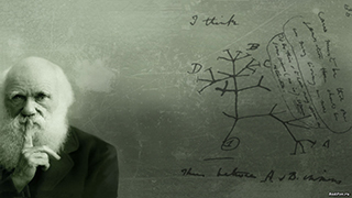
Treating Disease with Fecal Transplantation
Fossils of miniature humans (hobbits) discovered in Indonesia
Paleontology
Dinosaur tail, complete with feathers, found preserved in amber.
Astronomy

Mysterious fast radio burst (FRB) detected in the distant universe.
Big Data & Informatics

Big Data: Buzzword or Big Deal?
Hacking the genome: Identifying anonymized human subjects using publicly available data.