Other Sites:

Bibliography Options Menu

Robert J. Robbins is a biologist, an educator, a science administrator, a publisher, an information technologist, and an IT leader and manager who specializes in advancing biomedical knowledge and supporting education through the application of information technology. More About: RJR | OUR TEAM | OUR SERVICES | THIS WEBSITE
RJR: Recommended Bibliography 16 May 2026 at 02:02 Created:
Squid-Vibrio Symbiosis
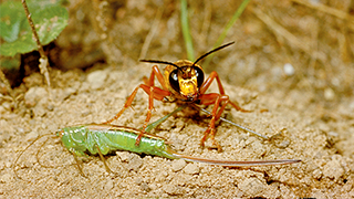
The small bobtail squid (Euprymna scolopes) has a mutually beneficial relationship with bacteria called Vibrio fischeri that live on the squid's underside. The bacteria allow the squid to produce light, which then allows the squid to escape from things that might want to eat it. "The squid emit ventral luminescence that is often very, very close to the quality of light coming from the moon and stars at night," explains Margaret McFall-Ngai, Margaret McFall-Ngai, professor of medical microbiology and immunology at the University of Wisconsin-Madison. For fish looking up from below for something to eat, the squid are camouflaged against the moon or the starlight because they don't cast a shadow. "It's like a 'Klingon' cloaking device," she notes. But the Vibrio fischeri don't stay in the squid continuously. Every day, in response to the light cue of dawn, the squid vents 90 percent of the bacteria back into the seawater. "And then, while it's sitting quiescent in the sand, the bacteria grow up in the crypt so that when [the squid] comes out in the evening, it will have a full complement of luminous Vibrio fischeri," says McFall-Ngai.
Created with PubMed® Query: ( (squid OR euprymna) AND (vibrio OR symbiosis OR symbiotic OR endosymbiont) ) NOT pmcbook NOT ispreviousversion
Citations The Papers (from PubMed®)
RevDate: 2026-05-15
CmpDate: 2026-05-15
Immunomodulatory effect of squid ink extract on lipoxygenase pathways and MIH gene in Litopenaeus vannamei under different salinity condition.
Brazilian journal of biology = Revista brasleira de biologia, 86:e301167 pii:S1519-69842026000100292.
Lipoxygenase (LOX)-derived eicosanoids (12-/15-HETE) and Molt-Inhibiting Hormone (MIH) are key mediators of inflammation and stress physiology in Litopenaeus vannamei during bacterial challenge. Squid ink (Loligo sp.) contains bioactives (betaine, choline, cinnamic acid) with potential immunomodulatory activity. This study aim to evaluate whether dietary squid ink extract modulates MIH responses, LOX-related metabolites, and survival of L. vannamei challenged with Vibrio harveyi under different salinities. Squid ink was methanol-macerated and evaporated to paste; 8 g paste was dissolved in 1 L water and spray-coated onto per kg commercial feed (fed at 5% biomass/day, 4 times/day). Shrimp were reared in 12 aquaria at 24, 27, and 30 ppt, with an infected positive control at 33 ppt, then challenged by immersion with V. harveyi (10^6 CFU/mL). MIH was identified by PCR, 12-HETE/15-HETE were quantified by LC-MS, and ligand-protein interactions were explored by interaction in silico analysis. Squid ink-related treatments reduced 12-HETE from 2.0 ± 0.3 to 0.2-0.5 nmol/mg (ANOVA F = 6.929; p = 0.006), whereas 15-HETE remained near the observed range (5.5-6.0 nmol/mg). MIH amplicons (250-283 bp) showed the strongest band intensity at 30 ppt. Survival improved in supplemented groups (80-90%) relative to the positive control (73.33 ± 5.77%), with the highest survival at 30 ppt (90 ± 10%) (post hoc, p < 0.05). Dietary squid ink extract attenuated the pro-inflammatory 12-LOX pathway and improved post-challenge survival, with responses depending on salinity, supporting squid ink as a natural immunostimulant candidate for shrimp aquaculture.
Additional Links: PMID-42138944
Publisher:
PubMed:
Citation:
show bibtex listing
hide bibtex listing
@article {pmid42138944,
year = {2026},
author = {Fadjar, M and Andayani, S and Kühn, H and Aisyah, D and Ramadhani, AW and Herlina, C},
title = {Immunomodulatory effect of squid ink extract on lipoxygenase pathways and MIH gene in Litopenaeus vannamei under different salinity condition.},
journal = {Brazilian journal of biology = Revista brasleira de biologia},
volume = {86},
number = {},
pages = {e301167},
doi = {10.1590/1519-6984.301167},
pmid = {42138944},
issn = {1678-4375},
mesh = {Animals ; *Penaeidae/microbiology/enzymology/drug effects/immunology/genetics ; *Decapodiformes/chemistry ; Vibrio ; Salinity ; *Lipoxygenase/metabolism/drug effects ; *Immunologic Factors/pharmacology ; },
abstract = {Lipoxygenase (LOX)-derived eicosanoids (12-/15-HETE) and Molt-Inhibiting Hormone (MIH) are key mediators of inflammation and stress physiology in Litopenaeus vannamei during bacterial challenge. Squid ink (Loligo sp.) contains bioactives (betaine, choline, cinnamic acid) with potential immunomodulatory activity. This study aim to evaluate whether dietary squid ink extract modulates MIH responses, LOX-related metabolites, and survival of L. vannamei challenged with Vibrio harveyi under different salinities. Squid ink was methanol-macerated and evaporated to paste; 8 g paste was dissolved in 1 L water and spray-coated onto per kg commercial feed (fed at 5% biomass/day, 4 times/day). Shrimp were reared in 12 aquaria at 24, 27, and 30 ppt, with an infected positive control at 33 ppt, then challenged by immersion with V. harveyi (10^6 CFU/mL). MIH was identified by PCR, 12-HETE/15-HETE were quantified by LC-MS, and ligand-protein interactions were explored by interaction in silico analysis. Squid ink-related treatments reduced 12-HETE from 2.0 ± 0.3 to 0.2-0.5 nmol/mg (ANOVA F = 6.929; p = 0.006), whereas 15-HETE remained near the observed range (5.5-6.0 nmol/mg). MIH amplicons (250-283 bp) showed the strongest band intensity at 30 ppt. Survival improved in supplemented groups (80-90%) relative to the positive control (73.33 ± 5.77%), with the highest survival at 30 ppt (90 ± 10%) (post hoc, p < 0.05). Dietary squid ink extract attenuated the pro-inflammatory 12-LOX pathway and improved post-challenge survival, with responses depending on salinity, supporting squid ink as a natural immunostimulant candidate for shrimp aquaculture.},
}
MeSH Terms:
show MeSH Terms
hide MeSH Terms
Animals
*Penaeidae/microbiology/enzymology/drug effects/immunology/genetics
*Decapodiformes/chemistry
Vibrio
Salinity
*Lipoxygenase/metabolism/drug effects
*Immunologic Factors/pharmacology
RevDate: 2019-12-10
CmpDate: 1978-08-14
A bacteriological study of some frozen and nonfrozen foods.
The Southeast Asian journal of tropical medicine and public health, 8(4):437-446.
Over a period of 19 months, a total of 331 food samples were submitted to the Food Section of the Bacteriology Division for bacteriological examination. These included 184 samples of frozen seafoods from exporters and 147 samples of fresh, nonfrozen foods from food caterers. The total bacterial count for frozen seafoods ranged from 1 x 10(2) to 2.98 x 10(6) per gm with a mean of 2.14 x 10(5) per gm. Coliforms, Escherichia coli and Staphylococcus aureus were present in 48.9%, 3.3% and 8.2% of the samples examined respectively. Two of the cooked prawn samples showed the presence of Vibrio parahaemolyticus. For the fresh, nonfrozen foods, the total bacterial count ranged from 1 x 10(2) to 3.87 x 10(6) per gm with a mean of 2.58 x 10(5) per gm. The examination also showed that 74.8% were coliform positive, 14.9% were E. coli positive, and 4.8% were S. aureus positive. V. parahaemolyticus was not isolated in any of the samples tested. Other pathogens, namely, Vibrio cholerae, Salmonella and Shigella were not isolated from any of the foods examined. The bacterial levels in these foods were determined and their sanitary and public health significance is discussed.
Additional Links: PMID-351818
PubMed:
Citation:
show bibtex listing
hide bibtex listing
@article {pmid351818,
year = {1977},
author = {Seng, LY and Jegathesan, M},
title = {A bacteriological study of some frozen and nonfrozen foods.},
journal = {The Southeast Asian journal of tropical medicine and public health},
volume = {8},
number = {4},
pages = {437-446},
pmid = {351818},
issn = {0125-1562},
mesh = {Animals ; Decapoda ; Decapodiformes ; Escherichia coli/isolation & purification ; Food Handling/*standards ; *Food Microbiology ; Frozen Foods ; Humans ; Public Health ; Staphylococcus aureus/isolation & purification ; Vibrio parahaemolyticus/isolation & purification ; },
abstract = {Over a period of 19 months, a total of 331 food samples were submitted to the Food Section of the Bacteriology Division for bacteriological examination. These included 184 samples of frozen seafoods from exporters and 147 samples of fresh, nonfrozen foods from food caterers. The total bacterial count for frozen seafoods ranged from 1 x 10(2) to 2.98 x 10(6) per gm with a mean of 2.14 x 10(5) per gm. Coliforms, Escherichia coli and Staphylococcus aureus were present in 48.9%, 3.3% and 8.2% of the samples examined respectively. Two of the cooked prawn samples showed the presence of Vibrio parahaemolyticus. For the fresh, nonfrozen foods, the total bacterial count ranged from 1 x 10(2) to 3.87 x 10(6) per gm with a mean of 2.58 x 10(5) per gm. The examination also showed that 74.8% were coliform positive, 14.9% were E. coli positive, and 4.8% were S. aureus positive. V. parahaemolyticus was not isolated in any of the samples tested. Other pathogens, namely, Vibrio cholerae, Salmonella and Shigella were not isolated from any of the foods examined. The bacterial levels in these foods were determined and their sanitary and public health significance is discussed.},
}
MeSH Terms:
show MeSH Terms
hide MeSH Terms
Animals
Decapoda
Decapodiformes
Escherichia coli/isolation & purification
Food Handling/*standards
*Food Microbiology
Frozen Foods
Humans
Public Health
Staphylococcus aureus/isolation & purification
Vibrio parahaemolyticus/isolation & purification
RevDate: 2019-05-08
CmpDate: 1992-08-07
Physical and functional maps of the luminescence gene cluster in an autoinducer-deficient Vibrio fischeri strain isolated from a squid light organ.
Journal of bacteriology, 174(13):4384-4390.
Vibrio fischeri ES114 is an isolate representing the specific bacterial light organ symbiont of the squid Euprymna scolopes. An interesting feature of this strain of V. fischeri is that it is visibly luminous within the light organ of the squid host but is nonluminous when grown under standard laboratory conditions. Luminescence can be restored in laboratory culture, however, by the addition of autoinducer, a species-specific inducer of the V. fischeri luminescence (lux) genes. Most other isolates of V. fischeri produce autoinducer in sufficient quantities to induce luminescence in laboratory culture. We have cloned an 8.8-kb DNA fragment from V. fischeri ES114 that encodes all of the functions necessary for luminescence in Escherichia coli in the absence of exogenous autoinducer. This DNA contains both of the recognized V. fischeri lux regulatory genes, one of which (luxI) directs E. coli to synthesize autoinducer. The organization of the individual lux genes within this DNA fragment appears to be the same as that in the other strains of V. fischeri studied; the restriction map of the V. fischeri ES114 lux DNA has diverged substantially, however, from the largely conserved maps of V. fischeri MJ1 and ATCC 7744. Although E. coli containing the V. fischeri ES114 lux DNA synthesizes considerable amounts of autoinducer, V. fischeri ES114 synthesizes autoinducer only in small amounts, even when transcription of the lux genes, including luxI, is activated by the addition of exogenous autoinducer. Nonetheless, transconjugants of V. fischeri ES114 that contain multicopy plasmids bearing the ES114 lux genes synthesize sufficient autoinducer to induce luminescence. These results suggest that V. fischeri ES11r does not lack a functional luxl, nor is it deficient in the ability to synthesize metabolic precursors for autoinducer synthesis.
Additional Links: PMID-1624432
PubMed:
Citation:
show bibtex listing
hide bibtex listing
@article {pmid1624432,
year = {1992},
author = {Gray, KM and Greenberg, EP},
title = {Physical and functional maps of the luminescence gene cluster in an autoinducer-deficient Vibrio fischeri strain isolated from a squid light organ.},
journal = {Journal of bacteriology},
volume = {174},
number = {13},
pages = {4384-4390},
pmid = {1624432},
issn = {0021-9193},
mesh = {4-Butyrolactone/*analogs & derivatives/metabolism/pharmacology ; Animals ; Base Sequence ; Chromosomes, Bacterial ; Cloning, Molecular ; DNA, Bacterial/genetics/isolation & purification ; Decapodiformes/microbiology ; Escherichia coli/genetics/growth & development/physiology ; *Genes, Bacterial/drug effects ; Luminescent Measurements ; Molecular Sequence Data ; *Multigene Family/drug effects ; Oligonucleotide Probes ; Plasmids ; Restriction Mapping ; Vibrio/*genetics/isolation & purification/physiology ; },
abstract = {Vibrio fischeri ES114 is an isolate representing the specific bacterial light organ symbiont of the squid Euprymna scolopes. An interesting feature of this strain of V. fischeri is that it is visibly luminous within the light organ of the squid host but is nonluminous when grown under standard laboratory conditions. Luminescence can be restored in laboratory culture, however, by the addition of autoinducer, a species-specific inducer of the V. fischeri luminescence (lux) genes. Most other isolates of V. fischeri produce autoinducer in sufficient quantities to induce luminescence in laboratory culture. We have cloned an 8.8-kb DNA fragment from V. fischeri ES114 that encodes all of the functions necessary for luminescence in Escherichia coli in the absence of exogenous autoinducer. This DNA contains both of the recognized V. fischeri lux regulatory genes, one of which (luxI) directs E. coli to synthesize autoinducer. The organization of the individual lux genes within this DNA fragment appears to be the same as that in the other strains of V. fischeri studied; the restriction map of the V. fischeri ES114 lux DNA has diverged substantially, however, from the largely conserved maps of V. fischeri MJ1 and ATCC 7744. Although E. coli containing the V. fischeri ES114 lux DNA synthesizes considerable amounts of autoinducer, V. fischeri ES114 synthesizes autoinducer only in small amounts, even when transcription of the lux genes, including luxI, is activated by the addition of exogenous autoinducer. Nonetheless, transconjugants of V. fischeri ES114 that contain multicopy plasmids bearing the ES114 lux genes synthesize sufficient autoinducer to induce luminescence. These results suggest that V. fischeri ES11r does not lack a functional luxl, nor is it deficient in the ability to synthesize metabolic precursors for autoinducer synthesis.},
}
MeSH Terms:
show MeSH Terms
hide MeSH Terms
4-Butyrolactone/*analogs & derivatives/metabolism/pharmacology
Animals
Base Sequence
Chromosomes, Bacterial
Cloning, Molecular
DNA, Bacterial/genetics/isolation & purification
Decapodiformes/microbiology
Escherichia coli/genetics/growth & development/physiology
*Genes, Bacterial/drug effects
Luminescent Measurements
Molecular Sequence Data
*Multigene Family/drug effects
Oligonucleotide Probes
Plasmids
Restriction Mapping
Vibrio/*genetics/isolation & purification/physiology
RevDate: 2019-05-08
CmpDate: 1992-08-14
A squid that glows in the night: development of an animal-bacterial mutualism.
Journal of bacteriology, 174(15):4865-4870.
Additional Links: PMID-1629148
PubMed:
Citation:
show bibtex listing
hide bibtex listing
@article {pmid1629148,
year = {1992},
author = {Ruby, EG and McFall-Ngai, MJ},
title = {A squid that glows in the night: development of an animal-bacterial mutualism.},
journal = {Journal of bacteriology},
volume = {174},
number = {15},
pages = {4865-4870},
pmid = {1629148},
issn = {0021-9193},
mesh = {Animals ; Decapodiformes/*physiology ; Light ; *Symbiosis ; Vibrio/*physiology ; },
}
MeSH Terms:
show MeSH Terms
hide MeSH Terms
Animals
Decapodiformes/*physiology
Light
*Symbiosis
Vibrio/*physiology
RevDate: 2019-06-18
CmpDate: 1992-01-09
Symbiont recognition and subsequent morphogenesis as early events in an animal-bacterial mutualism.
Science (New York, N.Y.), 254(5037):1491-1494.
Bacterial colonization of the developing light organ of the squid Euprymna scolopes is shown to be highly specific, with the establishment of a successful association resulting only when the juvenile host is exposed to seawater containing one of a subset of Vibrio fischeri strains. Before a symbiotic infection the organ has elaborate epithelial structures covered with cilia and microvilli that are involved in the transfer of bacteria to the incipient symbiotic tissue. These structures regressed within days following infection; however, they were retained in uninfected animals, suggesting that the initiation of symbiosis influences, and is perhaps a prerequisite for, the normal developmental program of the juvenile host.
Additional Links: PMID-1962208
Publisher:
PubMed:
Citation:
show bibtex listing
hide bibtex listing
@article {pmid1962208,
year = {1991},
author = {McFall-Ngai, MJ and Ruby, EG},
title = {Symbiont recognition and subsequent morphogenesis as early events in an animal-bacterial mutualism.},
journal = {Science (New York, N.Y.)},
volume = {254},
number = {5037},
pages = {1491-1494},
doi = {10.1126/science.1962208},
pmid = {1962208},
issn = {0036-8075},
mesh = {Animals ; Decapodiformes/anatomy & histology/growth & development/*microbiology ; Luminescence ; *Symbiosis ; Vibrio/*physiology ; },
abstract = {Bacterial colonization of the developing light organ of the squid Euprymna scolopes is shown to be highly specific, with the establishment of a successful association resulting only when the juvenile host is exposed to seawater containing one of a subset of Vibrio fischeri strains. Before a symbiotic infection the organ has elaborate epithelial structures covered with cilia and microvilli that are involved in the transfer of bacteria to the incipient symbiotic tissue. These structures regressed within days following infection; however, they were retained in uninfected animals, suggesting that the initiation of symbiosis influences, and is perhaps a prerequisite for, the normal developmental program of the juvenile host.},
}
MeSH Terms:
show MeSH Terms
hide MeSH Terms
Animals
Decapodiformes/anatomy & histology/growth & development/*microbiology
Luminescence
*Symbiosis
Vibrio/*physiology
RevDate: 2023-11-27
CmpDate: 1990-08-07
Depressed light emission by symbiotic Vibrio fischeri of the sepiolid squid Euprymna scolopes.
Journal of bacteriology, 172(7):3701-3706.
Bioluminescent marine bacteria of the species Vibrio fischeri are the specific light organ symbionts of the sepiolid squid Euprymna scolopes. Although they share morphological and physiological characteristics with other strains of V. fischeri, when cultured away from the light organ association the E. scolopes symbionts depress their maximal luminescence over 1,000-fold. The primary cause of this reduced luminescence is the underproduction by these bacteria of luciferase autoinducer, a molecule involved in the positive transcriptional regulation of the V. fischeri lux operon. Such an absence of visible light production outside of the symbiotic association has not been previously reported among light organ symbionts of this or any other species of luminous bacteria. Levels of luminescence approaching those of the E. scolopes bacteria in the intact association can be restored by the addition of exogenous autoinducer to bacteria in laboratory culture and are affected by the presence of cyclic AMP. We conclude that some condition(s) specific to the internal environment of the light organ is necessary for maximal autoinduction of luminescence in the symbionts of this squid-bacterial association.
Additional Links: PMID-2163384
PubMed:
Citation:
show bibtex listing
hide bibtex listing
@article {pmid2163384,
year = {1990},
author = {Boettcher, KJ and Ruby, EG},
title = {Depressed light emission by symbiotic Vibrio fischeri of the sepiolid squid Euprymna scolopes.},
journal = {Journal of bacteriology},
volume = {172},
number = {7},
pages = {3701-3706},
pmid = {2163384},
issn = {0021-9193},
mesh = {Animals ; Culture Media ; Cyclic AMP/pharmacology ; Decapodiformes/microbiology ; Light ; Luminescent Measurements ; Species Specificity ; Symbiosis ; Vibrio/growth & development/isolation & purification/*physiology ; },
abstract = {Bioluminescent marine bacteria of the species Vibrio fischeri are the specific light organ symbionts of the sepiolid squid Euprymna scolopes. Although they share morphological and physiological characteristics with other strains of V. fischeri, when cultured away from the light organ association the E. scolopes symbionts depress their maximal luminescence over 1,000-fold. The primary cause of this reduced luminescence is the underproduction by these bacteria of luciferase autoinducer, a molecule involved in the positive transcriptional regulation of the V. fischeri lux operon. Such an absence of visible light production outside of the symbiotic association has not been previously reported among light organ symbionts of this or any other species of luminous bacteria. Levels of luminescence approaching those of the E. scolopes bacteria in the intact association can be restored by the addition of exogenous autoinducer to bacteria in laboratory culture and are affected by the presence of cyclic AMP. We conclude that some condition(s) specific to the internal environment of the light organ is necessary for maximal autoinduction of luminescence in the symbionts of this squid-bacterial association.},
}
MeSH Terms:
show MeSH Terms
hide MeSH Terms
Animals
Culture Media
Cyclic AMP/pharmacology
Decapodiformes/microbiology
Light
Luminescent Measurements
Species Specificity
Symbiosis
Vibrio/growth & development/isolation & purification/*physiology
RevDate: 2021-05-26
CmpDate: 1986-03-10
Characterization and distribution of Vibrio alginolyticus and Vibrio parahaemolyticus isolated in Indonesia.
Applied and environmental microbiology, 50(6):1388-1394.
Previous studies have shown that Vibrio alginolyticus and Vibrio parahaemolyticus can be isolated from similar types of marine samples. In this report, the results of an examination of 567 V. alginolyticus and V. parahaemolyticus strains, isolated from seawater in Jakarta Bay and from more than 30 types of seafood from markets in Jakarta, Indonesia, are presented. Most isolates were from mackerel, shrimp, or squid. Numerical taxonomic analyses clustered 337 isolates and three V. alginolyticus reference strains at S greater than or equal to 80%. These strains produced acid from sucrose, but only approximately 80% produced acetoin or grew in the presence of 10% NaCl. The frequency of occurrence of V. alginolyticus in seawater samples ranged from 0% (in February and March 1972) to 100% (in September and December 1972) and was highest in seafood samples from August to December 1972. A second cluster of 230 isolates and seven V. parahaemolyticus reference strains was observed at S greater than or equal to 82%. These strains did not produce acetoin or acid from sucrose, and approximately 20% grew in the presence of 10% NaCl. V. parahaemolyticus was detected in seawater samples each month, with the highest frequency of occurrence (83.3%) in May 1972. Twenty-nine K antigen serotypes were demonstrated in V. parahaemolyticus isolates, and another 40% were untypable. The modal antibiotic resistance pattern for each species included five drugs. Only 12% of the V. parahaemolyticus strains were Kanagawa positive, and 10% elicited fluid accumulation in ligated rabbit ileal loops. All of the 7 V. alginolyticus strains and 94 (70%) of the V. parahaemolyticus strains tested killed mice when inoculated intraperitoneally.(ABSTRACT TRUNCATED AT 250 WORDS)
Additional Links: PMID-4091566
PubMed:
Citation:
show bibtex listing
hide bibtex listing
@article {pmid4091566,
year = {1985},
author = {Molitoris, E and Joseph, SW and Krichevsky, MI and Sindhuhardja, W and Colwell, RR},
title = {Characterization and distribution of Vibrio alginolyticus and Vibrio parahaemolyticus isolated in Indonesia.},
journal = {Applied and environmental microbiology},
volume = {50},
number = {6},
pages = {1388-1394},
pmid = {4091566},
issn = {0099-2240},
mesh = {Animals ; Anti-Bacterial Agents/pharmacology ; Drug Resistance, Microbial ; Fishes ; Geography ; Indonesia ; Mollusca/microbiology ; Seasons ; Species Specificity ; Vibrio/classification/drug effects/*isolation & purification ; },
abstract = {Previous studies have shown that Vibrio alginolyticus and Vibrio parahaemolyticus can be isolated from similar types of marine samples. In this report, the results of an examination of 567 V. alginolyticus and V. parahaemolyticus strains, isolated from seawater in Jakarta Bay and from more than 30 types of seafood from markets in Jakarta, Indonesia, are presented. Most isolates were from mackerel, shrimp, or squid. Numerical taxonomic analyses clustered 337 isolates and three V. alginolyticus reference strains at S greater than or equal to 80%. These strains produced acid from sucrose, but only approximately 80% produced acetoin or grew in the presence of 10% NaCl. The frequency of occurrence of V. alginolyticus in seawater samples ranged from 0% (in February and March 1972) to 100% (in September and December 1972) and was highest in seafood samples from August to December 1972. A second cluster of 230 isolates and seven V. parahaemolyticus reference strains was observed at S greater than or equal to 82%. These strains did not produce acetoin or acid from sucrose, and approximately 20% grew in the presence of 10% NaCl. V. parahaemolyticus was detected in seawater samples each month, with the highest frequency of occurrence (83.3%) in May 1972. Twenty-nine K antigen serotypes were demonstrated in V. parahaemolyticus isolates, and another 40% were untypable. The modal antibiotic resistance pattern for each species included five drugs. Only 12% of the V. parahaemolyticus strains were Kanagawa positive, and 10% elicited fluid accumulation in ligated rabbit ileal loops. All of the 7 V. alginolyticus strains and 94 (70%) of the V. parahaemolyticus strains tested killed mice when inoculated intraperitoneally.(ABSTRACT TRUNCATED AT 250 WORDS)},
}
MeSH Terms:
show MeSH Terms
hide MeSH Terms
Animals
Anti-Bacterial Agents/pharmacology
Drug Resistance, Microbial
Fishes
Geography
Indonesia
Mollusca/microbiology
Seasons
Species Specificity
Vibrio/classification/drug effects/*isolation & purification
RevDate: 2019-05-11
CmpDate: 1984-08-20
A common source foodborne outbreak of cholera in Singapore.
International journal of epidemiology, 13(2):210-215.
An epidemiological investigation of an outbreak of Vibrio cholerae 01, biotype El Tor, serotype Ogawa, phage type 1, confined to a group of foreign construction workers in Singapore is described. A total of 22 workers were confirmed to have cholera and another 15 had asymptomatic Vibrio cholerae 01 infection between 3 November and 11 November 1982. The source of infection was traced to contaminated seafood prepared at the construction site canteen where two food handlers were found to be infected with V. cholerae 01 (one symptomatic and the other asymptomatic). The incubation period of cholera in this outbreak ranged from 4 to 203 hours with a median of 38 hours. Only two workers had moderate to severe dehydration and required intravenous therapy. Early recognition of the outbreak and prompt implementation of control measures prevented the outbreak from spreading to other parts of Singapore.
Additional Links: PMID-6735567
Publisher:
PubMed:
Citation:
show bibtex listing
hide bibtex listing
@article {pmid6735567,
year = {1984},
author = {Goh, KT and Lam, S and Kumarapathy, S and Tan, JL},
title = {A common source foodborne outbreak of cholera in Singapore.},
journal = {International journal of epidemiology},
volume = {13},
number = {2},
pages = {210-215},
doi = {10.1093/ije/13.2.210},
pmid = {6735567},
issn = {0300-5771},
mesh = {Adolescent ; Adult ; Cholera/*epidemiology/transmission ; Decapodiformes/microbiology ; *Disease Outbreaks ; Female ; Food Handling ; *Food Microbiology ; Humans ; Male ; Singapore ; Transients and Migrants ; Vibrio cholerae ; },
abstract = {An epidemiological investigation of an outbreak of Vibrio cholerae 01, biotype El Tor, serotype Ogawa, phage type 1, confined to a group of foreign construction workers in Singapore is described. A total of 22 workers were confirmed to have cholera and another 15 had asymptomatic Vibrio cholerae 01 infection between 3 November and 11 November 1982. The source of infection was traced to contaminated seafood prepared at the construction site canteen where two food handlers were found to be infected with V. cholerae 01 (one symptomatic and the other asymptomatic). The incubation period of cholera in this outbreak ranged from 4 to 203 hours with a median of 38 hours. Only two workers had moderate to severe dehydration and required intravenous therapy. Early recognition of the outbreak and prompt implementation of control measures prevented the outbreak from spreading to other parts of Singapore.},
}
MeSH Terms:
show MeSH Terms
hide MeSH Terms
Adolescent
Adult
Cholera/*epidemiology/transmission
Decapodiformes/microbiology
*Disease Outbreaks
Female
Food Handling
*Food Microbiology
Humans
Male
Singapore
Transients and Migrants
Vibrio cholerae
RevDate: 2019-08-21
CmpDate: 1983-06-23
Immunochemical comparisons among lipopolysaccharides from symbiotic luminous bacteria isolated from several luminous marine animals.
Microbiology and immunology, 26(12):1181-1186.
Additional Links: PMID-6820471
Publisher:
PubMed:
Citation:
show bibtex listing
hide bibtex listing
@article {pmid6820471,
year = {1982},
author = {Kuwae, T and Fukasawa, S and Sasaki, T and Kurata, M},
title = {Immunochemical comparisons among lipopolysaccharides from symbiotic luminous bacteria isolated from several luminous marine animals.},
journal = {Microbiology and immunology},
volume = {26},
number = {12},
pages = {1181-1186},
doi = {10.1111/j.1348-0421.1982.tb00267.x},
pmid = {6820471},
issn = {0385-5600},
mesh = {Animals ; Chemical Precipitation ; Decapodiformes/microbiology ; Fishes/*microbiology ; Immune Sera/pharmacology ; Immunodiffusion ; Immunoelectrophoresis ; *Light ; Lipopolysaccharides/*analysis ; Photobacterium/analysis/immunology ; Rabbits ; Symbiosis ; Vibrio/analysis/immunology ; },
}
MeSH Terms:
show MeSH Terms
hide MeSH Terms
Animals
Chemical Precipitation
Decapodiformes/microbiology
Fishes/*microbiology
Immune Sera/pharmacology
Immunodiffusion
Immunoelectrophoresis
*Light
Lipopolysaccharides/*analysis
Photobacterium/analysis/immunology
Rabbits
Symbiosis
Vibrio/analysis/immunology
RevDate: 2019-08-21
CmpDate: 1983-08-11
Chemical and biological properties of Lipopolysaccharides from symbiotic luminous bacteria from several luminous marine animals.
Microbiology and immunology, 27(2):137-149.
The chemical and biological properties of lipopolysaccharides (LPS) in five strains of symbiotic luminous bacteria isolated from four species of luminous marine fishes, Coelorhynchus kishinouyei (CK-1), Chlorophthalmus albatrossis (CA-1), Ventrifossa garmani (VG-1), and Acropoma japonicum (AJ-1b), as well as from a luminous squid, Doryteuthis kensaki (DK-1) were examined. The LPS isolated from these symbiotic luminous bacteria were characterized by the absence of 2-keto-3-deoxyoctonate, known to be a basic component of the usual gram-negative bacterial LPS. All LPS from these symbiotic luminous bacteria upon electrophoresis in sodium dodecylsulfate polyacrylamide gel exhibited one or two clear main bands with high mobility, and one or two obscure minor bands with low mobility when stained with periodate-Schiff reagent. LPS from CA-1 and VG-1 exhibited similar electrophoretic patterns, whereas the electrophoretic patterns of the LPS from CK-1, AJ-1b, and DK-1 were easily distinguishable from each other. All these LPS also had similarly potent and diverse biological activities in regard to their adjuvanticity, immunosuppression, polyclonal effect, B-cell mitogenicity, and activation of the phagocytic function of macrophages.
Additional Links: PMID-6865804
Publisher:
PubMed:
Citation:
show bibtex listing
hide bibtex listing
@article {pmid6865804,
year = {1983},
author = {Kuwae, T and Kurata, M},
title = {Chemical and biological properties of Lipopolysaccharides from symbiotic luminous bacteria from several luminous marine animals.},
journal = {Microbiology and immunology},
volume = {27},
number = {2},
pages = {137-149},
doi = {10.1111/j.1348-0421.1983.tb03578.x},
pmid = {6865804},
issn = {0385-5600},
mesh = {Adjuvants, Immunologic ; Animals ; Antibody Formation ; Electrophoresis, Polyacrylamide Gel ; Enterobacteriaceae/*physiology ; Immune Tolerance ; Lipopolysaccharides/analysis/*physiology ; *Luminescent Measurements ; Lymphocyte Activation ; Macrophages/immunology ; Mice ; Phagocytosis ; Polysaccharides, Bacterial/*physiology ; },
abstract = {The chemical and biological properties of lipopolysaccharides (LPS) in five strains of symbiotic luminous bacteria isolated from four species of luminous marine fishes, Coelorhynchus kishinouyei (CK-1), Chlorophthalmus albatrossis (CA-1), Ventrifossa garmani (VG-1), and Acropoma japonicum (AJ-1b), as well as from a luminous squid, Doryteuthis kensaki (DK-1) were examined. The LPS isolated from these symbiotic luminous bacteria were characterized by the absence of 2-keto-3-deoxyoctonate, known to be a basic component of the usual gram-negative bacterial LPS. All LPS from these symbiotic luminous bacteria upon electrophoresis in sodium dodecylsulfate polyacrylamide gel exhibited one or two clear main bands with high mobility, and one or two obscure minor bands with low mobility when stained with periodate-Schiff reagent. LPS from CA-1 and VG-1 exhibited similar electrophoretic patterns, whereas the electrophoretic patterns of the LPS from CK-1, AJ-1b, and DK-1 were easily distinguishable from each other. All these LPS also had similarly potent and diverse biological activities in regard to their adjuvanticity, immunosuppression, polyclonal effect, B-cell mitogenicity, and activation of the phagocytic function of macrophages.},
}
MeSH Terms:
show MeSH Terms
hide MeSH Terms
Adjuvants, Immunologic
Animals
Antibody Formation
Electrophoresis, Polyacrylamide Gel
Enterobacteriaceae/*physiology
Immune Tolerance
Lipopolysaccharides/analysis/*physiology
*Luminescent Measurements
Lymphocyte Activation
Macrophages/immunology
Mice
Phagocytosis
Polysaccharides, Bacterial/*physiology
RevDate: 2019-06-16
CmpDate: 1981-08-20
Co-evolution of luminous bacteria and their eukaryotic hosts.
Annals of the New York Academy of Sciences, 361:76-91.
Several species of three genera of luminous bacteria from marine and soil environments are known to form specific symbioses with fish, squid, urochordates, and nematodes. These bacteria contain a unique and easily detectable enzyme, bacterial luciferase, which allows the detection of the bacteria even when they cannot be cultured from animal tissue. The bacteria-animal associations vary; they range from transient, nonspecific gut symbionts in many fishes to highly specific, nonculturable, intracellular symbionts in pyrosomes. The study of these microbe-animal symbioses may allow understanding of the alterations that occur in the partners during the establishment of intracellular organelles. These studies are incomplete, in particular the level of partner integration, the nature of the metabolic exchange, the mechanism of transmission of symbionts to offspring, and the identification of the cellular inclusions as modified bacteria are not always known. However, the outline of what is likely to be a continuous evolutionary sequence of luminous bacterial symbionts with their various animal hosts is becoming clear. As more of these symbioses are studied, we anticipate that new systems will be found that represent states between those described here, and that the luminous bacteria will provide a living model for the gradual evolution from free-living microbes in intracellular organelles.
Additional Links: PMID-6941738
Publisher:
PubMed:
Citation:
show bibtex listing
hide bibtex listing
@article {pmid6941738,
year = {1981},
author = {Nealson, K and Cohn, D and Leisman, G and Tebo, B},
title = {Co-evolution of luminous bacteria and their eukaryotic hosts.},
journal = {Annals of the New York Academy of Sciences},
volume = {361},
number = {},
pages = {76-91},
doi = {10.1111/j.1749-6632.1981.tb46512.x},
pmid = {6941738},
issn = {0077-8923},
mesh = {*Bacteria ; *Biological Evolution ; *Cells ; *Eukaryotic Cells ; Light ; Luminescent Measurements ; Organoids ; Symbiosis ; },
abstract = {Several species of three genera of luminous bacteria from marine and soil environments are known to form specific symbioses with fish, squid, urochordates, and nematodes. These bacteria contain a unique and easily detectable enzyme, bacterial luciferase, which allows the detection of the bacteria even when they cannot be cultured from animal tissue. The bacteria-animal associations vary; they range from transient, nonspecific gut symbionts in many fishes to highly specific, nonculturable, intracellular symbionts in pyrosomes. The study of these microbe-animal symbioses may allow understanding of the alterations that occur in the partners during the establishment of intracellular organelles. These studies are incomplete, in particular the level of partner integration, the nature of the metabolic exchange, the mechanism of transmission of symbionts to offspring, and the identification of the cellular inclusions as modified bacteria are not always known. However, the outline of what is likely to be a continuous evolutionary sequence of luminous bacterial symbionts with their various animal hosts is becoming clear. As more of these symbioses are studied, we anticipate that new systems will be found that represent states between those described here, and that the luminous bacteria will provide a living model for the gradual evolution from free-living microbes in intracellular organelles.},
}
MeSH Terms:
show MeSH Terms
hide MeSH Terms
*Bacteria
*Biological Evolution
*Cells
*Eukaryotic Cells
Light
Luminescent Measurements
Organoids
Symbiosis
RevDate: 2019-05-08
CmpDate: 1995-03-17
Detection and quantification of Vibrio fischeri autoinducer from symbiotic squid light organs.
Journal of bacteriology, 177(4):1053-1058.
Vibrio fischeri is the specific light organ symbiont of the sepiolid squid species Euprymna scolopes and Euprymna morsei. Both species of squid are luminescent by virtue of their bacterial symbionts, but the natural symbionts of E. scolopes do not produce visible luminescence in laboratory culture. The primary cause of this depressed luminescence by E. scolopes symbionts in culture was found to be the production of relatively low levels of V. fischeri autoinducer, a positive transcriptional coregulator of the lux regulon, identified as N-(3-oxohexanoyl) homoserine lactone. Concentrations of autoinducer activity produced by these symbionts in culture were quantified and found to be at least 10-fold lower than those produced by E. morsei isolates (which are visibly luminous outside the association) and perhaps 10,000-fold lower than those of the brightest V. fischeri strains. Despite the differences in their symbiont strains, the intact light organs of the two species of squid contained comparable amounts of extractable autoinducer activity (between 100 and 200 pg per adult animal). The chromatographic behavior of this autoinducer activity on reverse-phase high-performance liquid chromatography was consistent with its presumptive identification as V. fischeri autoinducer. Within the 5-microliter volume of the epithelial core of the light organ in which the symbiotic V. fischeri strains are housed, these amounts would result in an effective autoinducer concentration of at least 100 nM. Because these levels are over 40-fold higher than the concentration needed for the induction of luminescence of bacteria in culture, we conclude that the inherent degree of autoinducer production by strains of V. fischeri may not influence their effectiveness as light organ symbionts. Furthermore, this study provides the first direct evidence that the phenomenon of cell density-dependent autoinduction, discovered and described first for laboratory cultures of V. fischeri but believed to be a general phenomenon in many species of host-associated symbionts and pathogens, is in fact a consequence of bacterial colonizations of host tissues.
Additional Links: PMID-7860584
PubMed:
Citation:
show bibtex listing
hide bibtex listing
@article {pmid7860584,
year = {1995},
author = {Boettcher, KJ and Ruby, EG},
title = {Detection and quantification of Vibrio fischeri autoinducer from symbiotic squid light organs.},
journal = {Journal of bacteriology},
volume = {177},
number = {4},
pages = {1053-1058},
pmid = {7860584},
issn = {0021-9193},
mesh = {4-Butyrolactone/*analogs & derivatives/analysis ; Animals ; Biological Assay ; Decapodiformes/anatomy & histology/*microbiology ; Diffusion ; Epithelium/physiology ; *Luminescent Measurements ; Symbiosis/*physiology ; Vibrio/*physiology ; },
abstract = {Vibrio fischeri is the specific light organ symbiont of the sepiolid squid species Euprymna scolopes and Euprymna morsei. Both species of squid are luminescent by virtue of their bacterial symbionts, but the natural symbionts of E. scolopes do not produce visible luminescence in laboratory culture. The primary cause of this depressed luminescence by E. scolopes symbionts in culture was found to be the production of relatively low levels of V. fischeri autoinducer, a positive transcriptional coregulator of the lux regulon, identified as N-(3-oxohexanoyl) homoserine lactone. Concentrations of autoinducer activity produced by these symbionts in culture were quantified and found to be at least 10-fold lower than those produced by E. morsei isolates (which are visibly luminous outside the association) and perhaps 10,000-fold lower than those of the brightest V. fischeri strains. Despite the differences in their symbiont strains, the intact light organs of the two species of squid contained comparable amounts of extractable autoinducer activity (between 100 and 200 pg per adult animal). The chromatographic behavior of this autoinducer activity on reverse-phase high-performance liquid chromatography was consistent with its presumptive identification as V. fischeri autoinducer. Within the 5-microliter volume of the epithelial core of the light organ in which the symbiotic V. fischeri strains are housed, these amounts would result in an effective autoinducer concentration of at least 100 nM. Because these levels are over 40-fold higher than the concentration needed for the induction of luminescence of bacteria in culture, we conclude that the inherent degree of autoinducer production by strains of V. fischeri may not influence their effectiveness as light organ symbionts. Furthermore, this study provides the first direct evidence that the phenomenon of cell density-dependent autoinduction, discovered and described first for laboratory cultures of V. fischeri but believed to be a general phenomenon in many species of host-associated symbionts and pathogens, is in fact a consequence of bacterial colonizations of host tissues.},
}
MeSH Terms:
show MeSH Terms
hide MeSH Terms
4-Butyrolactone/*analogs & derivatives/analysis
Animals
Biological Assay
Decapodiformes/anatomy & histology/*microbiology
Diffusion
Epithelium/physiology
*Luminescent Measurements
Symbiosis/*physiology
Vibrio/*physiology
RevDate: 2022-02-15
CmpDate: 1994-11-01
Bacterial symbionts induce host organ morphogenesis during early postembryonic development of the squid Euprymna scolopes.
Development (Cambridge, England), 120(7):1719-1729.
The mutualistic association between the squid Euprymna scolopes and the bacterium Vibrio fischeri is an emerging experimental system for the study of the influence of bacteria on animal development. Taking advantage of the ability to raise both this host and its microbial partner independently under laboratory conditions, we describe the effects of bacterial interactions on morphogenesis of the juvenile host symbiotic organ. Our results show that bacteria are essential for normal postembryonic development of the symbiotic organ, which involves changes in both the surface epithelium and the epithelial tissue within the organ where the bacterial culture will take up residence. Cell death induced by exposure to symbiotic V. fischeri results in the regression of a complex ciliated surface epithelium, a tissue that apparently functions to facilitate inoculation of the juvenile organ with the appropriate specific bacterial species. Regression of this tissue begins within hours of exposure to symbiosis-competent bacteria and progresses over the next 5 days, at which time full regression is complete, resulting in a symbiotic organ whose epithelial surface resembles that of the fully mature organ. Moreover, symbiosis-competent bacteria induce modification of the epithelial cells of the crypts that will house these symbionts; these cells undergo significant changes in shape and size in response to interactions with symbiotic V. fischeri. In contrast, we find that when these tissues are not exposed to the proper bacterial symbionts they remain in a state of arrested morphogenesis, a condition that can be rescued by interactions with symbionts. The results of these studies are the first experimental data demonstrating that a specific bacterial symbiont can play an inductive role in animal development.
Additional Links: PMID-7924980
Publisher:
PubMed:
Citation:
show bibtex listing
hide bibtex listing
@article {pmid7924980,
year = {1994},
author = {Montgomery, MK and McFall-Ngai, M},
title = {Bacterial symbionts induce host organ morphogenesis during early postembryonic development of the squid Euprymna scolopes.},
journal = {Development (Cambridge, England)},
volume = {120},
number = {7},
pages = {1719-1729},
doi = {10.1242/dev.120.7.1719},
pmid = {7924980},
issn = {0950-1991},
mesh = {Animals ; Cell Death ; Cell Differentiation/physiology ; Decapodiformes/*physiology/ultrastructure ; Epithelium/physiology/ultrastructure ; Microscopy, Electron, Scanning ; Models, Biological ; Morphogenesis/physiology ; Symbiosis/*physiology ; Vibrio/*physiology ; },
abstract = {The mutualistic association between the squid Euprymna scolopes and the bacterium Vibrio fischeri is an emerging experimental system for the study of the influence of bacteria on animal development. Taking advantage of the ability to raise both this host and its microbial partner independently under laboratory conditions, we describe the effects of bacterial interactions on morphogenesis of the juvenile host symbiotic organ. Our results show that bacteria are essential for normal postembryonic development of the symbiotic organ, which involves changes in both the surface epithelium and the epithelial tissue within the organ where the bacterial culture will take up residence. Cell death induced by exposure to symbiotic V. fischeri results in the regression of a complex ciliated surface epithelium, a tissue that apparently functions to facilitate inoculation of the juvenile organ with the appropriate specific bacterial species. Regression of this tissue begins within hours of exposure to symbiosis-competent bacteria and progresses over the next 5 days, at which time full regression is complete, resulting in a symbiotic organ whose epithelial surface resembles that of the fully mature organ. Moreover, symbiosis-competent bacteria induce modification of the epithelial cells of the crypts that will house these symbionts; these cells undergo significant changes in shape and size in response to interactions with symbiotic V. fischeri. In contrast, we find that when these tissues are not exposed to the proper bacterial symbionts they remain in a state of arrested morphogenesis, a condition that can be rescued by interactions with symbionts. The results of these studies are the first experimental data demonstrating that a specific bacterial symbiont can play an inductive role in animal development.},
}
MeSH Terms:
show MeSH Terms
hide MeSH Terms
Animals
Cell Death
Cell Differentiation/physiology
Decapodiformes/*physiology/ultrastructure
Epithelium/physiology/ultrastructure
Microscopy, Electron, Scanning
Models, Biological
Morphogenesis/physiology
Symbiosis/*physiology
Vibrio/*physiology
RevDate: 2023-11-27
CmpDate: 1994-12-16
Effect of transposon-induced motility mutations on colonization of the host light organ by Vibrio fischeri.
Journal of bacteriology, 176(22):6986-6991.
Vibrio fischeri is found both as a free-living bacterium in seawater and as the specific, mutualistic light organ symbiont of several fish and squid species. To identify those characteristics of symbiosis-competent strains that are required for successful colonization of the nascent light organ of juvenile Euprymna scolopes squids, we generated a mutant pool by using the transposon Mu dI 1681 and screened this pool for strains that were no longer motile. Eighteen independently isolated nonmotile mutants that were either flagellated or nonflagellated were obtained. In contrast to the parent strain, none of these nonmotile mutants was able to colonize the juvenile squid light organ. The flagellated nonmotile mutant strain NM200 possessed a bundle of sheathed polar flagella indistinguishable from that of the wild-type strain, indicating that the presence of flagella alone is not sufficient for colonization and that it is motility itself that is required for successful light organ colonization. This study identifies motility as the first required symbiotic phenotype of V. fischeri.
Additional Links: PMID-7961462
PubMed:
Citation:
show bibtex listing
hide bibtex listing
@article {pmid7961462,
year = {1994},
author = {Graf, J and Dunlap, PV and Ruby, EG},
title = {Effect of transposon-induced motility mutations on colonization of the host light organ by Vibrio fischeri.},
journal = {Journal of bacteriology},
volume = {176},
number = {22},
pages = {6986-6991},
pmid = {7961462},
issn = {0021-9193},
mesh = {Animals ; Cell Movement/*genetics ; Crosses, Genetic ; Decapodiformes/anatomy & histology/*microbiology ; Flagella/genetics/ultrastructure ; Light ; Mutagenesis, Insertional ; Mutation ; Selection, Genetic ; Symbiosis/genetics/*physiology ; Vibrio/genetics/growth & development/*physiology/ultrastructure ; },
abstract = {Vibrio fischeri is found both as a free-living bacterium in seawater and as the specific, mutualistic light organ symbiont of several fish and squid species. To identify those characteristics of symbiosis-competent strains that are required for successful colonization of the nascent light organ of juvenile Euprymna scolopes squids, we generated a mutant pool by using the transposon Mu dI 1681 and screened this pool for strains that were no longer motile. Eighteen independently isolated nonmotile mutants that were either flagellated or nonflagellated were obtained. In contrast to the parent strain, none of these nonmotile mutants was able to colonize the juvenile squid light organ. The flagellated nonmotile mutant strain NM200 possessed a bundle of sheathed polar flagella indistinguishable from that of the wild-type strain, indicating that the presence of flagella alone is not sufficient for colonization and that it is motility itself that is required for successful light organ colonization. This study identifies motility as the first required symbiotic phenotype of V. fischeri.},
}
MeSH Terms:
show MeSH Terms
hide MeSH Terms
Animals
Cell Movement/*genetics
Crosses, Genetic
Decapodiformes/anatomy & histology/*microbiology
Flagella/genetics/ultrastructure
Light
Mutagenesis, Insertional
Mutation
Selection, Genetic
Symbiosis/genetics/*physiology
Vibrio/genetics/growth & development/*physiology/ultrastructure
RevDate: 2023-11-27
CmpDate: 1994-05-02
Competition between Vibrio fischeri strains during initiation and maintenance of a light organ symbiosis.
Journal of bacteriology, 176(7):1985-1991.
Colonization of the light-emitting organ of the Hawaiian squid Euprymna scolopes is initiated when the nascent organ of a newly hatched squid becomes inoculated with Vibrio fischeri cells present in the ambient seawater. Although they are induced for luminescence in the light organ, these symbiotic strains are characteristically non-visibly luminous (NVL) when grown in laboratory culture. The more typical visibly luminous (VL) type of V. fischeri co-occurs in Hawaiian seawater with these NVL strains; thus, two phenotypically distinct groups of this species potentially have access to the symbiotic niche, yet only the NVL ones are found there. In laboratory inoculation experiments, VL strains, when presented in pure culture, showed the same capability for colonizing the light organ as NVL strains. However, in experiments with mixed cultures composed of both VL and NVL strains, the VL ones were unable to compete with the NVL ones and did not persist within the light organ as the symbiosis became established. In addition, NVL strains entered light organs that had already been colonized by VL strains and displaced them. The mechanism underlying the symbiotic competitiveness exhibited by NVL strains remains unknown; however, it does not appear to be due to a higher potential for siderophore activity. While a difference in luminescence phenotype between VL and NVL strains in culture is not likely to be significant in the symbiosis, it has helped identify two distinct groups of V. fischeri that express different colonization capabilities in the squid light organ. This competitive difference provides a useful indication of important traits in light organ colonization.
Additional Links: PMID-8144466
PubMed:
Citation:
show bibtex listing
hide bibtex listing
@article {pmid8144466,
year = {1994},
author = {Lee, KH and Ruby, EG},
title = {Competition between Vibrio fischeri strains during initiation and maintenance of a light organ symbiosis.},
journal = {Journal of bacteriology},
volume = {176},
number = {7},
pages = {1985-1991},
pmid = {8144466},
issn = {0021-9193},
mesh = {Animals ; Decapodiformes/growth & development/*microbiology ; Genetic Variation ; *Luminescent Measurements ; *Selection, Genetic ; Siderophores/biosynthesis ; *Symbiosis ; Vibrio/classification/genetics/*growth & development/metabolism ; },
abstract = {Colonization of the light-emitting organ of the Hawaiian squid Euprymna scolopes is initiated when the nascent organ of a newly hatched squid becomes inoculated with Vibrio fischeri cells present in the ambient seawater. Although they are induced for luminescence in the light organ, these symbiotic strains are characteristically non-visibly luminous (NVL) when grown in laboratory culture. The more typical visibly luminous (VL) type of V. fischeri co-occurs in Hawaiian seawater with these NVL strains; thus, two phenotypically distinct groups of this species potentially have access to the symbiotic niche, yet only the NVL ones are found there. In laboratory inoculation experiments, VL strains, when presented in pure culture, showed the same capability for colonizing the light organ as NVL strains. However, in experiments with mixed cultures composed of both VL and NVL strains, the VL ones were unable to compete with the NVL ones and did not persist within the light organ as the symbiosis became established. In addition, NVL strains entered light organs that had already been colonized by VL strains and displaced them. The mechanism underlying the symbiotic competitiveness exhibited by NVL strains remains unknown; however, it does not appear to be due to a higher potential for siderophore activity. While a difference in luminescence phenotype between VL and NVL strains in culture is not likely to be significant in the symbiosis, it has helped identify two distinct groups of V. fischeri that express different colonization capabilities in the squid light organ. This competitive difference provides a useful indication of important traits in light organ colonization.},
}
MeSH Terms:
show MeSH Terms
hide MeSH Terms
Animals
Decapodiformes/growth & development/*microbiology
Genetic Variation
*Luminescent Measurements
*Selection, Genetic
Siderophores/biosynthesis
*Symbiosis
Vibrio/classification/genetics/*growth & development/metabolism
RevDate: 2019-05-08
CmpDate: 1993-08-25
Characterization of a periplasmic 3':5'-cyclic nucleotide phosphodiesterase gene, cpdP, from the marine symbiotic bacterium Vibrio fischeri.
Journal of bacteriology, 175(15):4615-4624.
Vibrio fischeri, a marine bacterium that forms a bioluminescent symbiosis with certain fish and squids, exhibits the unusual attribute of growth on 3':5'-cyclic AMP (cAMP), apparently through the activity of a 3':5'-cyclic nucleotide phosphodiesterase (3':5'-CNP) with exceptionally high activity. The V. fischeri 3':5'-CNP is located in the periplasm, a novel cellular location for this enzyme in bacteria. To gain insight into the physiological function of this enzyme, we cloned the gene (designated cpdP) encoding it from V. fischeri MJ-1. This is the first bacterial 3':5'-CNP gene to be cloned. Sequencing and analysis of the 1.26-kb cpdP locus revealed a single open reading frame specifying a protein of 330 amino acid residues, including a 22-amino-acid leader peptide. The putative cpdP promoter contained a reasonable -10 promoter region (TATTAT) but contained no obvious -35 region; instead, a 12-bp inverted repeat (TTAAATATTTAA) occurred just upstream of this location. A possible rho-independent transcriptional terminator with a calculated free energy of -21.2 kcal.mol-1 (ca. -88.7 kJ.mol-1) followed the CpdP protein coding sequence. The predicted subunit molecular weight of 33,636 for the mature CpdP protein (36,087 less 2,451 for the leader peptide) was consistent with the molecular weight of 34,000 estimated by sodium dodecyl sulfate-polyacrylamide gel electrophoresis. The deduced amino acid sequence of the CpdP protein exhibited 30.3% identity with that of the low-affinity 3':5'-CNP (PDE1) of Saccharomyces cerevisiae and 33.6% identity with that of the extracellular 3':5'-CNP of Dictyostelium discoideum. The residue identities clustered in two regions, residues 100 to 146 and 238 to 269, which contained 30 of the 33 amino acids conserved in all three proteins, 4 of which were histidines. A gene replacement mutant of V. fischeri MJ-1 containing a 0.45-kb BglII deletion within the cpdP gene lacked periplasmic 3':5'-CNP activity and did not grow on cAMP, confirming for V. fischeri the relationship among cpdP, synthesis of the periplasmic 3':5'-CNP, and growth on cAMP. The mutant exhibited no obvious sensitivity to high extracellular concentrations of cAMP (5 and 10 mM), suggesting that the enzyme does not play a role in defense against extracellular cAMP.
Additional Links: PMID-8393003
PubMed:
Citation:
show bibtex listing
hide bibtex listing
@article {pmid8393003,
year = {1993},
author = {Dunlap, PV and Callahan, SM},
title = {Characterization of a periplasmic 3':5'-cyclic nucleotide phosphodiesterase gene, cpdP, from the marine symbiotic bacterium Vibrio fischeri.},
journal = {Journal of bacteriology},
volume = {175},
number = {15},
pages = {4615-4624},
pmid = {8393003},
issn = {0021-9193},
mesh = {3',5'-Cyclic-AMP Phosphodiesterases/*genetics ; Amino Acid Sequence ; Base Sequence ; Cloning, Molecular ; Cyclic Nucleotide Phosphodiesterases, Type 1 ; Escherichia coli ; Genes, Bacterial/*genetics ; Molecular Sequence Data ; Mutation/genetics ; Phenotype ; Sequence Analysis ; Sequence Homology, Amino Acid ; Vibrio/*enzymology ; },
abstract = {Vibrio fischeri, a marine bacterium that forms a bioluminescent symbiosis with certain fish and squids, exhibits the unusual attribute of growth on 3':5'-cyclic AMP (cAMP), apparently through the activity of a 3':5'-cyclic nucleotide phosphodiesterase (3':5'-CNP) with exceptionally high activity. The V. fischeri 3':5'-CNP is located in the periplasm, a novel cellular location for this enzyme in bacteria. To gain insight into the physiological function of this enzyme, we cloned the gene (designated cpdP) encoding it from V. fischeri MJ-1. This is the first bacterial 3':5'-CNP gene to be cloned. Sequencing and analysis of the 1.26-kb cpdP locus revealed a single open reading frame specifying a protein of 330 amino acid residues, including a 22-amino-acid leader peptide. The putative cpdP promoter contained a reasonable -10 promoter region (TATTAT) but contained no obvious -35 region; instead, a 12-bp inverted repeat (TTAAATATTTAA) occurred just upstream of this location. A possible rho-independent transcriptional terminator with a calculated free energy of -21.2 kcal.mol-1 (ca. -88.7 kJ.mol-1) followed the CpdP protein coding sequence. The predicted subunit molecular weight of 33,636 for the mature CpdP protein (36,087 less 2,451 for the leader peptide) was consistent with the molecular weight of 34,000 estimated by sodium dodecyl sulfate-polyacrylamide gel electrophoresis. The deduced amino acid sequence of the CpdP protein exhibited 30.3% identity with that of the low-affinity 3':5'-CNP (PDE1) of Saccharomyces cerevisiae and 33.6% identity with that of the extracellular 3':5'-CNP of Dictyostelium discoideum. The residue identities clustered in two regions, residues 100 to 146 and 238 to 269, which contained 30 of the 33 amino acids conserved in all three proteins, 4 of which were histidines. A gene replacement mutant of V. fischeri MJ-1 containing a 0.45-kb BglII deletion within the cpdP gene lacked periplasmic 3':5'-CNP activity and did not grow on cAMP, confirming for V. fischeri the relationship among cpdP, synthesis of the periplasmic 3':5'-CNP, and growth on cAMP. The mutant exhibited no obvious sensitivity to high extracellular concentrations of cAMP (5 and 10 mM), suggesting that the enzyme does not play a role in defense against extracellular cAMP.},
}
MeSH Terms:
show MeSH Terms
hide MeSH Terms
3',5'-Cyclic-AMP Phosphodiesterases/*genetics
Amino Acid Sequence
Base Sequence
Cloning, Molecular
Cyclic Nucleotide Phosphodiesterases, Type 1
Escherichia coli
Genes, Bacterial/*genetics
Molecular Sequence Data
Mutation/genetics
Phenotype
Sequence Analysis
Sequence Homology, Amino Acid
Vibrio/*enzymology
RevDate: 2023-11-27
CmpDate: 1993-03-22
Growth and flagellation of Vibrio fischeri during initiation of the sepiolid squid light organ symbiosis.
Archives of microbiology, 159(2):160-167.
A pure culture of the luminous bacterium Vibrio fischeri is maintained in the light-emitting organ of the sepiolid squid Euprymna scolopes. When the juvenile squid emerges from its egg it is symbiont-free and, because bioluminescence is part of an anti-predatory behavior, therefore must obtain a bacterial inoculum from the surrounding environment. We document here the kinetics of the process by which newly hatched juvenile squids become infected by symbiosis-competent V. fischeri. When placed in seawater containing as few as 240 colony-forming-units (CFU) per ml, the juvenile became detectably bioluminescent within a few hours. Colonization of the nascent light organ was initiated with as few as 1 to 10 bacteria, which rapidly began to grow at an exponential rate until they reached a population size of approximately 10(5) cells by 12 h after the initial infection. Subsequently, the number of bacteria in the established symbiosis was maintained essentially constant by a combination of both a > 20-fold reduction in bacterial growth rate, and an expulsion of excess bacteria into the surrounding seawater. While V. fischeri cells are normally flagellated and motile, these bacteria did not elaborate these appendages once the symbiosis was established; however, they quickly began to synthesize flagella when they were removed from the light organ environment. Thus, two important biological characteristics, growth rate and flagellation, were modulated during establishment of the association, perhaps as part of a coordinated series of symbiotic responses.
Additional Links: PMID-8439236
PubMed:
Citation:
show bibtex listing
hide bibtex listing
@article {pmid8439236,
year = {1993},
author = {Ruby, EG and Asato, LM},
title = {Growth and flagellation of Vibrio fischeri during initiation of the sepiolid squid light organ symbiosis.},
journal = {Archives of microbiology},
volume = {159},
number = {2},
pages = {160-167},
pmid = {8439236},
issn = {0302-8933},
mesh = {Animals ; Decapodiformes/anatomy & histology/growth & development/*microbiology ; Flagella/*ultrastructure ; Luminescent Measurements ; Microscopy, Electron ; *Symbiosis ; Vibrio/classification/cytology/*growth & development/physiology ; },
abstract = {A pure culture of the luminous bacterium Vibrio fischeri is maintained in the light-emitting organ of the sepiolid squid Euprymna scolopes. When the juvenile squid emerges from its egg it is symbiont-free and, because bioluminescence is part of an anti-predatory behavior, therefore must obtain a bacterial inoculum from the surrounding environment. We document here the kinetics of the process by which newly hatched juvenile squids become infected by symbiosis-competent V. fischeri. When placed in seawater containing as few as 240 colony-forming-units (CFU) per ml, the juvenile became detectably bioluminescent within a few hours. Colonization of the nascent light organ was initiated with as few as 1 to 10 bacteria, which rapidly began to grow at an exponential rate until they reached a population size of approximately 10(5) cells by 12 h after the initial infection. Subsequently, the number of bacteria in the established symbiosis was maintained essentially constant by a combination of both a > 20-fold reduction in bacterial growth rate, and an expulsion of excess bacteria into the surrounding seawater. While V. fischeri cells are normally flagellated and motile, these bacteria did not elaborate these appendages once the symbiosis was established; however, they quickly began to synthesize flagella when they were removed from the light organ environment. Thus, two important biological characteristics, growth rate and flagellation, were modulated during establishment of the association, perhaps as part of a coordinated series of symbiotic responses.},
}
MeSH Terms:
show MeSH Terms
hide MeSH Terms
Animals
Decapodiformes/anatomy & histology/growth & development/*microbiology
Flagella/*ultrastructure
Luminescent Measurements
Microscopy, Electron
*Symbiosis
Vibrio/classification/cytology/*growth & development/physiology
RevDate: 2026-01-28
CmpDate: 1996-08-05
Evolutionary and systematic biologists converge.
Science (New York, N.Y.), 273(5272):181-182.
Additional Links: PMID-8668993
Publisher:
PubMed:
Citation:
show bibtex listing
hide bibtex listing
@article {pmid8668993,
year = {1996},
author = {Pennisi, E},
title = {Evolutionary and systematic biologists converge.},
journal = {Science (New York, N.Y.)},
volume = {273},
number = {5272},
pages = {181-182},
doi = {10.1126/science.273.5272.181},
pmid = {8668993},
issn = {0036-8075},
mesh = {Animals ; *Biological Evolution ; *Biology ; *Classification ; Decapodiformes/microbiology ; Societies, Scientific ; Symbiosis ; Vibrio/physiology ; },
}
MeSH Terms:
show MeSH Terms
hide MeSH Terms
Animals
*Biological Evolution
*Biology
*Classification
Decapodiformes/microbiology
Societies, Scientific
Symbiosis
Vibrio/physiology
RevDate: 2006-11-15
CmpDate: 1997-02-18
Lessons from a cooperative, bacterial-animal association: the Vibrio fischeri-Euprymna scolopes light organ symbiosis.
Annual review of microbiology, 50:591-624.
Although the study of microbe-host interactions has been traditionally dominated by an interest in pathogenic associations, there is an increasing awareness of the importance of cooperative symbiotic interactions in the biology of many bacteria and their animal and plant hosts. This review examines a model system for the study of such symbioses, the light organ association between the bobtail squid Euprymna scolopes and the marine luminous bacterium Vibrio fischeri. Specifically, the initiation, establishment, and persistence of the benign bacterial infection of the juvenile host light organ are described, as are efforts to understand the mechanisms underlying this specific colonization program. Using molecular genetic techniques, mutant strains of V. fischeri have been constructed that are defective at specific stages of the development of the association. Some of the lessons that these mutants have begun to teach us about the complex and long-term nature of this cooperative venture are summarized.
Additional Links: PMID-8905092
Publisher:
PubMed:
Citation:
show bibtex listing
hide bibtex listing
@article {pmid8905092,
year = {1996},
author = {Ruby, EG},
title = {Lessons from a cooperative, bacterial-animal association: the Vibrio fischeri-Euprymna scolopes light organ symbiosis.},
journal = {Annual review of microbiology},
volume = {50},
number = {},
pages = {591-624},
doi = {10.1146/annurev.micro.50.1.591},
pmid = {8905092},
issn = {0066-4227},
mesh = {Animals ; Cell Communication ; Cell Differentiation ; Decapodiformes/anatomy & histology/growth & development/*microbiology ; *Luminescent Measurements ; Molecular Biology ; Species Specificity ; Symbiosis/*physiology ; Vibrio/*physiology ; },
abstract = {Although the study of microbe-host interactions has been traditionally dominated by an interest in pathogenic associations, there is an increasing awareness of the importance of cooperative symbiotic interactions in the biology of many bacteria and their animal and plant hosts. This review examines a model system for the study of such symbioses, the light organ association between the bobtail squid Euprymna scolopes and the marine luminous bacterium Vibrio fischeri. Specifically, the initiation, establishment, and persistence of the benign bacterial infection of the juvenile host light organ are described, as are efforts to understand the mechanisms underlying this specific colonization program. Using molecular genetic techniques, mutant strains of V. fischeri have been constructed that are defective at specific stages of the development of the association. Some of the lessons that these mutants have begun to teach us about the complex and long-term nature of this cooperative venture are summarized.},
}
MeSH Terms:
show MeSH Terms
hide MeSH Terms
Animals
Cell Communication
Cell Differentiation
Decapodiformes/anatomy & histology/growth & development/*microbiology
*Luminescent Measurements
Molecular Biology
Species Specificity
Symbiosis/*physiology
Vibrio/*physiology
RevDate: 2019-07-07
CmpDate: 1996-12-16
Construction and symbiotic competence of a luxA-deletion mutant of Vibrio fischeri.
Gene, 175(1-2):89-94.
Bioluminescence by the squid Euprymna scolopes requires colonization of its light organ by the symbiotic luminous bacterium Vibrio fischeri. Investigation of the genetic determinants underlying bacterial symbiotic competence in this system has necessitated the continuing establishment and application of molecular genetic techniques in V. fischeri. We developed a procedure for the introduction of plasmid DNA into V. fischeri by electroporation, and isolated a mutant strain that overcame the apparent restriction barrier between V. fischeri and Escherichia coli. Using the technique of electroporation in combination with that of gene replacement, we constructed a non-luminous strain of V. fischeri (delta luxA::erm). In addition, we used the transducing phage rp-1 for the first time to transfer a chromosomal antibiotic resistance marker to another strain of V. fischeri. The luxA mutant was able to colonize E. scolopes as quickly and to the same extent as wild type. This result suggested that, at least during the initial stages of colonization, luminescence per se is not an essential factor for the symbiotic infection.
Additional Links: PMID-8917081
Publisher:
PubMed:
Citation:
show bibtex listing
hide bibtex listing
@article {pmid8917081,
year = {1996},
author = {Visick, KG and Ruby, EG},
title = {Construction and symbiotic competence of a luxA-deletion mutant of Vibrio fischeri.},
journal = {Gene},
volume = {175},
number = {1-2},
pages = {89-94},
doi = {10.1016/0378-1119(96)00129-1},
pmid = {8917081},
issn = {0378-1119},
mesh = {Animals ; Decapodiformes/*microbiology/physiology ; Electroporation ; Escherichia coli/*genetics ; Genetic Vectors ; Luciferases/*genetics ; *Luminescent Measurements ; Mutagenesis, Insertional/*methods ; Symbiosis/*genetics ; Transfection ; Transformation, Bacterial/*genetics ; Vibrio/*genetics/physiology ; },
abstract = {Bioluminescence by the squid Euprymna scolopes requires colonization of its light organ by the symbiotic luminous bacterium Vibrio fischeri. Investigation of the genetic determinants underlying bacterial symbiotic competence in this system has necessitated the continuing establishment and application of molecular genetic techniques in V. fischeri. We developed a procedure for the introduction of plasmid DNA into V. fischeri by electroporation, and isolated a mutant strain that overcame the apparent restriction barrier between V. fischeri and Escherichia coli. Using the technique of electroporation in combination with that of gene replacement, we constructed a non-luminous strain of V. fischeri (delta luxA::erm). In addition, we used the transducing phage rp-1 for the first time to transfer a chromosomal antibiotic resistance marker to another strain of V. fischeri. The luxA mutant was able to colonize E. scolopes as quickly and to the same extent as wild type. This result suggested that, at least during the initial stages of colonization, luminescence per se is not an essential factor for the symbiotic infection.},
}
MeSH Terms:
show MeSH Terms
hide MeSH Terms
Animals
Decapodiformes/*microbiology/physiology
Electroporation
Escherichia coli/*genetics
Genetic Vectors
Luciferases/*genetics
*Luminescent Measurements
Mutagenesis, Insertional/*methods
Symbiosis/*genetics
Transfection
Transformation, Bacterial/*genetics
Vibrio/*genetics/physiology
RevDate: 2023-11-27
CmpDate: 1997-01-16
A peroxidase related to the mammalian antimicrobial protein myeloperoxidase in the Euprymna-Vibrio mutualism.
Proceedings of the National Academy of Sciences of the United States of America, 93(24):13683-13688.
Many animal-bacteria cooperative associations occur in highly modified host organs that create a unique environment for housing and maintaining the symbionts. It has been assumed that these specialized organs develop through a program of symbiosis-specific or -enhanced gene expression in one or both partners, but a clear example of this process has been lacking. In this study, we provide evidence for the enhanced production of an enzyme in the symbiotic organ of the squid Euprymna scolopes, which harbors a culture of the luminous bacterium Vibrio fischeri. Our data show that this enzyme has a striking biochemical similarity to mammalian myeloperoxidase (MPO; EC 1.11.17), an antimicrobial dianisidine peroxidase that occurs in neutrophils. MPO and the squid peroxidase catalyze the same reaction, have similar apparent subunit molecular masses, and a polyclonal antibody to native human MPO specifically localized a peroxidase-like protein to the bacteria-containing regions of the symbiotic organ. We also provide evidence that a previously described squid cDNA encodes the protein (LO4) that is responsible for the observed dianisidine peroxidase activity. An antibody made against a fragment of LO4 immunoprecipiated dianisidine peroxidase activity from extracts of the symbiotic organ, and reacted against these extracts and human MPO in Western blot analysis. These data suggest that related biochemical mechanisms for the control of bacterial number and growth operate in associations that are as functionally diverse as pathogenesis and mutualism, and as phylogenetically distant as molluscs and mammals.
Additional Links: PMID-8942994
PubMed:
Citation:
show bibtex listing
hide bibtex listing
@article {pmid8942994,
year = {1996},
author = {Weis, VM and Small, AL and McFall-Ngai, MJ},
title = {A peroxidase related to the mammalian antimicrobial protein myeloperoxidase in the Euprymna-Vibrio mutualism.},
journal = {Proceedings of the National Academy of Sciences of the United States of America},
volume = {93},
number = {24},
pages = {13683-13688},
pmid = {8942994},
issn = {0027-8424},
mesh = {Animals ; Antibodies ; Cross Reactions ; DNA, Complementary ; Decapodiformes/*enzymology/*microbiology ; Electrophoresis, Polyacrylamide Gel ; Humans ; Immunoblotting ; Mammals ; Neutrophils/enzymology ; Organ Specificity ; Peroxidase/isolation & purification/*metabolism ; Peroxidases/biosynthesis/isolation & purification/*metabolism ; Symbiosis ; Vibrio/*physiology ; },
abstract = {Many animal-bacteria cooperative associations occur in highly modified host organs that create a unique environment for housing and maintaining the symbionts. It has been assumed that these specialized organs develop through a program of symbiosis-specific or -enhanced gene expression in one or both partners, but a clear example of this process has been lacking. In this study, we provide evidence for the enhanced production of an enzyme in the symbiotic organ of the squid Euprymna scolopes, which harbors a culture of the luminous bacterium Vibrio fischeri. Our data show that this enzyme has a striking biochemical similarity to mammalian myeloperoxidase (MPO; EC 1.11.17), an antimicrobial dianisidine peroxidase that occurs in neutrophils. MPO and the squid peroxidase catalyze the same reaction, have similar apparent subunit molecular masses, and a polyclonal antibody to native human MPO specifically localized a peroxidase-like protein to the bacteria-containing regions of the symbiotic organ. We also provide evidence that a previously described squid cDNA encodes the protein (LO4) that is responsible for the observed dianisidine peroxidase activity. An antibody made against a fragment of LO4 immunoprecipiated dianisidine peroxidase activity from extracts of the symbiotic organ, and reacted against these extracts and human MPO in Western blot analysis. These data suggest that related biochemical mechanisms for the control of bacterial number and growth operate in associations that are as functionally diverse as pathogenesis and mutualism, and as phylogenetically distant as molluscs and mammals.},
}
MeSH Terms:
show MeSH Terms
hide MeSH Terms
Animals
Antibodies
Cross Reactions
DNA, Complementary
Decapodiformes/*enzymology/*microbiology
Electrophoresis, Polyacrylamide Gel
Humans
Immunoblotting
Mammals
Neutrophils/enzymology
Organ Specificity
Peroxidase/isolation & purification/*metabolism
Peroxidases/biosynthesis/isolation & purification/*metabolism
Symbiosis
Vibrio/*physiology
RevDate: 2019-07-25
CmpDate: 1997-09-23
Roles for motility in bacterial-host interactions.
Molecular microbiology, 24(6):1109-1117.
The ability to move in a directed manner may confer distinct advantages upon host-adapted prokaryotes. Potential benefits of motility include increased efficiency of nutrient acquisition, avoidance of toxic substances, the ability to translocate to preferred hosts and access optimal colonization sites within them, and dispersal in the environment during the course of transmission. The costs of motility also may be significant. These include the metabolic burden of synthesizing flagellar components, the energetic expense of fuelling flagellar motors and the presentation of polymeric and highly antigenic targets to the immune system. It is therefore not surprising that synthesis of the motility apparatus is usually subject to strict control. Studies of a variety of bacterial-host interactions demonstrate roles for motility, and its regulation, at points throughout the infectious cycle.
Additional Links: PMID-9218761
Publisher:
PubMed:
Citation:
show bibtex listing
hide bibtex listing
@article {pmid9218761,
year = {1997},
author = {Ottemann, KM and Miller, JF},
title = {Roles for motility in bacterial-host interactions.},
journal = {Molecular microbiology},
volume = {24},
number = {6},
pages = {1109-1117},
doi = {10.1046/j.1365-2958.1997.4281787.x},
pmid = {9218761},
issn = {0950-382X},
support = {AI38417/AI/NIAID NIH HHS/United States ; },
mesh = {Animals ; *Bacterial Physiological Phenomena ; Decapodiformes/microbiology ; Humans ; Intestinal Mucosa/microbiology ; Trout/microbiology ; Urinary Tract/microbiology ; Vibrio/physiology ; },
abstract = {The ability to move in a directed manner may confer distinct advantages upon host-adapted prokaryotes. Potential benefits of motility include increased efficiency of nutrient acquisition, avoidance of toxic substances, the ability to translocate to preferred hosts and access optimal colonization sites within them, and dispersal in the environment during the course of transmission. The costs of motility also may be significant. These include the metabolic burden of synthesizing flagellar components, the energetic expense of fuelling flagellar motors and the presentation of polymeric and highly antigenic targets to the immune system. It is therefore not surprising that synthesis of the motility apparatus is usually subject to strict control. Studies of a variety of bacterial-host interactions demonstrate roles for motility, and its regulation, at points throughout the infectious cycle.},
}
MeSH Terms:
show MeSH Terms
hide MeSH Terms
Animals
*Bacterial Physiological Phenomena
Decapodiformes/microbiology
Humans
Intestinal Mucosa/microbiology
Trout/microbiology
Urinary Tract/microbiology
Vibrio/physiology
RevDate: 2011-11-17
CmpDate: 1997-11-13
Shewanella woodyi sp. nov., an exclusively respiratory luminous bacterium isolated from the Alboran Sea.
International journal of systematic bacteriology, 47(4):1034-1039.
Thirty-four strains of nonfermentative, respiratory, luminous bacteria were isolated from samples of squid ink and seawater from depths of 200 to 300 m in the Alboran Sea. Although these strains had a few properties similar to properties of Shewanella (Alteromonas) hanedai, they did not cluster phenotypically with any previously described bacterium. The nucleotide sequence of a 740-bp segment of luxA was not homologous with other known luxA sequences but clustered with the luxA sequences of Shewanella hanedai, Vibrio logei, Vibrio fischeri, and Photobacterium species. The 16S RNA gene from two strains was sequenced and was found to be most closely related to the S. hanedai 16S RNA gene. Based on the differences observed, we describe the new isolates as members of new species, Shewanella woodyi sp. nov. Strain ATCC 51908 (= MS32) is the type strain of this new species.
Additional Links: PMID-9336902
Publisher:
PubMed:
Citation:
show bibtex listing
hide bibtex listing
@article {pmid9336902,
year = {1997},
author = {Makemson, JC and Fulayfil, NR and Landry, W and Van Ert, LM and Wimpee, CF and Widder, EA and Case, JF},
title = {Shewanella woodyi sp. nov., an exclusively respiratory luminous bacterium isolated from the Alboran Sea.},
journal = {International journal of systematic bacteriology},
volume = {47},
number = {4},
pages = {1034-1039},
doi = {10.1099/00207713-47-4-1034},
pmid = {9336902},
issn = {0020-7713},
mesh = {Base Composition ; DNA, Bacterial/*analysis ; Fatty Acids/analysis ; Gram-Negative Facultatively Anaerobic Rods/chemistry/*classification/*genetics/physiology/ultrastructure ; Humans ; Luminescent Measurements ; Phylogeny ; Polymerase Chain Reaction ; RNA, Ribosomal, 16S/*analysis ; *Water Microbiology ; },
abstract = {Thirty-four strains of nonfermentative, respiratory, luminous bacteria were isolated from samples of squid ink and seawater from depths of 200 to 300 m in the Alboran Sea. Although these strains had a few properties similar to properties of Shewanella (Alteromonas) hanedai, they did not cluster phenotypically with any previously described bacterium. The nucleotide sequence of a 740-bp segment of luxA was not homologous with other known luxA sequences but clustered with the luxA sequences of Shewanella hanedai, Vibrio logei, Vibrio fischeri, and Photobacterium species. The 16S RNA gene from two strains was sequenced and was found to be most closely related to the S. hanedai 16S RNA gene. Based on the differences observed, we describe the new isolates as members of new species, Shewanella woodyi sp. nov. Strain ATCC 51908 (= MS32) is the type strain of this new species.},
}
MeSH Terms:
show MeSH Terms
hide MeSH Terms
Base Composition
DNA, Bacterial/*analysis
Fatty Acids/analysis
Gram-Negative Facultatively Anaerobic Rods/chemistry/*classification/*genetics/physiology/ultrastructure
Humans
Luminescent Measurements
Phylogeny
Polymerase Chain Reaction
RNA, Ribosomal, 16S/*analysis
*Water Microbiology
RevDate: 2024-03-22
CmpDate: 1998-02-03
A new niche for Vibrio logei, the predominant light organ symbiont of squids in the genus Sepiola.
Journal of bacteriology, 180(1):59-64.
Two genera of sepiolid squids--Euprymna, found primarily in shallow, coastal waters of Hawaii and the Western Pacific, and Sepiola, the deeper-, colder-water-dwelling Mediterranean and Atlantic squids--are known to recruit luminous bacteria into light organ symbioses. The light organ symbiont of Euprymna spp. is Vibrio fischeri, but until now, the light organ symbionts of Sepiola spp. have remained inadequately identified. We used a combination of molecular and physiological characteristics to reveal that the light organs of Sepiola affinis and Sepiola robusta contain a mixed population of Vibrio logei and V. fischeri, with V. logei comprising between 63 and 100% of the bacteria in the light organs that we analyzed. V. logei had not previously been known to exist in such symbioses. In addition, this is the first report of two different species of luminous bacteria co-occurring within a single light organ. The luminescence of these symbiotic V. logei strains, as well as that of other isolates of V. logei tested, is reduced when they are grown at temperatures above 20 degrees C, partly due to a limitation in the synthesis of aliphatic aldehyde, a substrate of the luminescence reaction. In contrast, the luminescence of the V. fischeri symbionts is optimal above 24 degrees C and is not enhanced by aldehyde addition. Also, V. fischeri strains were markedly more successful than V. logei at colonizing the light organs of juvenile Euprymna scolopes, especially at 26 degrees C. These findings have important implications for our understanding of the ecological dynamics and evolution of cooperative, and perhaps pathogenic, associations of Vibrio spp. with their animal hosts.
Additional Links: PMID-9422593
PubMed:
Citation:
show bibtex listing
hide bibtex listing
@article {pmid9422593,
year = {1998},
author = {Fidopiastis, PM and von Boletzky, S and Ruby, EG},
title = {A new niche for Vibrio logei, the predominant light organ symbiont of squids in the genus Sepiola.},
journal = {Journal of bacteriology},
volume = {180},
number = {1},
pages = {59-64},
pmid = {9422593},
issn = {0021-9193},
support = {R01 RR012294/RR/NCRR NIH HHS/United States ; RR12294/RR/NCRR NIH HHS/United States ; },
mesh = {Aldehydes/pharmacology ; Animals ; Biological Evolution ; Colony Count, Microbial ; DNA, Ribosomal/genetics ; Decapodiformes/*microbiology ; *Luminescent Measurements ; Polymerase Chain Reaction/methods ; RNA, Bacterial/genetics ; RNA, Ribosomal, 16S/genetics ; Sequence Homology, Nucleic Acid ; Symbiosis/*physiology ; Temperature ; Vibrio/genetics/*growth & development/pathogenicity ; },
abstract = {Two genera of sepiolid squids--Euprymna, found primarily in shallow, coastal waters of Hawaii and the Western Pacific, and Sepiola, the deeper-, colder-water-dwelling Mediterranean and Atlantic squids--are known to recruit luminous bacteria into light organ symbioses. The light organ symbiont of Euprymna spp. is Vibrio fischeri, but until now, the light organ symbionts of Sepiola spp. have remained inadequately identified. We used a combination of molecular and physiological characteristics to reveal that the light organs of Sepiola affinis and Sepiola robusta contain a mixed population of Vibrio logei and V. fischeri, with V. logei comprising between 63 and 100% of the bacteria in the light organs that we analyzed. V. logei had not previously been known to exist in such symbioses. In addition, this is the first report of two different species of luminous bacteria co-occurring within a single light organ. The luminescence of these symbiotic V. logei strains, as well as that of other isolates of V. logei tested, is reduced when they are grown at temperatures above 20 degrees C, partly due to a limitation in the synthesis of aliphatic aldehyde, a substrate of the luminescence reaction. In contrast, the luminescence of the V. fischeri symbionts is optimal above 24 degrees C and is not enhanced by aldehyde addition. Also, V. fischeri strains were markedly more successful than V. logei at colonizing the light organs of juvenile Euprymna scolopes, especially at 26 degrees C. These findings have important implications for our understanding of the ecological dynamics and evolution of cooperative, and perhaps pathogenic, associations of Vibrio spp. with their animal hosts.},
}
MeSH Terms:
show MeSH Terms
hide MeSH Terms
Aldehydes/pharmacology
Animals
Biological Evolution
Colony Count, Microbial
DNA, Ribosomal/genetics
Decapodiformes/*microbiology
*Luminescent Measurements
Polymerase Chain Reaction/methods
RNA, Bacterial/genetics
RNA, Ribosomal, 16S/genetics
Sequence Homology, Nucleic Acid
Symbiosis/*physiology
Temperature
Vibrio/genetics/*growth & development/pathogenicity
RevDate: 2020-07-24
CmpDate: 1998-02-12
Induction of a gradual, reversible morphogenesis of its host's epithelial brush border by Vibrio fischeri.
Infection and immunity, 66(2):777-785.
Bacteria exert a variety of influences on the morphology and physiology of animal cells whether they are pathogens or cooperative partners. The association between the luminous bacterium Vibrio fischeri and the sepiolid squid Euprymna scolopes provides an experimental model for the study of the influence of extracellular bacteria on the development of host epithelia. In this study, we analyzed bacterium-induced changes in the brush borders of the light organ crypt epithelia during the initial hours following colonization of this tissue. Transmission electron microscopy of the brush border morphology in colonized and uncolonized hosts revealed that the bacteria effect a fourfold increase in microvillar density over the first 4 days of the association. Estimates of the proportions of bacterial cells in contact with host microvilli showed that the intimacy of the bacterial cells with animal cell surfaces increases significantly during this time. Antibiotic curing of the organ following colonization showed that sustained interaction with bacteria is essential for the retention of the induced morphological changes. Bacteria that are defective in either light production or colonization efficiency produced changes similar to those by the parent strain. Conventional fluorescence and confocal scanning laser microscopy revealed that the brush border is supported by abundant filamentous actin. However, in situ hybridization with beta-actin probes did not show marked bacterium-induced increases in beta-actin gene expression. These experiments demonstrate that the E. scolopes-V. fischeri system is a viable model for the experimental study of bacterium-induced changes in host brush border morphology.
Additional Links: PMID-9453641
PubMed:
Citation:
show bibtex listing
hide bibtex listing
@article {pmid9453641,
year = {1998},
author = {Lamarcq, LH and McFall-Ngai, MJ},
title = {Induction of a gradual, reversible morphogenesis of its host's epithelial brush border by Vibrio fischeri.},
journal = {Infection and immunity},
volume = {66},
number = {2},
pages = {777-785},
pmid = {9453641},
issn = {0019-9567},
mesh = {Actins/analysis/genetics ; Animals ; Decapodiformes ; Epithelium/ultrastructure ; In Situ Hybridization ; Microvilli/physiology/*ultrastructure ; Morphogenesis ; RNA, Messenger/analysis ; Symbiosis ; Vibrio/*physiology ; },
abstract = {Bacteria exert a variety of influences on the morphology and physiology of animal cells whether they are pathogens or cooperative partners. The association between the luminous bacterium Vibrio fischeri and the sepiolid squid Euprymna scolopes provides an experimental model for the study of the influence of extracellular bacteria on the development of host epithelia. In this study, we analyzed bacterium-induced changes in the brush borders of the light organ crypt epithelia during the initial hours following colonization of this tissue. Transmission electron microscopy of the brush border morphology in colonized and uncolonized hosts revealed that the bacteria effect a fourfold increase in microvillar density over the first 4 days of the association. Estimates of the proportions of bacterial cells in contact with host microvilli showed that the intimacy of the bacterial cells with animal cell surfaces increases significantly during this time. Antibiotic curing of the organ following colonization showed that sustained interaction with bacteria is essential for the retention of the induced morphological changes. Bacteria that are defective in either light production or colonization efficiency produced changes similar to those by the parent strain. Conventional fluorescence and confocal scanning laser microscopy revealed that the brush border is supported by abundant filamentous actin. However, in situ hybridization with beta-actin probes did not show marked bacterium-induced increases in beta-actin gene expression. These experiments demonstrate that the E. scolopes-V. fischeri system is a viable model for the experimental study of bacterium-induced changes in host brush border morphology.},
}
MeSH Terms:
show MeSH Terms
hide MeSH Terms
Actins/analysis/genetics
Animals
Decapodiformes
Epithelium/ultrastructure
In Situ Hybridization
Microvilli/physiology/*ultrastructure
Morphogenesis
RNA, Messenger/analysis
Symbiosis
Vibrio/*physiology
RevDate: 2024-09-14
CmpDate: 1998-03-19
Host-derived amino acids support the proliferation of symbiotic bacteria.
Proceedings of the National Academy of Sciences of the United States of America, 95(4):1818-1822.
Animals are typically colonized by diverse bacterial symbionts, many of which are commensal and, in numerous cases, even essential for their host's proper development and growth. In exchange, the host must supply a sufficient array and quantity of nutrients to support the proliferation and persistence of its microbial community. In this investigation, we have examined such a nutritional environment by determining the symbiotic competence of auxotrophic mutants of the bioluminescent bacterium Vibrio fischeri, and have demonstrated that the host squid Euprymna scolopes provides at least 9 aa to the growing culture of symbiotic V. fischeri present in its light-emitting organ. We also collected and analyzed the extracellular fluid from this organ, in which the symbionts reside, and confirmed that it contained significant amounts of amino acids. The combined results suggested that host-derived free amino acids, as well as peptides or proteins, are a source of the amino acids that support the growth of the symbionts. This work describes a technique to sample the symbionts and their surrounding environment without contamination by host tissue components and, in combination with molecular genetic studies, allows the characterization of the nutritional conditions that support a cooperative animal-bacterial symbiosis.
Additional Links: PMID-9465100
PubMed:
Citation:
show bibtex listing
hide bibtex listing
@article {pmid9465100,
year = {1998},
author = {Graf, J and Ruby, EG},
title = {Host-derived amino acids support the proliferation of symbiotic bacteria.},
journal = {Proceedings of the National Academy of Sciences of the United States of America},
volume = {95},
number = {4},
pages = {1818-1822},
pmid = {9465100},
issn = {0027-8424},
mesh = {Amino Acids/*metabolism ; Animals ; Decapodiformes/microbiology ; Luminescent Measurements ; Microscopy, Electron ; Mutagenesis ; *Symbiosis ; Vibrio/*physiology ; },
abstract = {Animals are typically colonized by diverse bacterial symbionts, many of which are commensal and, in numerous cases, even essential for their host's proper development and growth. In exchange, the host must supply a sufficient array and quantity of nutrients to support the proliferation and persistence of its microbial community. In this investigation, we have examined such a nutritional environment by determining the symbiotic competence of auxotrophic mutants of the bioluminescent bacterium Vibrio fischeri, and have demonstrated that the host squid Euprymna scolopes provides at least 9 aa to the growing culture of symbiotic V. fischeri present in its light-emitting organ. We also collected and analyzed the extracellular fluid from this organ, in which the symbionts reside, and confirmed that it contained significant amounts of amino acids. The combined results suggested that host-derived free amino acids, as well as peptides or proteins, are a source of the amino acids that support the growth of the symbionts. This work describes a technique to sample the symbionts and their surrounding environment without contamination by host tissue components and, in combination with molecular genetic studies, allows the characterization of the nutritional conditions that support a cooperative animal-bacterial symbiosis.},
}
MeSH Terms:
show MeSH Terms
hide MeSH Terms
Amino Acids/*metabolism
Animals
Decapodiformes/microbiology
Luminescent Measurements
Microscopy, Electron
Mutagenesis
*Symbiosis
Vibrio/*physiology
RevDate: 2023-11-27
CmpDate: 1998-05-18
The periplasmic, group III catalase of Vibrio fischeri is required for normal symbiotic competence and is induced both by oxidative stress and by approach to stationary phase.
Journal of bacteriology, 180(8):2087-2092.
The catalase gene, katA, of the sepiolid squid symbiont Vibrio fischeri has been cloned and sequenced. The predicted amino acid sequence of KatA has a high degree of similarity to the recently defined group III catalases, including those found in Haemophilus influenzae, Bacteroides fragilis, and Proteus mirabilis. Upstream of the predicted start codon of katA is a sequence that closely matches the consensus sequence for promoters regulated in Escherichia coli by the alternative sigma factor encoded by rpoS. Further, the level of expression of the cloned katA gene in an E. coli rpoS mutant is much lower than in wild-type E. coli. Catalase activity is induced three- to fourfold both as growing V. fischeri cells approach stationary phase and upon the addition of a small amount of hydrogen peroxide during logarithmic growth. The catalase activity was localized in the periplasm of wild-type V. fischeri cells, where its role could be to detoxify hydrogen peroxide coming from the external environment. No significant catalase activity could be detected in a katA null mutant strain, demonstrating that KatA is the predominately expressed catalase in V. fischeri and indicating that V. fischeri carries only a single catalase gene. The catalase mutant was defective in its ability to competitively colonize the light organs of juvenile squids in coinoculation experiments with the parent strain, suggesting that the catalase enzyme plays an important role in the symbiosis between V. fischeri and its squid host.
Additional Links: PMID-9555890
PubMed:
Citation:
show bibtex listing
hide bibtex listing
@article {pmid9555890,
year = {1998},
author = {Visick, KL and Ruby, EG},
title = {The periplasmic, group III catalase of Vibrio fischeri is required for normal symbiotic competence and is induced both by oxidative stress and by approach to stationary phase.},
journal = {Journal of bacteriology},
volume = {180},
number = {8},
pages = {2087-2092},
pmid = {9555890},
issn = {0021-9193},
support = {F32 GM017424/GM/NIGMS NIH HHS/United States ; 1F32GM174724-01A1/GM/NIGMS NIH HHS/United States ; },
mesh = {Animals ; Base Sequence ; Catalase/*biosynthesis/genetics ; Decapodiformes/microbiology ; Enzyme Induction ; Escherichia coli/enzymology ; Gene Expression Regulation, Bacterial ; *Genes, Bacterial ; Kinetics ; Luminescent Measurements ; Molecular Sequence Data ; *Oxidative Stress ; Photoreceptor Cells/microbiology ; Plasmids ; Recombinant Proteins/biosynthesis ; Restriction Mapping ; Symbiosis ; Vibrio/genetics/growth & development/*physiology ; },
abstract = {The catalase gene, katA, of the sepiolid squid symbiont Vibrio fischeri has been cloned and sequenced. The predicted amino acid sequence of KatA has a high degree of similarity to the recently defined group III catalases, including those found in Haemophilus influenzae, Bacteroides fragilis, and Proteus mirabilis. Upstream of the predicted start codon of katA is a sequence that closely matches the consensus sequence for promoters regulated in Escherichia coli by the alternative sigma factor encoded by rpoS. Further, the level of expression of the cloned katA gene in an E. coli rpoS mutant is much lower than in wild-type E. coli. Catalase activity is induced three- to fourfold both as growing V. fischeri cells approach stationary phase and upon the addition of a small amount of hydrogen peroxide during logarithmic growth. The catalase activity was localized in the periplasm of wild-type V. fischeri cells, where its role could be to detoxify hydrogen peroxide coming from the external environment. No significant catalase activity could be detected in a katA null mutant strain, demonstrating that KatA is the predominately expressed catalase in V. fischeri and indicating that V. fischeri carries only a single catalase gene. The catalase mutant was defective in its ability to competitively colonize the light organs of juvenile squids in coinoculation experiments with the parent strain, suggesting that the catalase enzyme plays an important role in the symbiosis between V. fischeri and its squid host.},
}
MeSH Terms:
show MeSH Terms
hide MeSH Terms
Animals
Base Sequence
Catalase/*biosynthesis/genetics
Decapodiformes/microbiology
Enzyme Induction
Escherichia coli/enzymology
Gene Expression Regulation, Bacterial
*Genes, Bacterial
Kinetics
Luminescent Measurements
Molecular Sequence Data
*Oxidative Stress
Photoreceptor Cells/microbiology
Plasmids
Recombinant Proteins/biosynthesis
Restriction Mapping
Symbiosis
Vibrio/genetics/growth & development/*physiology
RevDate: 2006-11-15
CmpDate: 1998-07-27
Host-microbial symbiosis in the mammalian intestine: exploring an internal ecosystem.
BioEssays : news and reviews in molecular, cellular and developmental biology, 20(4):336-343.
The mammalian intestine contains a complex, dynamic, and spatially diversified society of nonpathogenic bacteria. Very little is known about the factors that help establish host-microbial symbiosis in this open ecosystem. By introducing single genetically manipulatable components of the microflora into germfree mice, simplified model systems have been created that will allow conversations between host and microbe to be heard and understood. Other paradigms of host-microbial symbiosis suggest that these interactions will involve an exchange of biochemical signals between host and symbionts as well as among the bacteria themselves. The integration of molecular microbiology, cell biology, and gnotobiology should provide new insights about how we adapt to a microbial world and reveal the roles played by our indigenous, 'nonpathogenic' flora.
Additional Links: PMID-9619105
Publisher:
PubMed:
Citation:
show bibtex listing
hide bibtex listing
@article {pmid9619105,
year = {1998},
author = {Hooper, LV and Bry, L and Falk, PG and Gordon, JI},
title = {Host-microbial symbiosis in the mammalian intestine: exploring an internal ecosystem.},
journal = {BioEssays : news and reviews in molecular, cellular and developmental biology},
volume = {20},
number = {4},
pages = {336-343},
doi = {10.1002/(SICI)1521-1878(199804)20:4<336::AID-BIES10>3.0.CO;2-3},
pmid = {9619105},
issn = {0265-9247},
mesh = {Adaptation, Physiological ; Animals ; Decapodiformes/microbiology ; Fabaceae/microbiology ; Germ-Free Life ; Humans ; Intestines/*microbiology ; Luminescent Measurements ; Mammals/*microbiology ; Mice ; Models, Biological ; Plants, Medicinal ; Rhizobium/physiology ; Symbiosis/*physiology ; Vibrio/physiology ; },
abstract = {The mammalian intestine contains a complex, dynamic, and spatially diversified society of nonpathogenic bacteria. Very little is known about the factors that help establish host-microbial symbiosis in this open ecosystem. By introducing single genetically manipulatable components of the microflora into germfree mice, simplified model systems have been created that will allow conversations between host and microbe to be heard and understood. Other paradigms of host-microbial symbiosis suggest that these interactions will involve an exchange of biochemical signals between host and symbionts as well as among the bacteria themselves. The integration of molecular microbiology, cell biology, and gnotobiology should provide new insights about how we adapt to a microbial world and reveal the roles played by our indigenous, 'nonpathogenic' flora.},
}
MeSH Terms:
show MeSH Terms
hide MeSH Terms
Adaptation, Physiological
Animals
Decapodiformes/microbiology
Fabaceae/microbiology
Germ-Free Life
Humans
Intestines/*microbiology
Luminescent Measurements
Mammals/*microbiology
Mice
Models, Biological
Plants, Medicinal
Rhizobium/physiology
Symbiosis/*physiology
Vibrio/physiology
RevDate: 2019-11-02
CmpDate: 1998-11-18
Induction of apoptosis by cooperative bacteria in the morphogenesis of host epithelial tissues.
Development genes and evolution, 208(6):295-303.
Associations with pathogenic bacteria have recently been shown to initiate apoptotic programs in the cells of their animal hosts, where host cell death is hypothesized to be a response of the immune system, either initiated as a mechanism of host defense or bacterial offense. In this study, we present evidence that bacterial initiation of apoptosis is neither restricted to pathogenesis nor to the initation of an immune response. In the cooperative association between the sepiolid squid Euprymna scolopes and the luminous bacterium Vibrio fischeri, the bacteria induce a dramatic morphogenesis of the host tissues during the first few days of interaction between these partners. The most striking change is the bacteria-triggered loss of an extensive superficial epithelium that potentiates the infection process. Our analyses of these tissues revealed that the bacteria induce apoptosis in the cells that comprise this epithelium within hours of the interaction with bacteria. Ultrastructural analysis revealed that after 24 h the integrity of the epithelium had been lost, i.e., the basement membrane had degenerated and the majority of the cells exhibited signs of apoptosis, most notably chromatin condensation. Analysis of these tissues with probes that reveal intracellular acidification showed that the cells first undergo an initial acidification beginning about 6-8 h after exposure to V. fischeri. As determined by end-labeling of DNA fragments, extensive endonuclease activity was detected at approximately 16-20 h post-infection. These data provide evidence that cooperative bacteria can participate in the remodeling of host tissues through the induction of host apoptotic programs.
Additional Links: PMID-9716720
Publisher:
PubMed:
Citation:
show bibtex listing
hide bibtex listing
@article {pmid9716720,
year = {1998},
author = {Foster, JS and McFall-Ngai, MJ},
title = {Induction of apoptosis by cooperative bacteria in the morphogenesis of host epithelial tissues.},
journal = {Development genes and evolution},
volume = {208},
number = {6},
pages = {295-303},
doi = {10.1007/s004270050185},
pmid = {9716720},
issn = {0949-944X},
support = {R01-RR12294/RR/NCRR NIH HHS/United States ; },
mesh = {Animals ; *Apoptosis ; Decapodiformes/*microbiology ; Endonucleases/metabolism ; Epithelium/anatomy & histology ; In Situ Nick-End Labeling ; Microscopy, Electron, Scanning ; Vibrio/enzymology/*physiology ; },
abstract = {Associations with pathogenic bacteria have recently been shown to initiate apoptotic programs in the cells of their animal hosts, where host cell death is hypothesized to be a response of the immune system, either initiated as a mechanism of host defense or bacterial offense. In this study, we present evidence that bacterial initiation of apoptosis is neither restricted to pathogenesis nor to the initation of an immune response. In the cooperative association between the sepiolid squid Euprymna scolopes and the luminous bacterium Vibrio fischeri, the bacteria induce a dramatic morphogenesis of the host tissues during the first few days of interaction between these partners. The most striking change is the bacteria-triggered loss of an extensive superficial epithelium that potentiates the infection process. Our analyses of these tissues revealed that the bacteria induce apoptosis in the cells that comprise this epithelium within hours of the interaction with bacteria. Ultrastructural analysis revealed that after 24 h the integrity of the epithelium had been lost, i.e., the basement membrane had degenerated and the majority of the cells exhibited signs of apoptosis, most notably chromatin condensation. Analysis of these tissues with probes that reveal intracellular acidification showed that the cells first undergo an initial acidification beginning about 6-8 h after exposure to V. fischeri. As determined by end-labeling of DNA fragments, extensive endonuclease activity was detected at approximately 16-20 h post-infection. These data provide evidence that cooperative bacteria can participate in the remodeling of host tissues through the induction of host apoptotic programs.},
}
MeSH Terms:
show MeSH Terms
hide MeSH Terms
Animals
*Apoptosis
Decapodiformes/*microbiology
Endonucleases/metabolism
Epithelium/anatomy & histology
In Situ Nick-End Labeling
Microscopy, Electron, Scanning
Vibrio/enzymology/*physiology
RevDate: 2024-03-22
CmpDate: 1998-10-30
Competitive dominance among strains of luminous bacteria provides an unusual form of evidence for parallel evolution in Sepiolid squid-vibrio symbioses.
Applied and environmental microbiology, 64(9):3209-3213.
One of the principal assumptions in symbiosis research is that associated partners have evolved in parallel. We report here experimental evidence for parallel speciation patterns among several partners of the sepiolid squid-luminous bacterial symbioses. Molecular phylogenies for 14 species of host squids were derived from sequences of both the nuclear internal transcribed spacer region and the mitochondrial cytochrome oxidase subunit I; the glyceraldehyde phosphate dehydrogenase locus was sequenced for phylogenetic determinations of 7 strains of bacterial symbionts. Comparisons of trees constructed for each of the three loci revealed a parallel phylogeny between the sepiolids and their respective symbionts. Because both the squids and their bacterial partners can be easily cultured independently in the laboratory, we were able to couple these phylogenetic analyses with experiments to examine the ability of the different symbiont strains to compete with each other during the colonization of one of the host species. Our results not only indicate a pronounced dominance of native symbiont strains over nonnative strains, but also reveal a hierarchy of symbiont competency that reflects the phylogenetic relationships of the partners. For the first time, molecular systematics has been coupled with experimental colonization assays to provide evidence for the existence of parallel speciation among a set of animal-bacterial associations.
Additional Links: PMID-9726861
PubMed:
Citation:
show bibtex listing
hide bibtex listing
@article {pmid9726861,
year = {1998},
author = {Nishiguchi, MK and Ruby, EG and McFall-Ngai, MJ},
title = {Competitive dominance among strains of luminous bacteria provides an unusual form of evidence for parallel evolution in Sepiolid squid-vibrio symbioses.},
journal = {Applied and environmental microbiology},
volume = {64},
number = {9},
pages = {3209-3213},
pmid = {9726861},
issn = {0099-2240},
support = {R01 RR10926/RR/NCRR NIH HHS/United States ; },
mesh = {Animals ; Biological Evolution ; DNA, Bacterial ; Decapodiformes/genetics/*microbiology ; Electron Transport Complex IV/genetics ; Glyceraldehyde-3-Phosphate Dehydrogenases/genetics ; Luminescent Measurements ; Molecular Sequence Data ; *Phylogeny ; Sequence Analysis, DNA ; Species Specificity ; *Symbiosis ; Vibrio/*genetics/*growth & development ; },
abstract = {One of the principal assumptions in symbiosis research is that associated partners have evolved in parallel. We report here experimental evidence for parallel speciation patterns among several partners of the sepiolid squid-luminous bacterial symbioses. Molecular phylogenies for 14 species of host squids were derived from sequences of both the nuclear internal transcribed spacer region and the mitochondrial cytochrome oxidase subunit I; the glyceraldehyde phosphate dehydrogenase locus was sequenced for phylogenetic determinations of 7 strains of bacterial symbionts. Comparisons of trees constructed for each of the three loci revealed a parallel phylogeny between the sepiolids and their respective symbionts. Because both the squids and their bacterial partners can be easily cultured independently in the laboratory, we were able to couple these phylogenetic analyses with experiments to examine the ability of the different symbiont strains to compete with each other during the colonization of one of the host species. Our results not only indicate a pronounced dominance of native symbiont strains over nonnative strains, but also reveal a hierarchy of symbiont competency that reflects the phylogenetic relationships of the partners. For the first time, molecular systematics has been coupled with experimental colonization assays to provide evidence for the existence of parallel speciation among a set of animal-bacterial associations.},
}
MeSH Terms:
show MeSH Terms
hide MeSH Terms
Animals
Biological Evolution
DNA, Bacterial
Decapodiformes/genetics/*microbiology
Electron Transport Complex IV/genetics
Glyceraldehyde-3-Phosphate Dehydrogenases/genetics
Luminescent Measurements
Molecular Sequence Data
*Phylogeny
Sequence Analysis, DNA
Species Specificity
*Symbiosis
Vibrio/*genetics/*growth & development
RevDate: 2023-11-27
CmpDate: 1998-12-03
Sampling the light-organ microenvironment of Euprymna scolopes: description of a population of host cells in association with the bacterial symbiont Vibrio fischeri.
The Biological bulletin, 195(2):89-97.
The symbiosis between the squid Euprymna scolopes and the luminous bacterium Vibrio fischeri has a pronounced diel rhythm, one component of which is the venting of the contents of the light organ into the surrounding seawater each day at dawn. In this study, we explored the use of this behavior to sample the microenvironment of the light-organ crypts. Intact crypt contents, which emerge from the lateral pores of the organ as a thick paste-like exudate, were collected from anesthetized host animals that had been exposed to a light cue. Microscopy revealed that the expelled material is composed of a conspicuous population of host cells in association with the bacterial symbionts, all of which are embedded in a dense acellular matrix that strongly resembles the bacteria-based biofilms described in other systems. Assays of the viability of expelled crypt cells revealed no dead bacterial symbionts and a mixture of live and dead host cells. Analyses of the ultrastructure, biochemistry, and phagocytic activity of a subset of the host cell population suggested that some of these cells are macrophage-like molluscan hemocytes.
Additional Links: PMID-9818359
Publisher:
PubMed:
Citation:
show bibtex listing
hide bibtex listing
@article {pmid9818359,
year = {1998},
author = {Nyholm, SV and McFall-Ngai, MJ},
title = {Sampling the light-organ microenvironment of Euprymna scolopes: description of a population of host cells in association with the bacterial symbiont Vibrio fischeri.},
journal = {The Biological bulletin},
volume = {195},
number = {2},
pages = {89-97},
doi = {10.2307/1542815},
pmid = {9818359},
issn = {0006-3185},
support = {R01 RR10926-O1A1/RR/NCRR NIH HHS/United States ; },
mesh = {Animals ; Circadian Rhythm ; Colony Count, Microbial ; Decapodiformes/cytology/*microbiology/physiology ; Environment ; Fluorescent Dyes/chemistry ; Hemocytes/physiology/ultrastructure ; Hydrogen-Ion Concentration ; Light ; Microscopy, Confocal ; Microscopy, Electron, Scanning ; Microscopy, Fluorescence ; Microscopy, Phase-Contrast ; Symbiosis/*physiology ; Vibrio/physiology/*ultrastructure ; },
abstract = {The symbiosis between the squid Euprymna scolopes and the luminous bacterium Vibrio fischeri has a pronounced diel rhythm, one component of which is the venting of the contents of the light organ into the surrounding seawater each day at dawn. In this study, we explored the use of this behavior to sample the microenvironment of the light-organ crypts. Intact crypt contents, which emerge from the lateral pores of the organ as a thick paste-like exudate, were collected from anesthetized host animals that had been exposed to a light cue. Microscopy revealed that the expelled material is composed of a conspicuous population of host cells in association with the bacterial symbionts, all of which are embedded in a dense acellular matrix that strongly resembles the bacteria-based biofilms described in other systems. Assays of the viability of expelled crypt cells revealed no dead bacterial symbionts and a mixture of live and dead host cells. Analyses of the ultrastructure, biochemistry, and phagocytic activity of a subset of the host cell population suggested that some of these cells are macrophage-like molluscan hemocytes.},
}
MeSH Terms:
show MeSH Terms
hide MeSH Terms
Animals
Circadian Rhythm
Colony Count, Microbial
Decapodiformes/cytology/*microbiology/physiology
Environment
Fluorescent Dyes/chemistry
Hemocytes/physiology/ultrastructure
Hydrogen-Ion Concentration
Light
Microscopy, Confocal
Microscopy, Electron, Scanning
Microscopy, Fluorescence
Microscopy, Phase-Contrast
Symbiosis/*physiology
Vibrio/physiology/*ultrastructure
RevDate: 2017-11-16
CmpDate: 1999-03-29
Bioluminescence.
Annual review of cell and developmental biology, 14:197-230.
Bioluminescence has evolved independently many times; thus the responsible genes are unrelated in bacteria, unicellular algae, coelenterates, beetles, fishes, and others. Chemically, all involve exergonic reactions of molecular oxygen with different substrates (luciferins) and enzymes (luciferases), resulting in photons of visible light (approximately 50 kcal). In addition to the structure of luciferan, several factors determine the color of the emissions, such as the amino acid sequence of the luciferase (as in beetles, for example) or the presence of accessory proteins, notably GFP, discovered in coelenterates and now used as a reporter of gene expression and a cellular marker. The mechanisms used to control the intensity and kinetics of luminescence, often emitted as flashes, also vary. Bioluminescence is credited with the discovery of how some bacteria, luminous or not, sense their density and regulate specific genes by chemical communication, as in the fascinating example of symbiosis between luminous bacteria and squid.
Additional Links: PMID-9891783
Publisher:
PubMed:
Citation:
show bibtex listing
hide bibtex listing
@article {pmid9891783,
year = {1998},
author = {Wilson, T and Hastings, JW},
title = {Bioluminescence.},
journal = {Annual review of cell and developmental biology},
volume = {14},
number = {},
pages = {197-230},
doi = {10.1146/annurev.cellbio.14.1.197},
pmid = {9891783},
issn = {1081-0706},
mesh = {Animals ; Bacteria/metabolism ; Cnidaria/metabolism ; Coleoptera ; Eukaryota/metabolism ; Firefly Luciferin/*metabolism ; Fishes ; Green Fluorescent Proteins ; Light ; Luciferases/*metabolism ; *Luminescent Measurements ; Luminescent Proteins/metabolism ; },
abstract = {Bioluminescence has evolved independently many times; thus the responsible genes are unrelated in bacteria, unicellular algae, coelenterates, beetles, fishes, and others. Chemically, all involve exergonic reactions of molecular oxygen with different substrates (luciferins) and enzymes (luciferases), resulting in photons of visible light (approximately 50 kcal). In addition to the structure of luciferan, several factors determine the color of the emissions, such as the amino acid sequence of the luciferase (as in beetles, for example) or the presence of accessory proteins, notably GFP, discovered in coelenterates and now used as a reporter of gene expression and a cellular marker. The mechanisms used to control the intensity and kinetics of luminescence, often emitted as flashes, also vary. Bioluminescence is credited with the discovery of how some bacteria, luminous or not, sense their density and regulate specific genes by chemical communication, as in the fascinating example of symbiosis between luminous bacteria and squid.},
}
MeSH Terms:
show MeSH Terms
hide MeSH Terms
Animals
Bacteria/metabolism
Cnidaria/metabolism
Coleoptera
Eukaryota/metabolism
Firefly Luciferin/*metabolism
Fishes
Green Fluorescent Proteins
Light
Luciferases/*metabolism
*Luminescent Measurements
Luminescent Proteins/metabolism
RevDate: 2016-11-03
CmpDate: 1999-02-19
Late postembryonic development of the symbiotic light organ of Euprymna scolopes (Cephalopoda: Sepiolidae).
The Biological bulletin, 195(3):326-336.
The symbiotic light organ of the sepiolid squid Euprymna scolopes undergoes significant anatomical, morphological, and biochemical changes during development. Previously we described the embryonic organogenesis and early postembryonic development of the light organ. During embryogenesis, tissues are developed that will promote the onset of an association with Vibrio fischeri, the light organ symbiont. Upon inoculation, and in response to the first interactions with the bacterial symbionts, the light organ undergoes a dramatic morphogenesis during the first 4-5 days of postembryonic development. Here we describe the final developmental stage of the light organ system, a period of late postembryonic development in which particular tissues of the light organ mature that eventually mediate the functional symbiosis. The maturation of the light organ occurs within 1 to 2 weeks posthatch and entails two principal processes: (1) changes in the shape of the organ and elaboration of the accessory tissues that modify the bacterially produced light; and (2) branching of the epithelial crypts, where the bacterial symbionts reside, and restriction of epithelial cell proliferation to the deepest branches of the crypts. The gross morphological changes of the organ occur in the absence of V. fischeri, although rudiments of the ciliated field of the hatchling remain in animals not exposed to the microbial symbiont.
Additional Links: PMID-9924775
Publisher:
PubMed:
Citation:
show bibtex listing
hide bibtex listing
@article {pmid9924775,
year = {1998},
author = {Montgomery, MK and McFall-Ngai, MJ},
title = {Late postembryonic development of the symbiotic light organ of Euprymna scolopes (Cephalopoda: Sepiolidae).},
journal = {The Biological bulletin},
volume = {195},
number = {3},
pages = {326-336},
doi = {10.2307/1543144},
pmid = {9924775},
issn = {0006-3185},
support = {R01-RR12294-02/RR/NCRR NIH HHS/United States ; RR01024/RR/NCRR NIH HHS/United States ; },
mesh = {Animal Structures/*growth & development/ultrastructure ; Animals ; Crystallins/analysis ; Decapodiformes/*growth & development ; Immunohistochemistry ; *Light ; Microscopy, Electron, Scanning ; *Symbiosis ; Time Factors ; },
abstract = {The symbiotic light organ of the sepiolid squid Euprymna scolopes undergoes significant anatomical, morphological, and biochemical changes during development. Previously we described the embryonic organogenesis and early postembryonic development of the light organ. During embryogenesis, tissues are developed that will promote the onset of an association with Vibrio fischeri, the light organ symbiont. Upon inoculation, and in response to the first interactions with the bacterial symbionts, the light organ undergoes a dramatic morphogenesis during the first 4-5 days of postembryonic development. Here we describe the final developmental stage of the light organ system, a period of late postembryonic development in which particular tissues of the light organ mature that eventually mediate the functional symbiosis. The maturation of the light organ occurs within 1 to 2 weeks posthatch and entails two principal processes: (1) changes in the shape of the organ and elaboration of the accessory tissues that modify the bacterially produced light; and (2) branching of the epithelial crypts, where the bacterial symbionts reside, and restriction of epithelial cell proliferation to the deepest branches of the crypts. The gross morphological changes of the organ occur in the absence of V. fischeri, although rudiments of the ciliated field of the hatchling remain in animals not exposed to the microbial symbiont.},
}
MeSH Terms:
show MeSH Terms
hide MeSH Terms
Animal Structures/*growth & development/ultrastructure
Animals
Crystallins/analysis
Decapodiformes/*growth & development
Immunohistochemistry
*Light
Microscopy, Electron, Scanning
*Symbiosis
Time Factors
RevDate: 2024-01-09
CmpDate: 1999-04-23
Halide peroxidase in tissues that interact with bacteria in the host squid Euprymna scolopes.
Journal of cellular biochemistry, 72(4):445-457.
An enzyme with similarities to myeloperoxidase, the antimicrobial halide peroxidase in mammalian neutrophils, occurs abundantly in the light organ tissue of Euprymna scolopes, a squid that maintains a beneficial association with the luminous bacterium Vibrio fischeri. Using three independent assays typically applied to the analysis of halide peroxidase enzymes, we directly compared the activity of the squid enzyme with that of human myeloperoxidase. One of these methods, the diethanolamine assay, confirmed that the squid peroxidase requires halide ions for its activity. The identification of a halide peroxidase in a cooperative bacterial association suggested that this type of enzyme can function not only to control pathogens, but also to modulate the interactions of host animals with their beneficial partners. To determine whether the squid peroxidase functions under both circumstances, we examined its distribution in a variety of host tissues, including those that typically interact with bacteria and those that do not. Tissues interacting with bacteria included those that have specific cooperative associations with bacteria (i.e., the light organ and accessory nidamental gland) and those that have transient nonspecific interactions with bacteria (i.e., the gills, which clear the cephalopod circulatory system of invading microorganisms). These bacteria-associated tissues were compared with the eye, digestive gland, white body, and ink-producing tissues, which do not typically interact directly with bacteria. Peroxidase enzyme assays, immunocytochemical localization, and DNA-RNA hybridizations showed that the halide-dependent peroxidase is consistently expressed in high concentration in tissues that interact bacteria. Elevated levels of the peroxidase were also found in the ink-producing tissues, which are known to have enzymatic pathways associated with antimicrobial activity. Taken together, these data suggest that the host uses a common biochemical response to the variety of types of associations that it forms with microorganisms.
Additional Links: PMID-10022605
PubMed:
Citation:
show bibtex listing
hide bibtex listing
@article {pmid10022605,
year = {1999},
author = {Small, AL and McFall-Ngai, MJ},
title = {Halide peroxidase in tissues that interact with bacteria in the host squid Euprymna scolopes.},
journal = {Journal of cellular biochemistry},
volume = {72},
number = {4},
pages = {445-457},
pmid = {10022605},
issn = {0730-2312},
support = {R01-RR10926-01A1/RR/NCRR NIH HHS/United States ; },
mesh = {Animals ; Decapodiformes/*metabolism/microbiology ; Epithelial Cells/enzymology ; Immunohistochemistry ; Peroxidase/analysis/*metabolism ; RNA, Messenger/metabolism ; Vibrio/*chemistry ; },
abstract = {An enzyme with similarities to myeloperoxidase, the antimicrobial halide peroxidase in mammalian neutrophils, occurs abundantly in the light organ tissue of Euprymna scolopes, a squid that maintains a beneficial association with the luminous bacterium Vibrio fischeri. Using three independent assays typically applied to the analysis of halide peroxidase enzymes, we directly compared the activity of the squid enzyme with that of human myeloperoxidase. One of these methods, the diethanolamine assay, confirmed that the squid peroxidase requires halide ions for its activity. The identification of a halide peroxidase in a cooperative bacterial association suggested that this type of enzyme can function not only to control pathogens, but also to modulate the interactions of host animals with their beneficial partners. To determine whether the squid peroxidase functions under both circumstances, we examined its distribution in a variety of host tissues, including those that typically interact with bacteria and those that do not. Tissues interacting with bacteria included those that have specific cooperative associations with bacteria (i.e., the light organ and accessory nidamental gland) and those that have transient nonspecific interactions with bacteria (i.e., the gills, which clear the cephalopod circulatory system of invading microorganisms). These bacteria-associated tissues were compared with the eye, digestive gland, white body, and ink-producing tissues, which do not typically interact directly with bacteria. Peroxidase enzyme assays, immunocytochemical localization, and DNA-RNA hybridizations showed that the halide-dependent peroxidase is consistently expressed in high concentration in tissues that interact bacteria. Elevated levels of the peroxidase were also found in the ink-producing tissues, which are known to have enzymatic pathways associated with antimicrobial activity. Taken together, these data suggest that the host uses a common biochemical response to the variety of types of associations that it forms with microorganisms.},
}
MeSH Terms:
show MeSH Terms
hide MeSH Terms
Animals
Decapodiformes/*metabolism/microbiology
Epithelial Cells/enzymology
Immunohistochemistry
Peroxidase/analysis/*metabolism
RNA, Messenger/metabolism
Vibrio/*chemistry
RevDate: 2023-11-27
CmpDate: 1999-12-21
Oxygen-utilizing reactions and symbiotic colonization of the squid light organ by Vibrio fischeri.
Trends in microbiology, 7(10):414-420.
A major goal in microbiology is to understand the processes by which bacteria successfully colonize host tissue. Although a wealth of studies focusing on pathogenic microorganisms has revealed much about the rare interactions that result in disease, far less is known about the regulation of the ubiquitous, long-term, cooperative associations of bacteria with their animal hosts.
Additional Links: PMID-10498950
Publisher:
PubMed:
Citation:
show bibtex listing
hide bibtex listing
@article {pmid10498950,
year = {1999},
author = {Ruby, EG and McFall-Ngai, MJ},
title = {Oxygen-utilizing reactions and symbiotic colonization of the squid light organ by Vibrio fischeri.},
journal = {Trends in microbiology},
volume = {7},
number = {10},
pages = {414-420},
doi = {10.1016/s0966-842x(99)01588-7},
pmid = {10498950},
issn = {0966-842X},
support = {R01-RR12294/RR/NCRR NIH HHS/United States ; },
mesh = {Animals ; Decapodiformes/*microbiology/physiology ; Light ; Oxygen/*metabolism ; *Symbiosis ; Vibrio/*growth & development ; },
abstract = {A major goal in microbiology is to understand the processes by which bacteria successfully colonize host tissue. Although a wealth of studies focusing on pathogenic microorganisms has revealed much about the rare interactions that result in disease, far less is known about the regulation of the ubiquitous, long-term, cooperative associations of bacteria with their animal hosts.},
}
MeSH Terms:
show MeSH Terms
hide MeSH Terms
Animals
Decapodiformes/*microbiology/physiology
Light
Oxygen/*metabolism
*Symbiosis
Vibrio/*growth & development
RevDate: 2011-11-17
CmpDate: 2000-03-09
Aposymbiotic culture of the sepiolid squid Euprymna scolopes: role of the symbiotic bacterium Vibrio fischeri in host animal growth, development, and light organ morphogenesis.
The Journal of experimental zoology, 286(3):280-296.
The sepiolid squid Euprymna scolopes forms a bioluminescent mutualism with the luminous bacterium Vibrio fischeri, harboring V. fischeri cells in a complex ventral light organ and using the bacterial light in predator avoidance. To characterize the contribution of V. fischeri to the growth and development of E. scolopes and to define the long-term effects of bacterial colonization on light organ morphogenesis, we developed a mariculture system for the culture of E. scolopes from hatching to adulthood, employing artificial seawater, lighting that mimicked that of the natural environment, and provision of prey sized to match the developmental stage of E. scolopes. Animals colonized by V. fischeri and animals cultured in the absence of V. fischeri (aposymbiotic) grew and survived equally well, developed similarly, and reached sexual maturity at a similar age. Development of the light organ accessory tissues (lens, reflectors, and ink sac) was similar in colonized and aposymbiotic animals with no obvious morphometric or histological differences. Colonization by V. fischeri influenced regression of the ciliated epithelial appendages (CEAs), the long-term growth of the light organ epithelial tubules, and the appearance of the cells composing the ciliated ducts, which exhibit characteristics of secretory tissue. In certain cases, aposymbiotic animals retained the CEAs in a partially regressed state and remained competent to initiate symbiosis with V. fischeri into adulthood. In other cases, the CEAs regressed fully in aposymbiotic animals, and these animals were not colonizable. The results demonstrate that V. fischeri is not required for normal growth and development of the animal or for development of the accessory light organ tissues and that morphogenesis of only those tissues coming in contact with the bacteria (CEAs, ciliated ducts, and light organ epithelium) is altered by bacterial colonization of the light organ. Therefore, V. fischeri apparently makes no major metabolic contribution to E. scolopes beyond light production, and post-embryonic development of the light organ is essentially symbiont independent. J. Exp. Zool. 286:280-296, 2000.
Additional Links: PMID-10653967
PubMed:
Citation:
show bibtex listing
hide bibtex listing
@article {pmid10653967,
year = {2000},
author = {Claes, MF and Dunlap, PV},
title = {Aposymbiotic culture of the sepiolid squid Euprymna scolopes: role of the symbiotic bacterium Vibrio fischeri in host animal growth, development, and light organ morphogenesis.},
journal = {The Journal of experimental zoology},
volume = {286},
number = {3},
pages = {280-296},
pmid = {10653967},
issn = {0022-104X},
mesh = {Animal Structures/*growth & development ; Animals ; Behavior, Animal/physiology ; Cilia ; Decapodiformes/*embryology/microbiology ; Epithelium/growth & development ; Female ; Luminescent Measurements ; Male ; Morphogenesis ; Symbiosis/*physiology ; Vibrio/*physiology ; },
abstract = {The sepiolid squid Euprymna scolopes forms a bioluminescent mutualism with the luminous bacterium Vibrio fischeri, harboring V. fischeri cells in a complex ventral light organ and using the bacterial light in predator avoidance. To characterize the contribution of V. fischeri to the growth and development of E. scolopes and to define the long-term effects of bacterial colonization on light organ morphogenesis, we developed a mariculture system for the culture of E. scolopes from hatching to adulthood, employing artificial seawater, lighting that mimicked that of the natural environment, and provision of prey sized to match the developmental stage of E. scolopes. Animals colonized by V. fischeri and animals cultured in the absence of V. fischeri (aposymbiotic) grew and survived equally well, developed similarly, and reached sexual maturity at a similar age. Development of the light organ accessory tissues (lens, reflectors, and ink sac) was similar in colonized and aposymbiotic animals with no obvious morphometric or histological differences. Colonization by V. fischeri influenced regression of the ciliated epithelial appendages (CEAs), the long-term growth of the light organ epithelial tubules, and the appearance of the cells composing the ciliated ducts, which exhibit characteristics of secretory tissue. In certain cases, aposymbiotic animals retained the CEAs in a partially regressed state and remained competent to initiate symbiosis with V. fischeri into adulthood. In other cases, the CEAs regressed fully in aposymbiotic animals, and these animals were not colonizable. The results demonstrate that V. fischeri is not required for normal growth and development of the animal or for development of the accessory light organ tissues and that morphogenesis of only those tissues coming in contact with the bacteria (CEAs, ciliated ducts, and light organ epithelium) is altered by bacterial colonization of the light organ. Therefore, V. fischeri apparently makes no major metabolic contribution to E. scolopes beyond light production, and post-embryonic development of the light organ is essentially symbiont independent. J. Exp. Zool. 286:280-296, 2000.},
}
MeSH Terms:
show MeSH Terms
hide MeSH Terms
Animal Structures/*growth & development
Animals
Behavior, Animal/physiology
Cilia
Decapodiformes/*embryology/microbiology
Epithelium/growth & development
Female
Luminescent Measurements
Male
Morphogenesis
Symbiosis/*physiology
Vibrio/*physiology
RevDate: 2023-11-27
CmpDate: 2000-04-11
An exclusive contract: specificity in the Vibrio fischeri-Euprymna scolopes partnership.
Journal of bacteriology, 182(7):1779-1787.
Additional Links: PMID-10714980
PubMed:
Citation:
show bibtex listing
hide bibtex listing
@article {pmid10714980,
year = {2000},
author = {Visick, KL and McFall-Ngai, MJ},
title = {An exclusive contract: specificity in the Vibrio fischeri-Euprymna scolopes partnership.},
journal = {Journal of bacteriology},
volume = {182},
number = {7},
pages = {1779-1787},
pmid = {10714980},
issn = {0021-9193},
support = {R01 RR10926/RR/NCRR NIH HHS/United States ; },
mesh = {Animals ; Decapodiformes/cytology/growth & development/*microbiology/*physiology ; Ecosystem ; Evolution, Molecular ; *Luminescent Measurements ; Macrophages/cytology/microbiology/physiology ; Species Specificity ; Symbiosis/*physiology ; Vibrio/cytology/growth & development/*physiology ; },
}
MeSH Terms:
show MeSH Terms
hide MeSH Terms
Animals
Decapodiformes/cytology/growth & development/*microbiology/*physiology
Ecosystem
Evolution, Molecular
*Luminescent Measurements
Macrophages/cytology/microbiology/physiology
Species Specificity
Symbiosis/*physiology
Vibrio/cytology/growth & development/*physiology
RevDate: 2024-11-18
CmpDate: 2000-06-12
LuxR- and acyl-homoserine-lactone-controlled non-lux genes define a quorum-sensing regulon in Vibrio fischeri.
Journal of bacteriology, 182(10):2811-2822.
The luminescence (lux) operon (luxICDABEG) of the symbiotic bacterium Vibrio fischeri is regulated by the transcriptional activator LuxR and two acyl-homoserine lactone (acyl-HSL) autoinducers (the luxI-dependent 3-oxo-hexanoyl-HSL [3-oxo-C6-HSL] and the ainS-dependent octanoyl-HSL [C8-HSL]) in a population density-responsive manner called quorum sensing. To identify quorum-sensing-regulated (QSR) proteins different from those encoded by lux genes, we examined the protein patterns of V. fischeri quorum-sensing mutants defective in luxI, ainS, and luxR by two-dimensional polyacrylamide gel electrophoresis. Five non-Lux QSR proteins, QsrP, RibB, AcfA, QsrV, and QSR 7, were identified; their production occurred preferentially at high population density, required both LuxR and 3-oxo-C6-HSL, and was inhibited by C8-HSL at low population density. The genes encoding two of the QSR proteins were characterized: qsrP directs cells to synthesize an apparently novel periplasmic protein, and ribB is a homolog of the Escherichia coli gene for 3,4-dihydroxy-2-butanone 4-phosphate synthase, a key enzyme for riboflavin synthesis. The qsrP and ribB promoter regions each contained a sequence similar to the lux operon lux box, a 20-bp region of dyad symmetry necessary for LuxR/3-oxo-C6-HSL-dependent activation of lux operon transcription. V. fischeri qsrP and ribB mutants exhibited no distinct phenotype in culture. However, a qsrP mutant, in competition with its parent strain, was less successful in colonizing Euprymna scolopes, the symbiotic host of V. fischeri. The newly identified QSR genes, together with the lux operon, define a LuxR/acyl-HSL-responsive quorum-sensing regulon in V. fischeri.
Additional Links: PMID-10781550
PubMed:
Citation:
show bibtex listing
hide bibtex listing
@article {pmid10781550,
year = {2000},
author = {Callahan, SM and Dunlap, PV},
title = {LuxR- and acyl-homoserine-lactone-controlled non-lux genes define a quorum-sensing regulon in Vibrio fischeri.},
journal = {Journal of bacteriology},
volume = {182},
number = {10},
pages = {2811-2822},
pmid = {10781550},
issn = {0021-9193},
mesh = {4-Butyrolactone/*analogs & derivatives/metabolism ; Amino Acid Sequence ; Animals ; Bacterial Proteins/*genetics/*metabolism ; Base Sequence ; DNA, Bacterial ; Decapodiformes/microbiology ; Gene Expression Regulation, Bacterial ; Intramolecular Transferases/*genetics ; Molecular Sequence Data ; *Regulon ; Repressor Proteins/genetics/*metabolism ; Symbiosis ; Trans-Activators/genetics/*metabolism ; Vibrio/*genetics ; },
abstract = {The luminescence (lux) operon (luxICDABEG) of the symbiotic bacterium Vibrio fischeri is regulated by the transcriptional activator LuxR and two acyl-homoserine lactone (acyl-HSL) autoinducers (the luxI-dependent 3-oxo-hexanoyl-HSL [3-oxo-C6-HSL] and the ainS-dependent octanoyl-HSL [C8-HSL]) in a population density-responsive manner called quorum sensing. To identify quorum-sensing-regulated (QSR) proteins different from those encoded by lux genes, we examined the protein patterns of V. fischeri quorum-sensing mutants defective in luxI, ainS, and luxR by two-dimensional polyacrylamide gel electrophoresis. Five non-Lux QSR proteins, QsrP, RibB, AcfA, QsrV, and QSR 7, were identified; their production occurred preferentially at high population density, required both LuxR and 3-oxo-C6-HSL, and was inhibited by C8-HSL at low population density. The genes encoding two of the QSR proteins were characterized: qsrP directs cells to synthesize an apparently novel periplasmic protein, and ribB is a homolog of the Escherichia coli gene for 3,4-dihydroxy-2-butanone 4-phosphate synthase, a key enzyme for riboflavin synthesis. The qsrP and ribB promoter regions each contained a sequence similar to the lux operon lux box, a 20-bp region of dyad symmetry necessary for LuxR/3-oxo-C6-HSL-dependent activation of lux operon transcription. V. fischeri qsrP and ribB mutants exhibited no distinct phenotype in culture. However, a qsrP mutant, in competition with its parent strain, was less successful in colonizing Euprymna scolopes, the symbiotic host of V. fischeri. The newly identified QSR genes, together with the lux operon, define a LuxR/acyl-HSL-responsive quorum-sensing regulon in V. fischeri.},
}
MeSH Terms:
show MeSH Terms
hide MeSH Terms
4-Butyrolactone/*analogs & derivatives/metabolism
Amino Acid Sequence
Animals
Bacterial Proteins/*genetics/*metabolism
Base Sequence
DNA, Bacterial
Decapodiformes/microbiology
Gene Expression Regulation, Bacterial
Intramolecular Transferases/*genetics
Molecular Sequence Data
*Regulon
Repressor Proteins/genetics/*metabolism
Symbiosis
Trans-Activators/genetics/*metabolism
Vibrio/*genetics
RevDate: 2022-03-30
CmpDate: 2000-08-18
Isolation of a pandemic O3:K6 clone of a Vibrio parahaemolyticus strain from environmental and clinical sources in Thailand.
Applied and environmental microbiology, 66(6):2685-2689.
Application of an immunomagnetic enrichment method selective for Vibrio parahaemolyticus serovar K6 allowed isolation of a strain belonging to the pandemic O3:K6 clone of V. parahaemolyticus from fresh shellfish not implicated in a clinical case in southern Thailand. Arbitrarily primed PCR profiles of this strain, clinical O3:K6 strains isolated from sporadic diarrhea cases in the same area, and a standard pandemic O3:K6 strain were indistinguishable.
Additional Links: PMID-10831459
PubMed:
Citation:
show bibtex listing
hide bibtex listing
@article {pmid10831459,
year = {2000},
author = {Vuddhakul, V and Chowdhury, A and Laohaprertthisan, V and Pungrasamee, P and Patararungrong, N and Thianmontri, P and Ishibashi, M and Matsumoto, C and Nishibuchi, M},
title = {Isolation of a pandemic O3:K6 clone of a Vibrio parahaemolyticus strain from environmental and clinical sources in Thailand.},
journal = {Applied and environmental microbiology},
volume = {66},
number = {6},
pages = {2685-2689},
pmid = {10831459},
issn = {0099-2240},
mesh = {Animals ; Bacteriological Techniques ; Colony Count, Microbial ; Decapoda/microbiology ; Decapodiformes/microbiology ; Diarrhea/microbiology ; *Disease Outbreaks ; Fishes/microbiology ; Humans ; Immunomagnetic Separation ; Polymerase Chain Reaction/methods ; Seafood/*microbiology ; Thailand/epidemiology ; Vibrio Infections/epidemiology/*microbiology ; Vibrio parahaemolyticus/classification/*genetics/*isolation & purification ; },
abstract = {Application of an immunomagnetic enrichment method selective for Vibrio parahaemolyticus serovar K6 allowed isolation of a strain belonging to the pandemic O3:K6 clone of V. parahaemolyticus from fresh shellfish not implicated in a clinical case in southern Thailand. Arbitrarily primed PCR profiles of this strain, clinical O3:K6 strains isolated from sporadic diarrhea cases in the same area, and a standard pandemic O3:K6 strain were indistinguishable.},
}
MeSH Terms:
show MeSH Terms
hide MeSH Terms
Animals
Bacteriological Techniques
Colony Count, Microbial
Decapoda/microbiology
Decapodiformes/microbiology
Diarrhea/microbiology
*Disease Outbreaks
Fishes/microbiology
Humans
Immunomagnetic Separation
Polymerase Chain Reaction/methods
Seafood/*microbiology
Thailand/epidemiology
Vibrio Infections/epidemiology/*microbiology
Vibrio parahaemolyticus/classification/*genetics/*isolation & purification
RevDate: 2023-11-27
CmpDate: 2000-08-28
Vibrio fischeri lux genes play an important role in colonization and development of the host light organ.
Journal of bacteriology, 182(16):4578-4586.
The bioluminescent bacterium Vibrio fischeri and juveniles of the squid Euprymna scolopes specifically recognize and respond to one another during the formation of a persistent colonization within the host's nascent light-emitting organ. The resulting fully developed light organ contains brightly luminescing bacteria and has undergone a bacterium-induced program of tissue differentiation, one component of which is a swelling of the epithelial cells that line the symbiont-containing crypts. While the luminescence (lux) genes of symbiotic V. fischeri have been shown to be highly induced within the crypts, the role of these genes in the initiation and persistence of the symbiosis has not been rigorously examined. We have constructed and examined three mutants (luxA, luxI, and luxR), defective in either luciferase enzymatic or regulatory proteins. All three are unable to induce normal luminescence levels in the host and, 2 days after initiating the association, had a three- to fourfold defect in the extent of colonization. Surprisingly, these lux mutants also were unable to induce swelling in the crypt epithelial cells. Complementing, in trans, the defect in light emission restored both normal colonization capability and induction of swelling. We hypothesize that a diminished level of oxygen consumption by a luciferase-deficient symbiotic population is responsible for the reduced fitness of lux mutants in the light organ crypts. This study is the first to show that the capacity for bioluminescence is critical for normal cell-cell interactions between a bacterium and its animal host and presents the first examples of V. fischeri genes that affect normal host tissue development.
Additional Links: PMID-10913092
PubMed:
Citation:
show bibtex listing
hide bibtex listing
@article {pmid10913092,
year = {2000},
author = {Visick, KL and Foster, J and Doino, J and McFall-Ngai, M and Ruby, EG},
title = {Vibrio fischeri lux genes play an important role in colonization and development of the host light organ.},
journal = {Journal of bacteriology},
volume = {182},
number = {16},
pages = {4578-4586},
pmid = {10913092},
issn = {0021-9193},
support = {F32 GM017424/GM/NIGMS NIH HHS/United States ; R01 RR012294/RR/NCRR NIH HHS/United States ; F32GM17424-02/GM/NIGMS NIH HHS/United States ; RR-12294/RR/NCRR NIH HHS/United States ; },
mesh = {Animals ; Bacterial Proteins/genetics ; Decapodiformes/microbiology ; Electric Organ/microbiology ; Epithelial Cells/cytology/*microbiology ; Luciferases/*genetics ; Luminescent Measurements ; Mutagenesis ; *Operon ; Plasmids ; Recombination, Genetic ; Repressor Proteins/genetics ; Symbiosis ; Trans-Activators/genetics ; Vibrio/enzymology/genetics/*physiology ; },
abstract = {The bioluminescent bacterium Vibrio fischeri and juveniles of the squid Euprymna scolopes specifically recognize and respond to one another during the formation of a persistent colonization within the host's nascent light-emitting organ. The resulting fully developed light organ contains brightly luminescing bacteria and has undergone a bacterium-induced program of tissue differentiation, one component of which is a swelling of the epithelial cells that line the symbiont-containing crypts. While the luminescence (lux) genes of symbiotic V. fischeri have been shown to be highly induced within the crypts, the role of these genes in the initiation and persistence of the symbiosis has not been rigorously examined. We have constructed and examined three mutants (luxA, luxI, and luxR), defective in either luciferase enzymatic or regulatory proteins. All three are unable to induce normal luminescence levels in the host and, 2 days after initiating the association, had a three- to fourfold defect in the extent of colonization. Surprisingly, these lux mutants also were unable to induce swelling in the crypt epithelial cells. Complementing, in trans, the defect in light emission restored both normal colonization capability and induction of swelling. We hypothesize that a diminished level of oxygen consumption by a luciferase-deficient symbiotic population is responsible for the reduced fitness of lux mutants in the light organ crypts. This study is the first to show that the capacity for bioluminescence is critical for normal cell-cell interactions between a bacterium and its animal host and presents the first examples of V. fischeri genes that affect normal host tissue development.},
}
MeSH Terms:
show MeSH Terms
hide MeSH Terms
Animals
Bacterial Proteins/genetics
Decapodiformes/microbiology
Electric Organ/microbiology
Epithelial Cells/cytology/*microbiology
Luciferases/*genetics
Luminescent Measurements
Mutagenesis
*Operon
Plasmids
Recombination, Genetic
Repressor Proteins/genetics
Symbiosis
Trans-Activators/genetics
Vibrio/enzymology/genetics/*physiology
RevDate: 2021-05-26
CmpDate: 2001-01-04
Temperature affects species distribution in symbiotic populations of Vibrio spp.
Applied and environmental microbiology, 66(8):3550-3555.
The genus Sepiola (Cephalopoda: Sepiolidae) contains 10 known species that occur in the Mediterranean Sea today. All Sepiola species have a light organ that contains at least one of two species of luminous bacteria, Vibrio fischeri and Vibrio logei. The two Vibrio species coexist in at least four Sepiola species (S. affinis, S. intermedia, S. ligulata, and S. robusta), and their concentrations in the light organ depend on changes in certain abiotic factors, including temperature. Strains of V. fischeri grew faster in vitro and in Sepiola juveniles when they were incubated at 26 degrees C. In contrast, strains of V. logei grew faster at 18 degrees C in culture and in Sepiola juveniles. When aposymbiotic S. affinis or S. ligulata juveniles were inoculated with one Vibrio species, all strains of V. fischeri and V. logei were capable of infecting both squid species at the optimum growth temperatures, regardless of the squid host from which the bacteria were initially isolated. However, when two different strains of V. fischeri and V. logei were placed in direct competition with each other at either 18 or 26 degrees C, strains of V. fischeri were present in sepiolid light organs in greater concentrations at 26 degrees C, whereas strains of V. logei were present in greater concentrations at 18 degrees C. In addition to the competition experiments, the ratios of the two bacterial species in adult Sepiola specimens caught throughout the season at various depths differed, and these differences were correlated with the temperature in the surrounding environment. My findings contribute additional data concerning the ecological and environmental factors that affect host-symbiont recognition and may provide insight into the evolution of animal-bacterium specificity.
Additional Links: PMID-10919820
PubMed:
Citation:
show bibtex listing
hide bibtex listing
@article {pmid10919820,
year = {2000},
author = {Nishiguchi, MK},
title = {Temperature affects species distribution in symbiotic populations of Vibrio spp.},
journal = {Applied and environmental microbiology},
volume = {66},
number = {8},
pages = {3550-3555},
pmid = {10919820},
issn = {0099-2240},
mesh = {Animals ; Colony Count, Microbial ; Decapodiformes/*classification/*microbiology ; Light ; Species Specificity ; *Symbiosis ; Temperature ; Vibrio/*growth & development/isolation & purification ; },
abstract = {The genus Sepiola (Cephalopoda: Sepiolidae) contains 10 known species that occur in the Mediterranean Sea today. All Sepiola species have a light organ that contains at least one of two species of luminous bacteria, Vibrio fischeri and Vibrio logei. The two Vibrio species coexist in at least four Sepiola species (S. affinis, S. intermedia, S. ligulata, and S. robusta), and their concentrations in the light organ depend on changes in certain abiotic factors, including temperature. Strains of V. fischeri grew faster in vitro and in Sepiola juveniles when they were incubated at 26 degrees C. In contrast, strains of V. logei grew faster at 18 degrees C in culture and in Sepiola juveniles. When aposymbiotic S. affinis or S. ligulata juveniles were inoculated with one Vibrio species, all strains of V. fischeri and V. logei were capable of infecting both squid species at the optimum growth temperatures, regardless of the squid host from which the bacteria were initially isolated. However, when two different strains of V. fischeri and V. logei were placed in direct competition with each other at either 18 or 26 degrees C, strains of V. fischeri were present in sepiolid light organs in greater concentrations at 26 degrees C, whereas strains of V. logei were present in greater concentrations at 18 degrees C. In addition to the competition experiments, the ratios of the two bacterial species in adult Sepiola specimens caught throughout the season at various depths differed, and these differences were correlated with the temperature in the surrounding environment. My findings contribute additional data concerning the ecological and environmental factors that affect host-symbiont recognition and may provide insight into the evolution of animal-bacterium specificity.},
}
MeSH Terms:
show MeSH Terms
hide MeSH Terms
Animals
Colony Count, Microbial
Decapodiformes/*classification/*microbiology
Light
Species Specificity
*Symbiosis
Temperature
Vibrio/*growth & development/isolation & purification
RevDate: 2023-11-27
CmpDate: 2000-10-10
Novel effects of a transposon insertion in the Vibrio fischeri glnD gene: defects in iron uptake and symbiotic persistence in addition to nitrogen utilization.
Molecular microbiology, 37(1):168-179.
Vibrio fischeri is the sole species colonizing the light-emitting organ of the Hawaiian squid, Euprymna scolopes. Upon entering the nascent light organ of a newly hatched juvenile squid, the bacteria undergo morphological and physiological changes that include the loss of flagellation and the induction of bioluminescence. These and other events reveal a pattern of genetic regulation that is a response to the colonization of host tissue. In this study, we isolated and characterized a glnD:mTn5Cm mutant of V. fischeri. In addition to the predicted defects in the efficiency of nitrogen utilization, this glnD mutant had an unexpected reduction in the ability to produce siderophore and grow under iron-limiting conditions. Although the glnD mutant could colonize juvenile squid normally over the first 24 h, it was subsequently unable to persist in the light organ to the usual extent. This persistence phenotype was more severe if the mutant was pregrown under iron-limiting conditions before inoculation, but could be ameliorated by the presence of excess iron. These results indicate that the ability to respond to iron limitation may be an important requirement in the developing symbiosis. Supplying the glnD gene in trans restored normal efficiency of nitrogen use, iron sequestration and colonization phenotypes to the glnD:mTn5Cm mutant; thus, there appears to be a genetic and/or metabolic linkage between nitrogen sensing, siderophore synthesis and symbiosis competence in V. fischeri that involves the glnD gene.
Additional Links: PMID-10931314
Publisher:
PubMed:
Citation:
show bibtex listing
hide bibtex listing
@article {pmid10931314,
year = {2000},
author = {Graf, J and Ruby, EG},
title = {Novel effects of a transposon insertion in the Vibrio fischeri glnD gene: defects in iron uptake and symbiotic persistence in addition to nitrogen utilization.},
journal = {Molecular microbiology},
volume = {37},
number = {1},
pages = {168-179},
doi = {10.1046/j.1365-2958.2000.01984.x},
pmid = {10931314},
issn = {0950-382X},
support = {RR12294/RR/NCRR NIH HHS/United States ; },
mesh = {Animals ; Carbon/metabolism ; Cloning, Molecular ; *DNA Transposable Elements ; Decapodiformes/microbiology ; Genetic Complementation Test ; Iron/*metabolism ; Nitrogen/*metabolism ; Nucleotidyltransferases/*genetics/metabolism ; PII Nitrogen Regulatory Proteins ; Phenotype ; Siderophores/metabolism ; *Symbiosis ; Vibrio/classification/*enzymology/genetics/growth & development ; },
abstract = {Vibrio fischeri is the sole species colonizing the light-emitting organ of the Hawaiian squid, Euprymna scolopes. Upon entering the nascent light organ of a newly hatched juvenile squid, the bacteria undergo morphological and physiological changes that include the loss of flagellation and the induction of bioluminescence. These and other events reveal a pattern of genetic regulation that is a response to the colonization of host tissue. In this study, we isolated and characterized a glnD:mTn5Cm mutant of V. fischeri. In addition to the predicted defects in the efficiency of nitrogen utilization, this glnD mutant had an unexpected reduction in the ability to produce siderophore and grow under iron-limiting conditions. Although the glnD mutant could colonize juvenile squid normally over the first 24 h, it was subsequently unable to persist in the light organ to the usual extent. This persistence phenotype was more severe if the mutant was pregrown under iron-limiting conditions before inoculation, but could be ameliorated by the presence of excess iron. These results indicate that the ability to respond to iron limitation may be an important requirement in the developing symbiosis. Supplying the glnD gene in trans restored normal efficiency of nitrogen use, iron sequestration and colonization phenotypes to the glnD:mTn5Cm mutant; thus, there appears to be a genetic and/or metabolic linkage between nitrogen sensing, siderophore synthesis and symbiosis competence in V. fischeri that involves the glnD gene.},
}
MeSH Terms:
show MeSH Terms
hide MeSH Terms
Animals
Carbon/metabolism
Cloning, Molecular
*DNA Transposable Elements
Decapodiformes/microbiology
Genetic Complementation Test
Iron/*metabolism
Nitrogen/*metabolism
Nucleotidyltransferases/*genetics/metabolism
PII Nitrogen Regulatory Proteins
Phenotype
Siderophores/metabolism
*Symbiosis
Vibrio/classification/*enzymology/genetics/growth & development
RevDate: 2007-11-14
CmpDate: 2000-09-12
The Euprymna scolopes-Vibrio fischeri symbiosis: a biomedical model for the study of bacterial colonization of animal tissue.
Journal of molecular microbiology and biotechnology, 1(1):13-21.
The diversity of microorganisms found in the marine environment reflects the immense size, range of physical conditions and energy sources, and evolutionary age of the sea. Because associations with living animal tissue are an important and ancient part of the ecology of many microorganisms, it is not surprising that the study of marine symbioses (including both cooperative and pathogenic interactions) has produced numerous discoveries of biotechnological and biomedical significance. The association between the bioluminescent bacterium Vibrio fischeri and the sepiolid squid Euprymna scolopes has emerged as a productive model system for the investigation of the mechanisms by which cooperative bacteria initiate colonization of specific host tissues. The results of the last decade of research on this system have begun to reveal surprising similarities between this association and the pathogenic associations of disease-causing Vibrio species, including those of interest to human health and aquaculture. Studies of the biochemical and molecular events underlying the development of the squid-vibrio symbiosis can be expected to continue to increase our understanding of the factors controlling both benign and pathogenic bacterial associations.
Additional Links: PMID-10941780
PubMed:
Citation:
show bibtex listing
hide bibtex listing
@article {pmid10941780,
year = {1999},
author = {Ruby, EG},
title = {The Euprymna scolopes-Vibrio fischeri symbiosis: a biomedical model for the study of bacterial colonization of animal tissue.},
journal = {Journal of molecular microbiology and biotechnology},
volume = {1},
number = {1},
pages = {13-21},
pmid = {10941780},
issn = {1464-1801},
support = {R01-RR12294/RR/NCRR NIH HHS/United States ; },
mesh = {Animals ; Decapodiformes/immunology/metabolism/*microbiology ; Humans ; Models, Biological ; Phagocytosis/immunology ; Reactive Oxygen Species/metabolism ; *Symbiosis ; Vibrio/*growth & development/immunology/metabolism ; },
abstract = {The diversity of microorganisms found in the marine environment reflects the immense size, range of physical conditions and energy sources, and evolutionary age of the sea. Because associations with living animal tissue are an important and ancient part of the ecology of many microorganisms, it is not surprising that the study of marine symbioses (including both cooperative and pathogenic interactions) has produced numerous discoveries of biotechnological and biomedical significance. The association between the bioluminescent bacterium Vibrio fischeri and the sepiolid squid Euprymna scolopes has emerged as a productive model system for the investigation of the mechanisms by which cooperative bacteria initiate colonization of specific host tissues. The results of the last decade of research on this system have begun to reveal surprising similarities between this association and the pathogenic associations of disease-causing Vibrio species, including those of interest to human health and aquaculture. Studies of the biochemical and molecular events underlying the development of the squid-vibrio symbiosis can be expected to continue to increase our understanding of the factors controlling both benign and pathogenic bacterial associations.},
}
MeSH Terms:
show MeSH Terms
hide MeSH Terms
Animals
Decapodiformes/immunology/metabolism/*microbiology
Humans
Models, Biological
Phagocytosis/immunology
Reactive Oxygen Species/metabolism
*Symbiosis
Vibrio/*growth & development/immunology/metabolism
RevDate: 2023-11-27
CmpDate: 2000-10-05
Establishment of an animal-bacterial association: recruiting symbiotic vibrios from the environment.
Proceedings of the National Academy of Sciences of the United States of America, 97(18):10231-10235.
While most animal-bacterial symbioses are reestablished each successive generation, the mechanisms by which the host and its potential microbial partners ensure tissue colonization remain largely undescribed. We used the model association between the squid Euprymna scolopes and Vibrio fischeri to examine this process. This light organ symbiosis is initiated when V. fischeri cells present in the surrounding seawater enter pores on the surface of the nascent organ and colonize deep epithelia-lined crypts. We discovered that when newly hatched squid were experimentally exposed to natural seawater, the animals responded by secreting a viscous material from the pores of the organ. Animals maintained in filtered seawater produced no secretions unless Gram-negative bacteria, either living or dead, were reintroduced. The viscous material bound only lectins that are specific for either N-acetylneuraminic acid or N-acetylgalactosamine, suggesting that it was composed of a mucus-containing matrix. Complex ciliated fields on the surface of the organ produced water currents that focused the matrix into a mass that was tethered to, and suspended above, the light organ pores. When V. fischeri cells were introduced into the seawater surrounding the squid, the bacteria were drawn into its fluid-filled body cavity during ventilation and were captured in the matrix. After residing as an aggregate for several hours, the symbionts migrated into the pores and colonized the crypt epithelia. This mode of infection may be an example of a widespread strategy by which aquatic hosts increase the likelihood of successful colonization by rarely encountered symbionts.
Additional Links: PMID-10963683
PubMed:
Citation:
show bibtex listing
hide bibtex listing
@article {pmid10963683,
year = {2000},
author = {Nyholm, SV and Stabb, EV and Ruby, EG and McFall-Ngai, MJ},
title = {Establishment of an animal-bacterial association: recruiting symbiotic vibrios from the environment.},
journal = {Proceedings of the National Academy of Sciences of the United States of America},
volume = {97},
number = {18},
pages = {10231-10235},
pmid = {10963683},
issn = {0027-8424},
support = {R01 RR012294/RR/NCRR NIH HHS/United States ; R01-RR12294/RR/NCRR NIH HHS/United States ; },
mesh = {Animals ; Cloning, Molecular ; Decapodiformes/*microbiology/*physiology ; Epithelium/microbiology/physiology ; Gram-Negative Bacteria/*physiology ; Gram-Positive Bacteria/*physiology ; Green Fluorescent Proteins ; Lectins ; Luminescent Proteins/analysis/genetics ; Recombinant Proteins/analysis ; Seawater/microbiology ; *Symbiosis ; Vibrio/*physiology ; },
abstract = {While most animal-bacterial symbioses are reestablished each successive generation, the mechanisms by which the host and its potential microbial partners ensure tissue colonization remain largely undescribed. We used the model association between the squid Euprymna scolopes and Vibrio fischeri to examine this process. This light organ symbiosis is initiated when V. fischeri cells present in the surrounding seawater enter pores on the surface of the nascent organ and colonize deep epithelia-lined crypts. We discovered that when newly hatched squid were experimentally exposed to natural seawater, the animals responded by secreting a viscous material from the pores of the organ. Animals maintained in filtered seawater produced no secretions unless Gram-negative bacteria, either living or dead, were reintroduced. The viscous material bound only lectins that are specific for either N-acetylneuraminic acid or N-acetylgalactosamine, suggesting that it was composed of a mucus-containing matrix. Complex ciliated fields on the surface of the organ produced water currents that focused the matrix into a mass that was tethered to, and suspended above, the light organ pores. When V. fischeri cells were introduced into the seawater surrounding the squid, the bacteria were drawn into its fluid-filled body cavity during ventilation and were captured in the matrix. After residing as an aggregate for several hours, the symbionts migrated into the pores and colonized the crypt epithelia. This mode of infection may be an example of a widespread strategy by which aquatic hosts increase the likelihood of successful colonization by rarely encountered symbionts.},
}
MeSH Terms:
show MeSH Terms
hide MeSH Terms
Animals
Cloning, Molecular
Decapodiformes/*microbiology/*physiology
Epithelium/microbiology/physiology
Gram-Negative Bacteria/*physiology
Gram-Positive Bacteria/*physiology
Green Fluorescent Proteins
Lectins
Luminescent Proteins/analysis/genetics
Recombinant Proteins/analysis
Seawater/microbiology
*Symbiosis
Vibrio/*physiology
RevDate: 2024-03-22
CmpDate: 2000-10-30
Alterations in the proteome of the Euprymna scolopes light organ in response to symbiotic Vibrio fischeri.
Applied and environmental microbiology, 66(9):4091-4097.
During the onset of the cooperative association between the Hawaiian sepiolid squid Euprymna scolopes and the marine luminous bacterium Vibrio fischeri, the anatomy and morphology of the host's symbiotic organ undergo dramatic changes that require interaction with the bacteria. This morphogenetic process involves an array of tissues, including those in direct contact with, as well as those remote from, the symbiotic bacteria. The bacteria induce the developmental program soon after colonization of the organ, although complete morphogenesis requires 96 h. In this study, to determine critical time points, we examined the biochemistry underlying bacterium-induced host development using two-dimensional polyacrylamide gel electrophoresis. Specifically, V. fischeri-induced changes in the soluble proteome of the symbiotic organ during the first 96 h of symbiosis were identified by comparing the protein profiles of symbiont-colonized and uncolonized organs. Both symbiosis-related changes and age-related changes were analyzed to determine what proportion of the differences in the proteomes was the result of specific responses to interaction with bacteria. Although no differences were detected over the first 24 h, numerous symbiosis-related changes became apparent at 48 and 96 h and were more abundant than age-related changes. In addition, many age-related protein changes occurred 48 h sooner in symbiotic animals, suggesting that the interaction of squid tissue with V. fischeri cells accelerates certain developmental processes of the symbiotic organ. These data suggest that V. fischeri-induced modifications in host tissues that occur in the first 24 h of the symbiosis are independent of marked alterations in the patterns of abundant proteins but that the full 4-day morphogenetic program requires significant alteration of the host soluble proteome.
Additional Links: PMID-10966433
PubMed:
Citation:
show bibtex listing
hide bibtex listing
@article {pmid10966433,
year = {2000},
author = {Doino Lemus, J and McFall-Ngai, MJ},
title = {Alterations in the proteome of the Euprymna scolopes light organ in response to symbiotic Vibrio fischeri.},
journal = {Applied and environmental microbiology},
volume = {66},
number = {9},
pages = {4091-4097},
pmid = {10966433},
issn = {0099-2240},
support = {R01 RR012294/RR/NCRR NIH HHS/United States ; R01RR12294/RR/NCRR NIH HHS/United States ; },
mesh = {Animal Structures/growth & development/metabolism/microbiology ; Animals ; Decapodiformes/growth & development/*microbiology/*physiology ; Electrophoresis, Gel, Two-Dimensional ; Light ; Proteome/*metabolism ; *Symbiosis ; Vibrio/*physiology ; },
abstract = {During the onset of the cooperative association between the Hawaiian sepiolid squid Euprymna scolopes and the marine luminous bacterium Vibrio fischeri, the anatomy and morphology of the host's symbiotic organ undergo dramatic changes that require interaction with the bacteria. This morphogenetic process involves an array of tissues, including those in direct contact with, as well as those remote from, the symbiotic bacteria. The bacteria induce the developmental program soon after colonization of the organ, although complete morphogenesis requires 96 h. In this study, to determine critical time points, we examined the biochemistry underlying bacterium-induced host development using two-dimensional polyacrylamide gel electrophoresis. Specifically, V. fischeri-induced changes in the soluble proteome of the symbiotic organ during the first 96 h of symbiosis were identified by comparing the protein profiles of symbiont-colonized and uncolonized organs. Both symbiosis-related changes and age-related changes were analyzed to determine what proportion of the differences in the proteomes was the result of specific responses to interaction with bacteria. Although no differences were detected over the first 24 h, numerous symbiosis-related changes became apparent at 48 and 96 h and were more abundant than age-related changes. In addition, many age-related protein changes occurred 48 h sooner in symbiotic animals, suggesting that the interaction of squid tissue with V. fischeri cells accelerates certain developmental processes of the symbiotic organ. These data suggest that V. fischeri-induced modifications in host tissues that occur in the first 24 h of the symbiosis are independent of marked alterations in the patterns of abundant proteins but that the full 4-day morphogenetic program requires significant alteration of the host soluble proteome.},
}
MeSH Terms:
show MeSH Terms
hide MeSH Terms
Animal Structures/growth & development/metabolism/microbiology
Animals
Decapodiformes/growth & development/*microbiology/*physiology
Electrophoresis, Gel, Two-Dimensional
Light
Proteome/*metabolism
*Symbiosis
Vibrio/*physiology
RevDate: 2019-10-25
CmpDate: 2000-11-28
Negotiations between animals and bacteria: the 'diplomacy' of the squid-vibrio symbiosis.
Comparative biochemistry and physiology. Part A, Molecular & integrative physiology, 126(4):471-480.
A shared characteristic among animals is their propensity to form stable, beneficial relationships with prokaryotes. Usually these associations occur in the form of consortia, i.e. a diverse assemblage of bacteria interacting with a single animal host. These complex communities, while common, have been difficult to characterize. The two-partner symbiosis between the squid Euprymna scolopes and the marine luminous bacterium Vibrio fischeri offers the opportunity to study the interaction between animal and bacterial cells, because both partners can be cultured in the laboratory and the symbiosis can be manipulated experimentally. This system is being used to characterize the mechanisms by which animals establish, develop and maintain stable alliances with bacteria. This review summarizes the progress to date on the development of this model.
Additional Links: PMID-10989339
Publisher:
PubMed:
Citation:
show bibtex listing
hide bibtex listing
@article {pmid10989339,
year = {2000},
author = {McFall-Ngai, MJ},
title = {Negotiations between animals and bacteria: the 'diplomacy' of the squid-vibrio symbiosis.},
journal = {Comparative biochemistry and physiology. Part A, Molecular & integrative physiology},
volume = {126},
number = {4},
pages = {471-480},
doi = {10.1016/s1095-6433(00)00233-6},
pmid = {10989339},
issn = {1095-6433},
mesh = {Animals ; Decapodiformes/*microbiology ; Symbiosis/*physiology ; Vibrio/*physiology ; },
abstract = {A shared characteristic among animals is their propensity to form stable, beneficial relationships with prokaryotes. Usually these associations occur in the form of consortia, i.e. a diverse assemblage of bacteria interacting with a single animal host. These complex communities, while common, have been difficult to characterize. The two-partner symbiosis between the squid Euprymna scolopes and the marine luminous bacterium Vibrio fischeri offers the opportunity to study the interaction between animal and bacterial cells, because both partners can be cultured in the laboratory and the symbiosis can be manipulated experimentally. This system is being used to characterize the mechanisms by which animals establish, develop and maintain stable alliances with bacteria. This review summarizes the progress to date on the development of this model.},
}
MeSH Terms:
show MeSH Terms
hide MeSH Terms
Animals
Decapodiformes/*microbiology
Symbiosis/*physiology
Vibrio/*physiology
RevDate: 2023-11-27
CmpDate: 2000-11-08
Vibrio fischeri lipopolysaccharide induces developmental apoptosis, but not complete morphogenesis, of the Euprymna scolopes symbiotic light organ.
Developmental biology, 226(2):242-254.
During initiation of the association between the squid host Euprymna scolopes and its bacterial partner Vibrio fischeri, the bacteria induce dramatic morphogenesis of the host symbiotic organ, a portion of which involves the signaling of widespread apoptosis of the cells in a superficial ciliated epithelium on the colonized organ. In this study, we investigated the role in this process of lipopolysaccharide (LPS), a bacterial cell-surface molecule implicated in the induction of animal cell apoptosis in other systems. Purified V. fischeri LPS, as well as the LPS of V. cholerae, Haemophilus influenzae, Escherichia coli, and Shigella flexneri, added in the concentration range of pg/ml to ng/ml, induced apoptosis in epithelial cells 10- to 100-fold above background levels. The absence of species specificity suggested that the conserved lipid A portion of the LPS was the responsible component of the LPS molecule. Lipid A from V. fischeri, E. coli, or S. flexneri induced apoptosis. In addition, strains of H. influenzae carrying a mutation in the htrB gene, which is involved in the synthesis of virulent lipid A, showed a diminished ability to induce apoptosis of host cells. Confocal microscopy using fluorescently labeled LPS indicated that the LPS behaves similar to intact bacterial symbionts, interacting with host cells in the internal crypt spaces and not directly with the superficial epithelium. Although LPS was able to induce apoptosis, it did not induce the full morphogenesis of the ciliated surface, suggesting that multiple signals are necessary to mediate the development of this animal-bacterial mutualism.
Additional Links: PMID-11023684
Publisher:
PubMed:
Citation:
show bibtex listing
hide bibtex listing
@article {pmid11023684,
year = {2000},
author = {Foster, JS and Apicella, MA and McFall-Ngai, MJ},
title = {Vibrio fischeri lipopolysaccharide induces developmental apoptosis, but not complete morphogenesis, of the Euprymna scolopes symbiotic light organ.},
journal = {Developmental biology},
volume = {226},
number = {2},
pages = {242-254},
doi = {10.1006/dbio.2000.9868},
pmid = {11023684},
issn = {0012-1606},
support = {AI24616/AI/NIAID NIH HHS/United States ; R01RR12294/RR/NCRR NIH HHS/United States ; },
mesh = {Animal Structures/*drug effects/growth & development/microbiology/ultrastructure ; Animals ; Apoptosis/*drug effects ; Decapodiformes/anatomy & histology/*growth & development/microbiology ; Epithelial Cells/drug effects ; Escherichia coli/chemistry ; Haemophilus influenzae/chemistry/genetics ; Lipid A/biosynthesis/pharmacology ; Lipopolysaccharides/isolation & purification/*pharmacology ; *Luminescent Measurements ; Microscopy, Electron, Scanning ; Morphogenesis/drug effects ; Shigella flexneri/chemistry ; Species Specificity ; *Symbiosis ; Vibrio/chemistry/*physiology ; },
abstract = {During initiation of the association between the squid host Euprymna scolopes and its bacterial partner Vibrio fischeri, the bacteria induce dramatic morphogenesis of the host symbiotic organ, a portion of which involves the signaling of widespread apoptosis of the cells in a superficial ciliated epithelium on the colonized organ. In this study, we investigated the role in this process of lipopolysaccharide (LPS), a bacterial cell-surface molecule implicated in the induction of animal cell apoptosis in other systems. Purified V. fischeri LPS, as well as the LPS of V. cholerae, Haemophilus influenzae, Escherichia coli, and Shigella flexneri, added in the concentration range of pg/ml to ng/ml, induced apoptosis in epithelial cells 10- to 100-fold above background levels. The absence of species specificity suggested that the conserved lipid A portion of the LPS was the responsible component of the LPS molecule. Lipid A from V. fischeri, E. coli, or S. flexneri induced apoptosis. In addition, strains of H. influenzae carrying a mutation in the htrB gene, which is involved in the synthesis of virulent lipid A, showed a diminished ability to induce apoptosis of host cells. Confocal microscopy using fluorescently labeled LPS indicated that the LPS behaves similar to intact bacterial symbionts, interacting with host cells in the internal crypt spaces and not directly with the superficial epithelium. Although LPS was able to induce apoptosis, it did not induce the full morphogenesis of the ciliated surface, suggesting that multiple signals are necessary to mediate the development of this animal-bacterial mutualism.},
}
MeSH Terms:
show MeSH Terms
hide MeSH Terms
Animal Structures/*drug effects/growth & development/microbiology/ultrastructure
Animals
Apoptosis/*drug effects
Decapodiformes/anatomy & histology/*growth & development/microbiology
Epithelial Cells/drug effects
Escherichia coli/chemistry
Haemophilus influenzae/chemistry/genetics
Lipid A/biosynthesis/pharmacology
Lipopolysaccharides/isolation & purification/*pharmacology
*Luminescent Measurements
Microscopy, Electron, Scanning
Morphogenesis/drug effects
Shigella flexneri/chemistry
Species Specificity
*Symbiosis
Vibrio/chemistry/*physiology
RevDate: 2020-09-30
Fundamental Concepts in Symbiotic Interactions: Light and Dark, Day and Night, Squid and Legume.
Journal of plant growth regulation, 19(2):113-130.
The legume-Rhizobium symbiosis and that between Euprymna scolopes and Vibrio fischeri show some surprising physiological similarities as well as differences. Both interactions rely on exchange of signal molecules, some of which are derived from bacterial cell surface molecules. Although the legume-Rhizobium symbiosis is nutritionally based as are many animal-microbe symbioses, it is not obligate because the plant initiates nodule formation only when the soil is deficient in nitrogen. In contrast, the squid-Vibrio symbiosis is obligate for the squid but is not nutritionally based. Rather, the bacteria produce light, which enables the animal to evade predators. These similarities and differences are described and discussed in term of the overall question of whether or not these two symbiotic relationships have evolved from commensal or pathogenic/parasitic interactions between prokaryotes and eukaryotes.
Additional Links: PMID-11038222
Publisher:
PubMed:
Citation:
show bibtex listing
hide bibtex listing
@article {pmid11038222,
year = {2000},
author = {Hirsch, AM and McFall-Ngai, MJ},
title = {Fundamental Concepts in Symbiotic Interactions: Light and Dark, Day and Night, Squid and Legume.},
journal = {Journal of plant growth regulation},
volume = {19},
number = {2},
pages = {113-130},
doi = {10.1007/s003440000025},
pmid = {11038222},
issn = {0721-7595},
abstract = {The legume-Rhizobium symbiosis and that between Euprymna scolopes and Vibrio fischeri show some surprising physiological similarities as well as differences. Both interactions rely on exchange of signal molecules, some of which are derived from bacterial cell surface molecules. Although the legume-Rhizobium symbiosis is nutritionally based as are many animal-microbe symbioses, it is not obligate because the plant initiates nodule formation only when the soil is deficient in nitrogen. In contrast, the squid-Vibrio symbiosis is obligate for the squid but is not nutritionally based. Rather, the bacteria produce light, which enables the animal to evade predators. These similarities and differences are described and discussed in term of the overall question of whether or not these two symbiotic relationships have evolved from commensal or pathogenic/parasitic interactions between prokaryotes and eukaryotes.},
}
RevDate: 2018-12-21
CmpDate: 2001-01-18
Vibrio fischeri genes hvnA and hvnB encode secreted NAD(+)-glycohydrolases.
Journal of bacteriology, 183(1):309-317.
HvnA and HvnB are proteins secreted by Vibrio fischeri ES114, an extracellular light organ symbiont of the squid Euprymna scolopes, that catalyze the transfer of ADP-ribose from NAD(+) to polyarginine. Based on this activity, HvnA and HvnB were presumptively designated mono-ADP-ribosyltransferases (ARTases), and it was hypothesized that they mediate bacterium-host signaling. We have cloned hvnA and hvnB from strain ES114. hvnA appears to be expressed as part of a four-gene operon, whereas hvnB is monocistronic. The predicted HvnA and HvnB amino acid sequences are 46% identical to one another and share 44% and 34% identity, respectively, with an open reading frame present in the Pseudomonas aeruginosa genome. Four lines of evidence indicate that HvnA and HvnB mediate polyarginine ADP-ribosylation not by ARTase activity, but indirectly through an NAD(+)-glycohydrolase (NADase) activity that releases free, reactive, ADP-ribose: (i) like other NADases, and in contrast to the ARTase cholera toxin, HvnA and HvnB catalyzed ribosylation of not only polyarginine but also polylysine and polyhistidine, and ribosylation was inhibited by hydroxylamine; (ii) HvnA and HvnB cleaved 1, N(6)-etheno-NAD(+) and NAD(+); (iii) incubation of HvnA and HvnB with [(32)P]NAD(+) resulted in the production of ADP-ribose; and (iv) purified HvnA displayed an NADase V(max) of 400 mol min(-1) mol(-1), which is within the range reported for other NADases and 10(2)- to 10(4)-fold higher than the minor NADase activity reported in bacterial ARTase toxins. Construction and analysis of an hvnA hvnB mutant revealed no other NADase activity in culture supernatants of V. fischeri, and this mutant initiated the light organ symbiosis and triggered regression of the light organ ciliated epithelium in a manner similar to that for the wild type.
Additional Links: PMID-11114931
PubMed:
Citation:
show bibtex listing
hide bibtex listing
@article {pmid11114931,
year = {2001},
author = {Stabb, EV and Reich, KA and Ruby, EG},
title = {Vibrio fischeri genes hvnA and hvnB encode secreted NAD(+)-glycohydrolases.},
journal = {Journal of bacteriology},
volume = {183},
number = {1},
pages = {309-317},
pmid = {11114931},
issn = {0021-9193},
support = {F32 GM020041/GM/NIGMS NIH HHS/United States ; R01 RR012294/RR/NCRR NIH HHS/United States ; GM20041/GM/NIGMS NIH HHS/United States ; RR12294/RR/NCRR NIH HHS/United States ; },
mesh = {ADP Ribose Transferases ; Amino Acid Sequence ; Animals ; *Bacterial Proteins ; Cloning, Molecular ; Decapodiformes/anatomy & histology/*microbiology ; Gene Deletion ; Genes, Bacterial ; Molecular Sequence Data ; NAD+ Nucleosidase/chemistry/*genetics/*metabolism ; Pentosyltransferases/chemistry/*genetics/metabolism ; Poly(ADP-ribose) Polymerases/metabolism ; Sequence Alignment ; Sequence Analysis, DNA ; Symbiosis ; Vibrio/*enzymology/*genetics ; },
abstract = {HvnA and HvnB are proteins secreted by Vibrio fischeri ES114, an extracellular light organ symbiont of the squid Euprymna scolopes, that catalyze the transfer of ADP-ribose from NAD(+) to polyarginine. Based on this activity, HvnA and HvnB were presumptively designated mono-ADP-ribosyltransferases (ARTases), and it was hypothesized that they mediate bacterium-host signaling. We have cloned hvnA and hvnB from strain ES114. hvnA appears to be expressed as part of a four-gene operon, whereas hvnB is monocistronic. The predicted HvnA and HvnB amino acid sequences are 46% identical to one another and share 44% and 34% identity, respectively, with an open reading frame present in the Pseudomonas aeruginosa genome. Four lines of evidence indicate that HvnA and HvnB mediate polyarginine ADP-ribosylation not by ARTase activity, but indirectly through an NAD(+)-glycohydrolase (NADase) activity that releases free, reactive, ADP-ribose: (i) like other NADases, and in contrast to the ARTase cholera toxin, HvnA and HvnB catalyzed ribosylation of not only polyarginine but also polylysine and polyhistidine, and ribosylation was inhibited by hydroxylamine; (ii) HvnA and HvnB cleaved 1, N(6)-etheno-NAD(+) and NAD(+); (iii) incubation of HvnA and HvnB with [(32)P]NAD(+) resulted in the production of ADP-ribose; and (iv) purified HvnA displayed an NADase V(max) of 400 mol min(-1) mol(-1), which is within the range reported for other NADases and 10(2)- to 10(4)-fold higher than the minor NADase activity reported in bacterial ARTase toxins. Construction and analysis of an hvnA hvnB mutant revealed no other NADase activity in culture supernatants of V. fischeri, and this mutant initiated the light organ symbiosis and triggered regression of the light organ ciliated epithelium in a manner similar to that for the wild type.},
}
MeSH Terms:
show MeSH Terms
hide MeSH Terms
ADP Ribose Transferases
Amino Acid Sequence
Animals
*Bacterial Proteins
Cloning, Molecular
Decapodiformes/anatomy & histology/*microbiology
Gene Deletion
Genes, Bacterial
Molecular Sequence Data
NAD+ Nucleosidase/chemistry/*genetics/*metabolism
Pentosyltransferases/chemistry/*genetics/metabolism
Poly(ADP-ribose) Polymerases/metabolism
Sequence Alignment
Sequence Analysis, DNA
Symbiosis
Vibrio/*enzymology/*genetics
RevDate: 2019-11-04
CmpDate: 2001-02-01
Developmental biology in marine invertebrate symbioses.
Current opinion in microbiology, 3(6):603-607.
Associations between marine invertebrates and their cooperative bacterial symbionts offer access to an understanding of the roots of host-microbe interaction; for example, several symbioses like the squid-vibrio light organ association serve as models for investigating how each partner affects the developmental biology of the other. Previous results have identified a program of specific developmental events that unfolds as the association is initiated. In the past year, published studies have focused primarily on describing the mechanisms underlying the signaling processes that occur between the juvenile squid and the luminous bacteria that colonize it.
Additional Links: PMID-11121780
Publisher:
PubMed:
Citation:
show bibtex listing
hide bibtex listing
@article {pmid11121780,
year = {2000},
author = {McFall-Ngai, MJ and Ruby, EG},
title = {Developmental biology in marine invertebrate symbioses.},
journal = {Current opinion in microbiology},
volume = {3},
number = {6},
pages = {603-607},
doi = {10.1016/s1369-5274(00)00147-8},
pmid = {11121780},
issn = {1369-5274},
support = {RR12294/RR/NCRR NIH HHS/United States ; },
mesh = {Animals ; Decapodiformes/*microbiology ; Luminescent Measurements ; Species Specificity ; Symbiosis/*physiology ; Vibrio/*physiology ; },
abstract = {Associations between marine invertebrates and their cooperative bacterial symbionts offer access to an understanding of the roots of host-microbe interaction; for example, several symbioses like the squid-vibrio light organ association serve as models for investigating how each partner affects the developmental biology of the other. Previous results have identified a program of specific developmental events that unfolds as the association is initiated. In the past year, published studies have focused primarily on describing the mechanisms underlying the signaling processes that occur between the juvenile squid and the luminous bacteria that colonize it.},
}
MeSH Terms:
show MeSH Terms
hide MeSH Terms
Animals
Decapodiformes/*microbiology
Luminescent Measurements
Species Specificity
Symbiosis/*physiology
Vibrio/*physiology
RevDate: 2024-03-14
CmpDate: 2001-04-19
Two-component sensor required for normal symbiotic colonization of euprymna scolopes by Vibrio fischeri.
Journal of bacteriology, 183(3):835-842.
The light organ of the squid Euprymna scolopes is specifically colonized to a high density by the marine bacterium Vibrio fischeri. To date, only a few factors contributing to the specificity of this symbiosis have been identified. Using a genetic screen for random transposon mutants defective in initiating the symbiotic association or in colonizing the light organ to high density, we identified a mutant of V. fischeri that exhibited an apparent defect in symbiosis initiation. This mutant was not defective in motility, luminescence, or growth in minimal medium, suggesting that it lacks an essential, previously unidentified symbiotic function. By sequence analysis, we showed that the locus inactivated in this mutant encodes a predicted 927-amino-acid protein with a high degree of similarity to the sensor component of hybrid two-component regulatory systems. We have therefore designated this locus rscS, for regulator of symbiotic colonization-sensor. Sequence analysis revealed two hydrophobic regions which may result in the formation of a periplasmic loop involved in signal recognition; PhoA fusion data supported this proposed membrane topology. We have investigated the start site of rscS transcription by primer extension and identified a putative promoter region. We hypothesize that RscS recognizes a signal associated with the light organ environment and responds by stimulating a putative response regulator that controls protein function or gene expression to coordinate early colonization events. Further studies on RscS, its cognate response regulator, and the signaling conditions will provide important insight into the interaction between V. fischeri and E. scolopes.
Additional Links: PMID-11208780
PubMed:
Citation:
show bibtex listing
hide bibtex listing
@article {pmid11208780,
year = {2001},
author = {Visick, KL and Skoufos, LM},
title = {Two-component sensor required for normal symbiotic colonization of euprymna scolopes by Vibrio fischeri.},
journal = {Journal of bacteriology},
volume = {183},
number = {3},
pages = {835-842},
pmid = {11208780},
issn = {0021-9193},
support = {R01 GM059690/GM/NIGMS NIH HHS/United States ; 1 RO1 GM59690-01A1/GM/NIGMS NIH HHS/United States ; },
mesh = {Amino Acid Sequence ; Animals ; *Bacterial Proteins ; Base Sequence ; Decapodiformes/*microbiology ; Genes, Bacterial ; *Genes, Regulator ; Genetic Complementation Test ; Membrane Proteins/*genetics ; Molecular Sequence Data ; Mutagenesis, Insertional ; Periplasm ; Promoter Regions, Genetic ; Protein Conformation ; *Symbiosis ; Vibrio/*growth & development ; },
abstract = {The light organ of the squid Euprymna scolopes is specifically colonized to a high density by the marine bacterium Vibrio fischeri. To date, only a few factors contributing to the specificity of this symbiosis have been identified. Using a genetic screen for random transposon mutants defective in initiating the symbiotic association or in colonizing the light organ to high density, we identified a mutant of V. fischeri that exhibited an apparent defect in symbiosis initiation. This mutant was not defective in motility, luminescence, or growth in minimal medium, suggesting that it lacks an essential, previously unidentified symbiotic function. By sequence analysis, we showed that the locus inactivated in this mutant encodes a predicted 927-amino-acid protein with a high degree of similarity to the sensor component of hybrid two-component regulatory systems. We have therefore designated this locus rscS, for regulator of symbiotic colonization-sensor. Sequence analysis revealed two hydrophobic regions which may result in the formation of a periplasmic loop involved in signal recognition; PhoA fusion data supported this proposed membrane topology. We have investigated the start site of rscS transcription by primer extension and identified a putative promoter region. We hypothesize that RscS recognizes a signal associated with the light organ environment and responds by stimulating a putative response regulator that controls protein function or gene expression to coordinate early colonization events. Further studies on RscS, its cognate response regulator, and the signaling conditions will provide important insight into the interaction between V. fischeri and E. scolopes.},
}
MeSH Terms:
show MeSH Terms
hide MeSH Terms
Amino Acid Sequence
Animals
*Bacterial Proteins
Base Sequence
Decapodiformes/*microbiology
Genes, Bacterial
*Genes, Regulator
Genetic Complementation Test
Membrane Proteins/*genetics
Molecular Sequence Data
Mutagenesis, Insertional
Periplasm
Promoter Regions, Genetic
Protein Conformation
*Symbiosis
Vibrio/*growth & development
RevDate: 2019-11-04
CmpDate: 2001-06-21
Anaerobic respiratory growth of Vibrio harveyi, Vibrio fischeri and Photobacterium leiognathi with trimethylamine N-oxide, nitrate and fumarate: ecological implications.
Environmental microbiology, 2(4):399-406.
Two symbiotic species, Photobacterium leiognathi and Vibrio fischeri, and one non-symbiotic species, Vibrio harveyi, of the Vibrionaceae were tested for their ability to grow by anaerobic respiration on various electron acceptors, including trimethylamine N-oxide (TMAO) and dimethylsulphoxide (DMSO), compounds common in the marine environment. Each species was able to grow anaerobically with TMAO, nitrate or fumarate, but not with DMSO, as an electron acceptor. Cell growth under microaerophilic growth conditions resulted in elevated levels of TMAO reductase, nitrate reductase and fumarate reductase activity in each strain, whereas growth in the presence of the respective substrate for each enzyme further elevated enzyme activity. TMAO reductase specific activity was the highest of all the reductases. Interestingly, the bacteria-colonized light organs from the two squids, Euprymna scolopes and Euprymna morsei, and the light organ of the ponyfish, Leiognathus equus, also had high levels of TMAO reductase enzyme activity, in contrast to non-symbiotic tissues. The ability of these bacterial symbionts to support cell growth by respiration with TMAO may conceivably eliminate the competition for oxygen needed for both bioluminescence and metabolism.
Additional Links: PMID-11234928
Publisher:
PubMed:
Citation:
show bibtex listing
hide bibtex listing
@article {pmid11234928,
year = {2000},
author = {Proctor, LM and Gunsalus, RP},
title = {Anaerobic respiratory growth of Vibrio harveyi, Vibrio fischeri and Photobacterium leiognathi with trimethylamine N-oxide, nitrate and fumarate: ecological implications.},
journal = {Environmental microbiology},
volume = {2},
number = {4},
pages = {399-406},
doi = {10.1046/j.1462-2920.2000.00121.x},
pmid = {11234928},
issn = {1462-2912},
mesh = {Anaerobiosis ; Animals ; Cell Respiration ; Decapodiformes/microbiology ; Dimethyl Sulfoxide/metabolism ; Ecosystem ; Fishes/microbiology ; Fumarates/*metabolism ; Methylamines/*metabolism ; Nitrate Reductases/metabolism ; Nitrates/*metabolism ; Oxidoreductases/metabolism ; *Oxidoreductases Acting on CH-CH Group Donors ; Oxidoreductases, N-Demethylating/metabolism ; Photobacterium/enzymology/*growth & development ; Substrate Specificity ; Symbiosis ; Vibrio/enzymology/*growth & development ; },
abstract = {Two symbiotic species, Photobacterium leiognathi and Vibrio fischeri, and one non-symbiotic species, Vibrio harveyi, of the Vibrionaceae were tested for their ability to grow by anaerobic respiration on various electron acceptors, including trimethylamine N-oxide (TMAO) and dimethylsulphoxide (DMSO), compounds common in the marine environment. Each species was able to grow anaerobically with TMAO, nitrate or fumarate, but not with DMSO, as an electron acceptor. Cell growth under microaerophilic growth conditions resulted in elevated levels of TMAO reductase, nitrate reductase and fumarate reductase activity in each strain, whereas growth in the presence of the respective substrate for each enzyme further elevated enzyme activity. TMAO reductase specific activity was the highest of all the reductases. Interestingly, the bacteria-colonized light organs from the two squids, Euprymna scolopes and Euprymna morsei, and the light organ of the ponyfish, Leiognathus equus, also had high levels of TMAO reductase enzyme activity, in contrast to non-symbiotic tissues. The ability of these bacterial symbionts to support cell growth by respiration with TMAO may conceivably eliminate the competition for oxygen needed for both bioluminescence and metabolism.},
}
MeSH Terms:
show MeSH Terms
hide MeSH Terms
Anaerobiosis
Animals
Cell Respiration
Decapodiformes/microbiology
Dimethyl Sulfoxide/metabolism
Ecosystem
Fishes/microbiology
Fumarates/*metabolism
Methylamines/*metabolism
Nitrate Reductases/metabolism
Nitrates/*metabolism
Oxidoreductases/metabolism
*Oxidoreductases Acting on CH-CH Group Donors
Oxidoreductases, N-Demethylating/metabolism
Photobacterium/enzymology/*growth & development
Substrate Specificity
Symbiosis
Vibrio/enzymology/*growth & development
RevDate: 2019-11-04
CmpDate: 2001-12-04
Phylogenetic characterization of epibiotic bacteria in the accessory nidamental gland and egg capsules of the squid Loligo pealei (Cephalopoda:Loliginidae).
Environmental microbiology, 3(3):151-167.
Sexually mature female squid Loligo pealei harbour dense bacterial communities in their accessory nidamental glands (ANGs) and in their egg capsules (ECs). This study describes a molecular approach using the 16S rRNA gene (rDNA) to identify bacterial populations within the ANG and the ECs of the North Atlantic squid species L. pealei. Fluorescent in situ hybridization (FISH) and 16S rDNA analysis showed that predominantly alpha- and, to a lesser extent, gamma-proteobacteria were the predominant components of the ANG and EC bacterial communities. Sequencing results showed the presence of alpha-proteobacterial populations affiliated with the Roseobacter group and additional deep-branching alpha-proteobacterial lineages. In contrast, isolates from the ANG and ECs contained only a few alpha-proteobacteria of the Roseobacter group compared with several gamma-proteobacterial isolates, mostly Shewanella and Pseudoalteromonas species. Most of the ANG-associated bacterial populations were also found within the ECs of L. pealei. The molecular approach allowed the visualization of alpha-proteobacteria as major constituents of a bacterial symbiosis within the reproductive system of the Loliginidae.
Additional Links: PMID-11321532
Publisher:
PubMed:
Citation:
show bibtex listing
hide bibtex listing
@article {pmid11321532,
year = {2001},
author = {Barbieri, E and Paster, BJ and Hughes, D and Zurek, L and Moser, DP and Teske, A and Sogin, ML},
title = {Phylogenetic characterization of epibiotic bacteria in the accessory nidamental gland and egg capsules of the squid Loligo pealei (Cephalopoda:Loliginidae).},
journal = {Environmental microbiology},
volume = {3},
number = {3},
pages = {151-167},
doi = {10.1046/j.1462-2920.2001.00172.x},
pmid = {11321532},
issn = {1462-2912},
mesh = {Anaerobiosis ; Animals ; Bacteria/*classification/genetics/*isolation & purification ; DNA, Bacterial/analysis/genetics ; DNA, Ribosomal/genetics ; Decapodiformes/*anatomy & histology/*microbiology ; Ecosystem ; Exocrine Glands/*microbiology ; Female ; Molecular Sequence Data ; Phylogeny ; Polymerase Chain Reaction ; Proteobacteria/*classification/genetics/*isolation & purification/physiology ; RNA, Ribosomal, 16S/genetics ; Reproduction ; Sequence Analysis, DNA ; Vibrio/classification/genetics/isolation & purification ; },
abstract = {Sexually mature female squid Loligo pealei harbour dense bacterial communities in their accessory nidamental glands (ANGs) and in their egg capsules (ECs). This study describes a molecular approach using the 16S rRNA gene (rDNA) to identify bacterial populations within the ANG and the ECs of the North Atlantic squid species L. pealei. Fluorescent in situ hybridization (FISH) and 16S rDNA analysis showed that predominantly alpha- and, to a lesser extent, gamma-proteobacteria were the predominant components of the ANG and EC bacterial communities. Sequencing results showed the presence of alpha-proteobacterial populations affiliated with the Roseobacter group and additional deep-branching alpha-proteobacterial lineages. In contrast, isolates from the ANG and ECs contained only a few alpha-proteobacteria of the Roseobacter group compared with several gamma-proteobacterial isolates, mostly Shewanella and Pseudoalteromonas species. Most of the ANG-associated bacterial populations were also found within the ECs of L. pealei. The molecular approach allowed the visualization of alpha-proteobacteria as major constituents of a bacterial symbiosis within the reproductive system of the Loliginidae.},
}
MeSH Terms:
show MeSH Terms
hide MeSH Terms
Anaerobiosis
Animals
Bacteria/*classification/genetics/*isolation & purification
DNA, Bacterial/analysis/genetics
DNA, Ribosomal/genetics
Decapodiformes/*anatomy & histology/*microbiology
Ecosystem
Exocrine Glands/*microbiology
Female
Molecular Sequence Data
Phylogeny
Polymerase Chain Reaction
Proteobacteria/*classification/genetics/*isolation & purification/physiology
RNA, Ribosomal, 16S/genetics
Reproduction
Sequence Analysis, DNA
Vibrio/classification/genetics/isolation & purification
RevDate: 2014-11-20
CmpDate: 2001-07-26
The evolution of bioluminescent oxygen consumption as an ancient oxygen detoxification mechanism.
Journal of molecular evolution, 52(4):321-332.
Endogenous reductants such as hydrogen sulfide and alkylthiols provided free radical scavenging systems during the early evolution of life. The development of oxygenic photosynthesis spectacularly increased oxygen levels, and ancient life forms were obliged to develop additional antioxidative systems. We develop here the hypothesis of how "prototypical" bioluminescent reactions had a plausible role as an ancient defense against oxygen toxicity through their "futile" consumption of oxygen. As oxygen concentrations increased, sufficient light would have been emitted from such systems for detection by primitive photosensors, and evolutionary pressures could then act upon the light emitting characteristics of such systems independently of their use as futile consumers of oxygen. Finally, an example of survival of this ancient mechanism in present-day bioluminescent bacteria (in the Euprymna scolopes-Vibrio fischeri mutualism) is discussed. Once increasing ambient oxygen levels reached sufficiently high levels, the use of "futile" oxygen consumption became too bioenergetically costly, so that from this time the evolution of bioluminescence via this role was made impossible, and other mechanisms must be developed to account for the evolution of bioluminescence by a wide range of organisms that patently occurred after this (e.g., by insects).
Additional Links: PMID-11343128
Publisher:
PubMed:
Citation:
show bibtex listing
hide bibtex listing
@article {pmid11343128,
year = {2001},
author = {Timmins, GS and Jackson, SK and Swartz, HM},
title = {The evolution of bioluminescent oxygen consumption as an ancient oxygen detoxification mechanism.},
journal = {Journal of molecular evolution},
volume = {52},
number = {4},
pages = {321-332},
doi = {10.1007/s002390010162},
pmid = {11343128},
issn = {0022-2844},
support = {P41 RR11602/RR/NCRR NIH HHS/United States ; R01 GM34250/GM/NIGMS NIH HHS/United States ; },
mesh = {Adaptation, Physiological ; Animals ; Antioxidants/*metabolism ; Biological Evolution ; Decapodiformes/physiology ; Inactivation, Metabolic/physiology ; Luminescent Measurements ; Oxygen/*metabolism/physiology/*poisoning ; Oxygen Consumption/*physiology ; Vibrio/physiology ; },
abstract = {Endogenous reductants such as hydrogen sulfide and alkylthiols provided free radical scavenging systems during the early evolution of life. The development of oxygenic photosynthesis spectacularly increased oxygen levels, and ancient life forms were obliged to develop additional antioxidative systems. We develop here the hypothesis of how "prototypical" bioluminescent reactions had a plausible role as an ancient defense against oxygen toxicity through their "futile" consumption of oxygen. As oxygen concentrations increased, sufficient light would have been emitted from such systems for detection by primitive photosensors, and evolutionary pressures could then act upon the light emitting characteristics of such systems independently of their use as futile consumers of oxygen. Finally, an example of survival of this ancient mechanism in present-day bioluminescent bacteria (in the Euprymna scolopes-Vibrio fischeri mutualism) is discussed. Once increasing ambient oxygen levels reached sufficiently high levels, the use of "futile" oxygen consumption became too bioenergetically costly, so that from this time the evolution of bioluminescence via this role was made impossible, and other mechanisms must be developed to account for the evolution of bioluminescence by a wide range of organisms that patently occurred after this (e.g., by insects).},
}
MeSH Terms:
show MeSH Terms
hide MeSH Terms
Adaptation, Physiological
Animals
Antioxidants/*metabolism
Biological Evolution
Decapodiformes/physiology
Inactivation, Metabolic/physiology
Luminescent Measurements
Oxygen/*metabolism/physiology/*poisoning
Oxygen Consumption/*physiology
Vibrio/physiology
RevDate: 2022-04-09
CmpDate: 2001-12-04
Vibrio fischeri outer membrane protein OmpU plays a role in normal symbiotic colonization.
Journal of bacteriology, 183(22):6590-6597.
The nascent light-emitting organ of newly hatched juveniles of the Hawaiian sepiolid squid Euprymna scolopes is specifically colonized by cells of Vibrio fischeri that are obtained from the ambient seawater. The mechanisms that promote this specific, cooperative colonization are likely to require a number of bacterial and host-derived factors and activities, only some of which have been described to date. A characteristic of many host-pathogen associations is the presence of bacterial mechanisms that allow attachment to specific tissues. These mechanisms have been well characterized and often involve bacterial fimbriae or outer membrane proteins (OMPs) that act as adhesins, the expression of which has been linked to virulence regulators such as ToxR in Vibrio cholerae. Analogous or even homologous mechanisms are probably operative in the initiation and persistence of cooperative bacterial associations, although considerably less is known about them. We report the presence in V. fischeri of ompU, a gene encoding a 32.5-kDa protein homolog of two other OMPs, OmpU of V. cholerae (50.8% amino acid sequence identity) and OmpL of Photobacterium profundum (45.5% identity). A null mutation introduced into the V. fischeri ompU resulted in the loss of an OMP with an estimated molecular mass of about 34 kDa; genetic complementation of the mutant strain with a DNA fragment containing only the ompU gene restored the production of this protein. The expression of the V. fischeri OmpU was not significantly affected by either (i) iron or phosphate limitation or (ii) a mutation that renders V. fischeri defective in the synthesis of a homolog of the OMP-regulatory protein ToxR. The ompU mutant grew normally in complex nutrient media but was more susceptible to growth inhibition in the presence of either anionic detergents or the antimicrobial peptide protamine sulfate. Interestingly, colonization experiments showed that the ompU null mutant initiated a symbiotic association with juvenile light organ tissue with only about 60% of the effectiveness of the parent strain. When colonization did occur, it proceeded more slowly and resulted in an approximately fourfold-smaller bacterial population. Surprisingly, there was no evidence that in a mixed infection with its parent, the ompU-defective strain had a competitive disadvantage, suggesting that the presence of the parent strain provided a shared compensatory activity. Thus, the OmpU protein appears to play a role in the normal process by which V. fischeri initiates its colonization of the nascent light organ of juvenile squids.
Additional Links: PMID-11673429
PubMed:
Citation:
show bibtex listing
hide bibtex listing
@article {pmid11673429,
year = {2001},
author = {Aeckersberg, F and Lupp, C and Feliciano, B and Ruby, EG},
title = {Vibrio fischeri outer membrane protein OmpU plays a role in normal symbiotic colonization.},
journal = {Journal of bacteriology},
volume = {183},
number = {22},
pages = {6590-6597},
pmid = {11673429},
issn = {0021-9193},
support = {R01 RR012294/RR/NCRR NIH HHS/United States ; RR-12294/RR/NCRR NIH HHS/United States ; },
mesh = {Adhesins, Bacterial/chemistry/genetics/*physiology ; Amino Acid Sequence ; Animals ; Animals, Newborn ; Bacterial Outer Membrane Proteins/chemistry/genetics/*physiology ; Decapodiformes/*microbiology ; Electric Organ/microbiology ; Molecular Sequence Data ; Mutation ; Sequence Alignment ; Sequence Homology, Amino Acid ; *Symbiosis ; Vibrio/chemistry/*physiology ; },
abstract = {The nascent light-emitting organ of newly hatched juveniles of the Hawaiian sepiolid squid Euprymna scolopes is specifically colonized by cells of Vibrio fischeri that are obtained from the ambient seawater. The mechanisms that promote this specific, cooperative colonization are likely to require a number of bacterial and host-derived factors and activities, only some of which have been described to date. A characteristic of many host-pathogen associations is the presence of bacterial mechanisms that allow attachment to specific tissues. These mechanisms have been well characterized and often involve bacterial fimbriae or outer membrane proteins (OMPs) that act as adhesins, the expression of which has been linked to virulence regulators such as ToxR in Vibrio cholerae. Analogous or even homologous mechanisms are probably operative in the initiation and persistence of cooperative bacterial associations, although considerably less is known about them. We report the presence in V. fischeri of ompU, a gene encoding a 32.5-kDa protein homolog of two other OMPs, OmpU of V. cholerae (50.8% amino acid sequence identity) and OmpL of Photobacterium profundum (45.5% identity). A null mutation introduced into the V. fischeri ompU resulted in the loss of an OMP with an estimated molecular mass of about 34 kDa; genetic complementation of the mutant strain with a DNA fragment containing only the ompU gene restored the production of this protein. The expression of the V. fischeri OmpU was not significantly affected by either (i) iron or phosphate limitation or (ii) a mutation that renders V. fischeri defective in the synthesis of a homolog of the OMP-regulatory protein ToxR. The ompU mutant grew normally in complex nutrient media but was more susceptible to growth inhibition in the presence of either anionic detergents or the antimicrobial peptide protamine sulfate. Interestingly, colonization experiments showed that the ompU null mutant initiated a symbiotic association with juvenile light organ tissue with only about 60% of the effectiveness of the parent strain. When colonization did occur, it proceeded more slowly and resulted in an approximately fourfold-smaller bacterial population. Surprisingly, there was no evidence that in a mixed infection with its parent, the ompU-defective strain had a competitive disadvantage, suggesting that the presence of the parent strain provided a shared compensatory activity. Thus, the OmpU protein appears to play a role in the normal process by which V. fischeri initiates its colonization of the nascent light organ of juvenile squids.},
}
MeSH Terms:
show MeSH Terms
hide MeSH Terms
Adhesins, Bacterial/chemistry/genetics/*physiology
Amino Acid Sequence
Animals
Animals, Newborn
Bacterial Outer Membrane Proteins/chemistry/genetics/*physiology
Decapodiformes/*microbiology
Electric Organ/microbiology
Molecular Sequence Data
Mutation
Sequence Alignment
Sequence Homology, Amino Acid
*Symbiosis
Vibrio/chemistry/*physiology
RevDate: 2019-11-05
CmpDate: 2002-09-18
The use of physiological data to corroborate cospeciation events in symbiosis.
EXS.
The symbiotic association between sepiolid squids (Family Sepiolidae) and luminous bacteria (Genus Vibrio) provides an unusually tractable model to study the evolution and speciation of mutualistic partnerships. Both host and symbiont can be cultured separately, providing a new avenue to test phylogenetic congruence through molecular and physiological techniques. Combining both molecular and morphological data as well as measuring the degree of infectivity between closely related pairs can help decipher not only patterns of co-speciation between these tightly linked associations, but can also shed new light on the evolution of specificity and recognition among animal-bacterial associations.
Additional Links: PMID-11924500
Publisher:
PubMed:
Citation:
show bibtex listing
hide bibtex listing
@article {pmid11924500,
year = {2002},
author = {Nishiguchi, MK},
title = {The use of physiological data to corroborate cospeciation events in symbiosis.},
journal = {EXS},
volume = {},
number = {92},
pages = {237-245},
doi = {10.1007/978-3-0348-8114-2_17},
pmid = {11924500},
issn = {1023-294X},
mesh = {Animals ; Decapodiformes/classification/microbiology/*physiology ; *Phylogeny ; *Symbiosis ; Vibrio/*physiology ; },
abstract = {The symbiotic association between sepiolid squids (Family Sepiolidae) and luminous bacteria (Genus Vibrio) provides an unusually tractable model to study the evolution and speciation of mutualistic partnerships. Both host and symbiont can be cultured separately, providing a new avenue to test phylogenetic congruence through molecular and physiological techniques. Combining both molecular and morphological data as well as measuring the degree of infectivity between closely related pairs can help decipher not only patterns of co-speciation between these tightly linked associations, but can also shed new light on the evolution of specificity and recognition among animal-bacterial associations.},
}
MeSH Terms:
show MeSH Terms
hide MeSH Terms
Animals
Decapodiformes/classification/microbiology/*physiology
*Phylogeny
*Symbiosis
Vibrio/*physiology
RevDate: 2024-03-22
CmpDate: 2002-07-12
Alterations in Vibrio fischeri motility correlate with a delay in symbiosis initiation and are associated with additional symbiotic colonization defects.
Applied and environmental microbiology, 68(5):2519-2528.
Motility is required for Vibrio fischeri cells to interact with and specifically colonize the light-emitting organ of their host, the squid Euprymna scolopes. To investigate the influence of motility on the expression of the symbiotic phenotype, we isolated mutants of the squid symbiont V. fischeri ES114 that had altered migration abilities. Spontaneous hyperswimmer (HS) mutants, which migrated more rapidly in soft agar and were hyperflagellated relative to the wild type, were isolated and grouped into three phenotypic classes. All of the HS strains tested, regardless of class, were delayed in symbiosis initiation. This result suggested that the hypermotile phenotype alone contributes to an inability to colonize squid normally. Class III HS strains showed the greatest colonization defect: they colonized squid to a level that was only 0.1 to 10% that achieved by ES114. In addition, class III strains were defective in two capabilities, hemagglutination and luminescence, that have been previously described as colonization factors in V. fischeri. Class II and III mutants also share a mucoid colony morphology; however, class II mutants can colonize E. scolopes to a level that was 40% of that achieved by ES114. Thus, the mucoid phenotype alone does not contribute to the greater defect exhibited by class III strains. When squid were exposed to ES114 and any one of the HS mutant strains as a coinoculation, the parent strain dominated the resulting symbiotic light-organ population. To further investigate the colonization defects of the HS strains, we used confocal laser-scanning microscopy to visualize V. fischeri cells in their initial interaction with E. scolopes tissue. Compared to ES114, HS strains from all three classes were delayed in two behaviors involved in colonization: (i) aggregation on host-derived mucus structures and (ii) migration to the crypts. These results suggest that, while motility is required to initiate colonization, the presence of multiple flagella may actually interfere with normal aggregation and attachment behavior. Furthermore, the pleiotropic nature of class III HS strains provides evidence that motility is coregulated with other symbiotic determinants in V. fischeri.
Additional Links: PMID-11976129
PubMed:
Citation:
show bibtex listing
hide bibtex listing
@article {pmid11976129,
year = {2002},
author = {Millikan, DS and Ruby, EG},
title = {Alterations in Vibrio fischeri motility correlate with a delay in symbiosis initiation and are associated with additional symbiotic colonization defects.},
journal = {Applied and environmental microbiology},
volume = {68},
number = {5},
pages = {2519-2528},
pmid = {11976129},
issn = {0099-2240},
support = {R01 RR012294/RR/NCRR NIH HHS/United States ; RR12294/RR/NCRR NIH HHS/United States ; },
mesh = {Colony Count, Microbial ; Flagella/physiology ; Luminescence ; Movement/*physiology ; Mutation ; Phenotype ; Symbiosis/*physiology ; Vibrio/genetics/*physiology ; },
abstract = {Motility is required for Vibrio fischeri cells to interact with and specifically colonize the light-emitting organ of their host, the squid Euprymna scolopes. To investigate the influence of motility on the expression of the symbiotic phenotype, we isolated mutants of the squid symbiont V. fischeri ES114 that had altered migration abilities. Spontaneous hyperswimmer (HS) mutants, which migrated more rapidly in soft agar and were hyperflagellated relative to the wild type, were isolated and grouped into three phenotypic classes. All of the HS strains tested, regardless of class, were delayed in symbiosis initiation. This result suggested that the hypermotile phenotype alone contributes to an inability to colonize squid normally. Class III HS strains showed the greatest colonization defect: they colonized squid to a level that was only 0.1 to 10% that achieved by ES114. In addition, class III strains were defective in two capabilities, hemagglutination and luminescence, that have been previously described as colonization factors in V. fischeri. Class II and III mutants also share a mucoid colony morphology; however, class II mutants can colonize E. scolopes to a level that was 40% of that achieved by ES114. Thus, the mucoid phenotype alone does not contribute to the greater defect exhibited by class III strains. When squid were exposed to ES114 and any one of the HS mutant strains as a coinoculation, the parent strain dominated the resulting symbiotic light-organ population. To further investigate the colonization defects of the HS strains, we used confocal laser-scanning microscopy to visualize V. fischeri cells in their initial interaction with E. scolopes tissue. Compared to ES114, HS strains from all three classes were delayed in two behaviors involved in colonization: (i) aggregation on host-derived mucus structures and (ii) migration to the crypts. These results suggest that, while motility is required to initiate colonization, the presence of multiple flagella may actually interfere with normal aggregation and attachment behavior. Furthermore, the pleiotropic nature of class III HS strains provides evidence that motility is coregulated with other symbiotic determinants in V. fischeri.},
}
MeSH Terms:
show MeSH Terms
hide MeSH Terms
Colony Count, Microbial
Flagella/physiology
Luminescence
Movement/*physiology
Mutation
Phenotype
Symbiosis/*physiology
Vibrio/genetics/*physiology
RevDate: 2023-11-27
CmpDate: 2002-09-27
Host-symbiont recognition in the environmentally transmitted sepiolid squid-Vibrio mutualism.
Microbial ecology, 44(1):10-18.
Associations between environmentally transmitted symbionts and their hosts provide a unique opportunity to study the evolution of specificity and subsequent radiation of tightly coupled host-symbiont assemblages [3, 8, 24]. The evidence provided here from the environmentally transmitted bacterial symbiont Vibrio fischeri and its sepiolid squid host (Sepiolidae: Euprymna) demonstrates how host-symbiont specificity can still evolve without vertical transmission of the symbiont [1]. Infection by intraspecific V. fischeri symbionts exhibited preferential colonization over interspecific V. fischeri symbionts, indicating a high degree of specificity for the native symbiotic strains. Inoculation with symbiotic bacteria from other taxa (monocentrid fish and loliginid squids) produced little or no colonization in two species of Euprymna, despite their presence in the same or similar habitats as these squids. These findings of host specificity between native Vibrios and sepiolid squids provides evidence that the presence of multiple strains of symbionts does not dictate the composition of bacterial symbionts in the host.
Additional Links: PMID-12019463
PubMed:
Citation:
show bibtex listing
hide bibtex listing
@article {pmid12019463,
year = {2002},
author = {Nishiguchi, MK},
title = {Host-symbiont recognition in the environmentally transmitted sepiolid squid-Vibrio mutualism.},
journal = {Microbial ecology},
volume = {44},
number = {1},
pages = {10-18},
pmid = {12019463},
issn = {0095-3628},
support = {SO6-GM08136-26/GM/NIGMS NIH HHS/United States ; },
mesh = {Animals ; Biological Evolution ; Decapodiformes/classification/genetics/*microbiology ; Environment ; Luminescent Measurements ; Photobacterium/genetics/*physiology ; Species Specificity ; *Symbiosis ; Time Factors ; Vibrio/genetics/*physiology ; },
abstract = {Associations between environmentally transmitted symbionts and their hosts provide a unique opportunity to study the evolution of specificity and subsequent radiation of tightly coupled host-symbiont assemblages [3, 8, 24]. The evidence provided here from the environmentally transmitted bacterial symbiont Vibrio fischeri and its sepiolid squid host (Sepiolidae: Euprymna) demonstrates how host-symbiont specificity can still evolve without vertical transmission of the symbiont [1]. Infection by intraspecific V. fischeri symbionts exhibited preferential colonization over interspecific V. fischeri symbionts, indicating a high degree of specificity for the native symbiotic strains. Inoculation with symbiotic bacteria from other taxa (monocentrid fish and loliginid squids) produced little or no colonization in two species of Euprymna, despite their presence in the same or similar habitats as these squids. These findings of host specificity between native Vibrios and sepiolid squids provides evidence that the presence of multiple strains of symbionts does not dictate the composition of bacterial symbionts in the host.},
}
MeSH Terms:
show MeSH Terms
hide MeSH Terms
Animals
Biological Evolution
Decapodiformes/classification/genetics/*microbiology
Environment
Luminescent Measurements
Photobacterium/genetics/*physiology
Species Specificity
*Symbiosis
Time Factors
Vibrio/genetics/*physiology
RevDate: 2023-11-27
CmpDate: 2003-01-28
LitR, a new transcriptional activator in Vibrio fischeri, regulates luminescence and symbiotic light organ colonization.
Molecular microbiology, 45(1):131-143.
Vibrio fischeri is the bacterial symbiont within the light-emitting organ of the sepiolid squid Euprymna scolopes. Upon colonizing juvenile squids, bacterial symbionts grow on host-supplied nutrients, while providing a bioluminescence that the host uses during its nocturnal activities. Mutant bacterial strains that are unable to emit light have been shown to be defective in normal colonization. A 606 bp open reading frame was cloned from V. fischeri that encoded a protein, which we named LitR, that had about 60% identity to four related regulator proteins: Vibrio cholerae HapR, Vibrio harveyi LuxR, Vibrio parahaemolyticus OpaR and Vibrio vulnificus SmcR. When grown in culture, cells of V. fischeri strain PMF8, in which litR was insertionally inactivated, were delayed in the onset of luminescence induction and emitted only about 20% as much light per cell as its parent. Protein-binding studies suggested that LitR enhances quorum sensing by regulating the transcription of the luxR gene. Interestingly, when competed against its parent in mixed inocula, PMF8 became the predominant symbiont present in 83% of light organs. Thus, the litR mutation appears to represent a novel class of mutations in which the loss of a regulatory gene function enhances the bacterium's competence in initiating a benign infection.
Additional Links: PMID-12100554
Publisher:
PubMed:
Citation:
show bibtex listing
hide bibtex listing
@article {pmid12100554,
year = {2002},
author = {Fidopiastis, PM and Miyamoto, CM and Jobling, MG and Meighen, EA and Ruby, EG},
title = {LitR, a new transcriptional activator in Vibrio fischeri, regulates luminescence and symbiotic light organ colonization.},
journal = {Molecular microbiology},
volume = {45},
number = {1},
pages = {131-143},
doi = {10.1046/j.1365-2958.2002.02996.x},
pmid = {12100554},
issn = {0950-382X},
support = {RR12294/RR/NCRR NIH HHS/United States ; },
mesh = {Amino Acid Sequence ; Animals ; Bacterial Proteins/chemistry/metabolism ; Decapodiformes/anatomy & histology/*microbiology ; *Gene Expression Regulation, Bacterial ; Light ; *Luminescent Measurements ; Molecular Sequence Data ; Repressor Proteins/genetics/metabolism ; Sequence Analysis, DNA ; *Symbiosis ; Trans-Activators/chemistry/genetics/*metabolism ; Vibrio/genetics/growth & development/metabolism/*physiology ; },
abstract = {Vibrio fischeri is the bacterial symbiont within the light-emitting organ of the sepiolid squid Euprymna scolopes. Upon colonizing juvenile squids, bacterial symbionts grow on host-supplied nutrients, while providing a bioluminescence that the host uses during its nocturnal activities. Mutant bacterial strains that are unable to emit light have been shown to be defective in normal colonization. A 606 bp open reading frame was cloned from V. fischeri that encoded a protein, which we named LitR, that had about 60% identity to four related regulator proteins: Vibrio cholerae HapR, Vibrio harveyi LuxR, Vibrio parahaemolyticus OpaR and Vibrio vulnificus SmcR. When grown in culture, cells of V. fischeri strain PMF8, in which litR was insertionally inactivated, were delayed in the onset of luminescence induction and emitted only about 20% as much light per cell as its parent. Protein-binding studies suggested that LitR enhances quorum sensing by regulating the transcription of the luxR gene. Interestingly, when competed against its parent in mixed inocula, PMF8 became the predominant symbiont present in 83% of light organs. Thus, the litR mutation appears to represent a novel class of mutations in which the loss of a regulatory gene function enhances the bacterium's competence in initiating a benign infection.},
}
MeSH Terms:
show MeSH Terms
hide MeSH Terms
Amino Acid Sequence
Animals
Bacterial Proteins/chemistry/metabolism
Decapodiformes/anatomy & histology/*microbiology
*Gene Expression Regulation, Bacterial
Light
*Luminescent Measurements
Molecular Sequence Data
Repressor Proteins/genetics/metabolism
Sequence Analysis, DNA
*Symbiosis
Trans-Activators/chemistry/genetics/*metabolism
Vibrio/genetics/growth & development/metabolism/*physiology
RevDate: 2023-09-17
CmpDate: 2002-10-04
Role for phosphoglucomutase in Vibrio fischeri-Euprymna scolopes symbiosis.
Journal of bacteriology, 184(18):5121-5129.
Vibrio fischeri, a luminescent marine bacterium, specifically colonizes the light organ of its symbiotic partner, the Hawaiian squid Euprymna scolopes. In a screen for V. fischeri colonization mutants, we identified a strain that exhibited on average a 10-fold decrease in colonization levels relative to that achieved by wild-type V. fischeri. Further characterization revealed that this defect did not result from reduced luminescence or motility, two processes required for normal colonization. We determined that the transposon in this mutant disrupted a gene with high sequence identity to the pgm (phosphoglucomutase) gene of Escherichia coli, which encodes an enzyme that functions in both galactose metabolism and the synthesis of UDP-glucose. The V. fischeri mutant grew poorly with galactose as a sole carbon source and was defective for phosphoglucomutase activity, suggesting functional identity between E. coli Pgm and the product of the V. fischeri gene, which was therefore designated pgm. In addition, lipopolysaccharide profiles of the mutant were distinct from that of the parent strain and the mutant exhibited increased sensitivity to various cationic agents and detergents. Chromosomal complementation with the wild-type pgm allele restored the colonization ability to the mutant and also complemented the other noted defects. Unlike the pgm mutant, a galactose-utilization mutant (galK) of V. fischeri colonized juvenile squid to wild-type levels, indicating that the symbiotic defect of the pgm mutant is not due to an inability to catabolize galactose. Thus, pgm represents a new gene required for promoting colonization of E. scolopes by V. fischeri.
Additional Links: PMID-12193629
PubMed:
Citation:
show bibtex listing
hide bibtex listing
@article {pmid12193629,
year = {2002},
author = {DeLoney, CR and Bartley, TM and Visick, KL},
title = {Role for phosphoglucomutase in Vibrio fischeri-Euprymna scolopes symbiosis.},
journal = {Journal of bacteriology},
volume = {184},
number = {18},
pages = {5121-5129},
pmid = {12193629},
issn = {0021-9193},
support = {R01 GM059690/GM/NIGMS NIH HHS/United States ; GM59690/GM/NIGMS NIH HHS/United States ; },
mesh = {Animals ; Culture Media ; DNA Transposable Elements ; Decapodiformes/*microbiology ; Gene Library ; Luminescent Measurements ; Molecular Sequence Data ; Mutation ; Phosphoglucomutase/chemistry/genetics/*metabolism ; *Symbiosis ; Vibrio/*enzymology/genetics/*growth & development/physiology ; },
abstract = {Vibrio fischeri, a luminescent marine bacterium, specifically colonizes the light organ of its symbiotic partner, the Hawaiian squid Euprymna scolopes. In a screen for V. fischeri colonization mutants, we identified a strain that exhibited on average a 10-fold decrease in colonization levels relative to that achieved by wild-type V. fischeri. Further characterization revealed that this defect did not result from reduced luminescence or motility, two processes required for normal colonization. We determined that the transposon in this mutant disrupted a gene with high sequence identity to the pgm (phosphoglucomutase) gene of Escherichia coli, which encodes an enzyme that functions in both galactose metabolism and the synthesis of UDP-glucose. The V. fischeri mutant grew poorly with galactose as a sole carbon source and was defective for phosphoglucomutase activity, suggesting functional identity between E. coli Pgm and the product of the V. fischeri gene, which was therefore designated pgm. In addition, lipopolysaccharide profiles of the mutant were distinct from that of the parent strain and the mutant exhibited increased sensitivity to various cationic agents and detergents. Chromosomal complementation with the wild-type pgm allele restored the colonization ability to the mutant and also complemented the other noted defects. Unlike the pgm mutant, a galactose-utilization mutant (galK) of V. fischeri colonized juvenile squid to wild-type levels, indicating that the symbiotic defect of the pgm mutant is not due to an inability to catabolize galactose. Thus, pgm represents a new gene required for promoting colonization of E. scolopes by V. fischeri.},
}
MeSH Terms:
show MeSH Terms
hide MeSH Terms
Animals
Culture Media
DNA Transposable Elements
Decapodiformes/*microbiology
Gene Library
Luminescent Measurements
Molecular Sequence Data
Mutation
Phosphoglucomutase/chemistry/genetics/*metabolism
*Symbiosis
Vibrio/*enzymology/genetics/*growth & development/physiology
RevDate: 2024-03-22
CmpDate: 2003-02-06
Roles of Vibrio fischeri and nonsymbiotic bacteria in the dynamics of mucus secretion during symbiont colonization of the Euprymna scolopes light organ.
Applied and environmental microbiology, 68(10):5113-5122.
During light organ colonization of the squid Euprymna scolopes by Vibrio fischeri, host-derived mucus provides a surface upon which environmental V. fischeri forms a biofilm and aggregates prior to colonization. In this study we defined the temporal and spatial characteristics of this process. Although permanent colonization is specific to certain strains of V. fischeri, confocal microscopy analyses revealed that light organ crypt spaces took up nonspecific bacteria and particles that were less than 2 micro m in diameter during the first hour after hatching. However, within 2 h after inoculation, these cells or particles were not detectable, and further entry by nonspecific bacteria or particles appeared to be blocked. Exposure to environmental gram-negative or -positive bacteria or bacterial peptidoglycan caused the cells of the organ's superficial ciliated epithelium to release dense mucin stores at 1 to 2 h after hatching that were used to form the substrate upon which V. fischeri formed a biofilm and aggregated. Whereas the uncolonized organ surface continued to shed mucus, within 48 h of symbiont colonization mucus shedding ceased and the formation of bacterial aggregations was no longer observed. Eliminating the symbiont from the crypts with antibiotics restored the ability of the ciliated fields to secrete mucus and aggregate bacteria. While colonization by V. fischeri inhibited mucus secretion by the surface epithelium, secretion of host-derived mucus was induced in the crypt spaces. Together, these data indicate that although initiation of mucus secretion from the superficial epithelium is nonspecific, the inhibition of mucus secretion in these cells and the concomitant induction of secretion in the crypt cells are specific to natural colonization by V. fischeri.
Additional Links: PMID-12324362
PubMed:
Citation:
show bibtex listing
hide bibtex listing
@article {pmid12324362,
year = {2002},
author = {Nyholm, SV and Deplancke, B and Gaskins, HR and Apicella, MA and McFall-Ngai, MJ},
title = {Roles of Vibrio fischeri and nonsymbiotic bacteria in the dynamics of mucus secretion during symbiont colonization of the Euprymna scolopes light organ.},
journal = {Applied and environmental microbiology},
volume = {68},
number = {10},
pages = {5113-5122},
pmid = {12324362},
issn = {0099-2240},
support = {R01 RR012294/RR/NCRR NIH HHS/United States ; RR12294/RR/NCRR NIH HHS/United States ; },
mesh = {Animals ; Behavior, Animal/physiology ; Cilia ; Decapodiformes/embryology/*microbiology ; Epithelium/growth & development ; Female ; Luminescent Measurements ; Male ; Morphogenesis ; Symbiosis/*physiology ; Vibrio/*physiology ; },
abstract = {During light organ colonization of the squid Euprymna scolopes by Vibrio fischeri, host-derived mucus provides a surface upon which environmental V. fischeri forms a biofilm and aggregates prior to colonization. In this study we defined the temporal and spatial characteristics of this process. Although permanent colonization is specific to certain strains of V. fischeri, confocal microscopy analyses revealed that light organ crypt spaces took up nonspecific bacteria and particles that were less than 2 micro m in diameter during the first hour after hatching. However, within 2 h after inoculation, these cells or particles were not detectable, and further entry by nonspecific bacteria or particles appeared to be blocked. Exposure to environmental gram-negative or -positive bacteria or bacterial peptidoglycan caused the cells of the organ's superficial ciliated epithelium to release dense mucin stores at 1 to 2 h after hatching that were used to form the substrate upon which V. fischeri formed a biofilm and aggregated. Whereas the uncolonized organ surface continued to shed mucus, within 48 h of symbiont colonization mucus shedding ceased and the formation of bacterial aggregations was no longer observed. Eliminating the symbiont from the crypts with antibiotics restored the ability of the ciliated fields to secrete mucus and aggregate bacteria. While colonization by V. fischeri inhibited mucus secretion by the surface epithelium, secretion of host-derived mucus was induced in the crypt spaces. Together, these data indicate that although initiation of mucus secretion from the superficial epithelium is nonspecific, the inhibition of mucus secretion in these cells and the concomitant induction of secretion in the crypt cells are specific to natural colonization by V. fischeri.},
}
MeSH Terms:
show MeSH Terms
hide MeSH Terms
Animals
Behavior, Animal/physiology
Cilia
Decapodiformes/embryology/*microbiology
Epithelium/growth & development
Female
Luminescent Measurements
Male
Morphogenesis
Symbiosis/*physiology
Vibrio/*physiology
RevDate: 2007-11-14
CmpDate: 2003-03-31
The Vibrio fischeri sapABCDF locus is required for normal growth, both in culture and in symbiosis.
Archives of microbiology, 179(1):57-65.
Inactivation of the sapABCDF genes results in a loss of virulence in several bacterial pathogens of animals and plants. The role of this locus in the growth physiology of Vibrio fischeri, and in the symbiotic colonization of the squid Euprymna scolopes was investigated. In rich medium, a V. fischeri sapA insertion mutant grew at only 85% the rate of its wild-type parent. While a similar effect has been attributed to a potassium-transport defect in sap mutants of enteric bacteria, the V. fischeri mutant grew more slowly regardless of the potassium concentration of the medium. Similarly, the growth-rate defect was independent of the source of either carbon, nitrogen, or phosphorous, indicating that the V. fischeri sap genes do not encode functions required for the transport of a specific form of any of these nutrients. Finally, while a delay in colonizing the nascent light organ of the squid could be accounted for by the lower growth rate of the mutant, a small but statistically significant reduction in its final population size in the host, but not in medium, suggests that the sap genes play another role in the symbiosis. All of these phenotypic defects could be genetically complemented in trans by the sapABCDF genes, but not by the sapA gene alone, indicating that the insertion in sapA is polar to the four downstream genes in the locus. Thus, while the sap locus is important to the normal growth of V. fischeri, it plays different physiological roles in growth and tissue colonization than it does in enteric pathogens.
Additional Links: PMID-12471505
Publisher:
PubMed:
Citation:
show bibtex listing
hide bibtex listing
@article {pmid12471505,
year = {2002},
author = {Lupp, C and Hancock, RE and Ruby, EG},
title = {The Vibrio fischeri sapABCDF locus is required for normal growth, both in culture and in symbiosis.},
journal = {Archives of microbiology},
volume = {179},
number = {1},
pages = {57-65},
doi = {10.1007/s00203-002-0502-7},
pmid = {12471505},
issn = {0302-8933},
support = {RR12294/RR/NCRR NIH HHS/United States ; },
mesh = {Animals ; Base Sequence ; Cell Membrane/metabolism ; Cloning, Molecular ; Culture Media ; Decapodiformes/*genetics/microbiology ; Models, Genetic ; Mutation ; Phenotype ; Quantitative Trait Loci ; Sequence Homology, Nucleic Acid ; *Symbiosis ; Vibrio/*genetics/*growth & development/physiology ; },
abstract = {Inactivation of the sapABCDF genes results in a loss of virulence in several bacterial pathogens of animals and plants. The role of this locus in the growth physiology of Vibrio fischeri, and in the symbiotic colonization of the squid Euprymna scolopes was investigated. In rich medium, a V. fischeri sapA insertion mutant grew at only 85% the rate of its wild-type parent. While a similar effect has been attributed to a potassium-transport defect in sap mutants of enteric bacteria, the V. fischeri mutant grew more slowly regardless of the potassium concentration of the medium. Similarly, the growth-rate defect was independent of the source of either carbon, nitrogen, or phosphorous, indicating that the V. fischeri sap genes do not encode functions required for the transport of a specific form of any of these nutrients. Finally, while a delay in colonizing the nascent light organ of the squid could be accounted for by the lower growth rate of the mutant, a small but statistically significant reduction in its final population size in the host, but not in medium, suggests that the sap genes play another role in the symbiosis. All of these phenotypic defects could be genetically complemented in trans by the sapABCDF genes, but not by the sapA gene alone, indicating that the insertion in sapA is polar to the four downstream genes in the locus. Thus, while the sap locus is important to the normal growth of V. fischeri, it plays different physiological roles in growth and tissue colonization than it does in enteric pathogens.},
}
MeSH Terms:
show MeSH Terms
hide MeSH Terms
Animals
Base Sequence
Cell Membrane/metabolism
Cloning, Molecular
Culture Media
Decapodiformes/*genetics/microbiology
Models, Genetic
Mutation
Phenotype
Quantitative Trait Loci
Sequence Homology, Nucleic Acid
*Symbiosis
Vibrio/*genetics/*growth & development/physiology
RevDate: 2021-05-26
CmpDate: 2003-04-15
Contribution of pilA to competitive colonization of the squid Euprymna scolopes by Vibrio fischeri.
Applied and environmental microbiology, 69(2):820-826.
Vibrio fischeri colonizes the squid Euprymna scolopes in a mutualistic symbiosis. Hatchling squid lack these bacterial symbionts, and V. fischeri strains must compete to occupy this privileged niche. We cloned a V. fischeri gene, designated pilA, that contributes to colonization competitiveness and encodes a protein similar to type IV-A pilins. Unlike its closest known relatives, Vibrio cholerae mshA and vcfA, pilA is monocistronic and not clustered with genes associated with pilin export or assembly. Using wild-type strain ES114 as the parent, we generated an in-frame pilA deletion mutant, as well as pilA mutants marked with a kanamycin resistance gene. In mixed inocula, marked mutants were repeatedly outcompeted by ES114 (P < 0.05) but not by an unmarked pilA mutant, for squid colonization. In contrast, the ratio of mutant to ES114 CFUs did not change during 70 generations of coculturing. The competitive defect of pilA mutants ranged from 1.7- to 10-fold and was more pronounced when inocula were within the range estimated for V. fischeri populations in Hawaiian seawater (200 to 2,000 cells/ml) than when higher densities were used. ES114 also outcompeted a pilA mutant by an average of twofold at lower inoculum densities, when only a fraction of the squid became infected, most by only one strain. V. fischeri strain ET101, which was isolated from Euprymna tasmanica and is outcompeted by ES114, lacks pilA; however, 11 other diverse V. fischeri isolates apparently possess pilA. The competitive defect of pilA mutants suggests that cell surface molecules may play important roles in the initiation of beneficial symbioses in which animals must acquire symbionts from a mixed community of environmental bacteria.
Additional Links: PMID-12571000
PubMed:
Citation:
show bibtex listing
hide bibtex listing
@article {pmid12571000,
year = {2003},
author = {Stabb, EV and Ruby, EG},
title = {Contribution of pilA to competitive colonization of the squid Euprymna scolopes by Vibrio fischeri.},
journal = {Applied and environmental microbiology},
volume = {69},
number = {2},
pages = {820-826},
pmid = {12571000},
issn = {0099-2240},
support = {F32 GM020041/GM/NIGMS NIH HHS/United States ; GM20041/GM/NIGMS NIH HHS/United States ; },
mesh = {Amino Acid Sequence ; Animal Structures/microbiology ; Animals ; Bacterial Proteins/*genetics/*metabolism ; DNA-Binding Proteins/*genetics/*metabolism ; Decapodiformes/*microbiology ; *Fimbriae Proteins ; Light ; Molecular Sequence Data ; Mutation ; Sequence Analysis, DNA ; *Symbiosis ; Vibrio/*growth & development/physiology ; },
abstract = {Vibrio fischeri colonizes the squid Euprymna scolopes in a mutualistic symbiosis. Hatchling squid lack these bacterial symbionts, and V. fischeri strains must compete to occupy this privileged niche. We cloned a V. fischeri gene, designated pilA, that contributes to colonization competitiveness and encodes a protein similar to type IV-A pilins. Unlike its closest known relatives, Vibrio cholerae mshA and vcfA, pilA is monocistronic and not clustered with genes associated with pilin export or assembly. Using wild-type strain ES114 as the parent, we generated an in-frame pilA deletion mutant, as well as pilA mutants marked with a kanamycin resistance gene. In mixed inocula, marked mutants were repeatedly outcompeted by ES114 (P < 0.05) but not by an unmarked pilA mutant, for squid colonization. In contrast, the ratio of mutant to ES114 CFUs did not change during 70 generations of coculturing. The competitive defect of pilA mutants ranged from 1.7- to 10-fold and was more pronounced when inocula were within the range estimated for V. fischeri populations in Hawaiian seawater (200 to 2,000 cells/ml) than when higher densities were used. ES114 also outcompeted a pilA mutant by an average of twofold at lower inoculum densities, when only a fraction of the squid became infected, most by only one strain. V. fischeri strain ET101, which was isolated from Euprymna tasmanica and is outcompeted by ES114, lacks pilA; however, 11 other diverse V. fischeri isolates apparently possess pilA. The competitive defect of pilA mutants suggests that cell surface molecules may play important roles in the initiation of beneficial symbioses in which animals must acquire symbionts from a mixed community of environmental bacteria.},
}
MeSH Terms:
show MeSH Terms
hide MeSH Terms
Amino Acid Sequence
Animal Structures/microbiology
Animals
Bacterial Proteins/*genetics/*metabolism
DNA-Binding Proteins/*genetics/*metabolism
Decapodiformes/*microbiology
*Fimbriae Proteins
Light
Molecular Sequence Data
Mutation
Sequence Analysis, DNA
*Symbiosis
Vibrio/*growth & development/physiology
RevDate: 2024-11-25
CmpDate: 2003-06-17
Nod factor elicits two separable calcium responses in Medicago truncatula root hair cells.
Plant physiology, 131(3):976-984.
Modulation of intracellular calcium levels plays a key role in the transduction of many biological signals. Here, we characterize early calcium responses of wild-type and mutant Medicago truncatula plants to nodulation factors produced by the bacterial symbiont Sinorhizobium meliloti using a dual-dye ratiometric imaging technique. When presented with 1 nM Nod factor, root hair cells exhibited only the previously described calcium spiking response initiating 10 min after application. Nod factor (10 nM) elicited an immediate increase in calcium levels that was temporally earlier and spatially distinct from calcium spikes occurring later in the same cell. Nod factor analogs that were structurally related, applied at 10 nM, failed to initiate this calcium flux response. Cells induced to spike with low Nod factor concentrations show a calcium flux response when Nod factor is raised from 1 to 10 nM. Plant mutants previously shown to be deficient for the calcium spiking response (dmi1 and dmi2) exhibited an immediate, truncated calcium flux with 10 nM Nod factor, demonstrating a competence to respond to Nod factor but an impaired ability to generate a full biphasic response. These results demonstrate that the legume root hair cell exhibits two independent calcium responses to Nod factor triggered at different agonist concentrations and suggests an early branch point in the Nod factor signal transduction pathway.
Additional Links: PMID-12644650
PubMed:
Citation:
show bibtex listing
hide bibtex listing
@article {pmid12644650,
year = {2003},
author = {Shaw, SL and Long, SR},
title = {Nod factor elicits two separable calcium responses in Medicago truncatula root hair cells.},
journal = {Plant physiology},
volume = {131},
number = {3},
pages = {976-984},
pmid = {12644650},
issn = {0032-0889},
mesh = {Biological Transport/drug effects ; Calcium/*metabolism ; Calcium Signaling/drug effects ; Cytoplasm/drug effects ; Lipopolysaccharides/*pharmacology ; Medicago/drug effects/genetics/*microbiology ; Mutation ; Plant Roots/cytology/*drug effects/microbiology ; Signal Transduction/drug effects/physiology ; Symbiosis/*drug effects/physiology ; },
abstract = {Modulation of intracellular calcium levels plays a key role in the transduction of many biological signals. Here, we characterize early calcium responses of wild-type and mutant Medicago truncatula plants to nodulation factors produced by the bacterial symbiont Sinorhizobium meliloti using a dual-dye ratiometric imaging technique. When presented with 1 nM Nod factor, root hair cells exhibited only the previously described calcium spiking response initiating 10 min after application. Nod factor (10 nM) elicited an immediate increase in calcium levels that was temporally earlier and spatially distinct from calcium spikes occurring later in the same cell. Nod factor analogs that were structurally related, applied at 10 nM, failed to initiate this calcium flux response. Cells induced to spike with low Nod factor concentrations show a calcium flux response when Nod factor is raised from 1 to 10 nM. Plant mutants previously shown to be deficient for the calcium spiking response (dmi1 and dmi2) exhibited an immediate, truncated calcium flux with 10 nM Nod factor, demonstrating a competence to respond to Nod factor but an impaired ability to generate a full biphasic response. These results demonstrate that the legume root hair cell exhibits two independent calcium responses to Nod factor triggered at different agonist concentrations and suggests an early branch point in the Nod factor signal transduction pathway.},
}
MeSH Terms:
show MeSH Terms
hide MeSH Terms
Biological Transport/drug effects
Calcium/*metabolism
Calcium Signaling/drug effects
Cytoplasm/drug effects
Lipopolysaccharides/*pharmacology
Medicago/drug effects/genetics/*microbiology
Mutation
Plant Roots/cytology/*drug effects/microbiology
Signal Transduction/drug effects/physiology
Symbiosis/*drug effects/physiology
RevDate: 2022-04-08
CmpDate: 2003-07-07
FlrA, a sigma54-dependent transcriptional activator in Vibrio fischeri, is required for motility and symbiotic light-organ colonization.
Journal of bacteriology, 185(12):3547-3557.
Flagellum-mediated motility of Vibrio fischeri is an essential factor in the bacterium's ability to colonize its host, the Hawaiian squid Euprymna scolopes. To begin characterizing the nature of the flagellar regulon, we have cloned a gene, designated flrA, from V. fischeri that encodes a putative sigma(54)-dependent transcriptional activator. Genetic arrangement of the flrA locus in V. fischeri is similar to motility master-regulator operons of Vibrio cholerae and Vibrio parahaemolyticus. In addition, examination of regulatory regions of a number of flagellar operons in V. fischeri revealed apparent sigma(54) recognition motifs, suggesting that the flagellar regulatory hierarchy is controlled by a similar mechanism to that described in V. cholerae. However, in contrast to its closest known relatives, flrA mutant strains of V. fischeri ES114 were completely abolished in swimming capability. Although flrA provided in trans restored motility to the flrA mutant, the complemented strain was unable to reach wild-type levels of symbiotic colonization in juvenile squid, suggesting a possible role for the proper expression of FlrA in regulating symbiotic colonization factors in addition to those required for motility. Comparative RNA arbitrarily primed PCR analysis of the flrA mutant and its wild-type parent revealed several differentially expressed transcripts. These results define a regulon that includes both flagellar structural genes and other genes apparently not involved in flagellum elaboration or function. Thus, the transcriptional activator FlrA plays an essential role in regulating motility, and apparently in modulating other symbiotic functions, in V. fischeri.
Additional Links: PMID-12775692
PubMed:
Citation:
show bibtex listing
hide bibtex listing
@article {pmid12775692,
year = {2003},
author = {Millikan, DS and Ruby, EG},
title = {FlrA, a sigma54-dependent transcriptional activator in Vibrio fischeri, is required for motility and symbiotic light-organ colonization.},
journal = {Journal of bacteriology},
volume = {185},
number = {12},
pages = {3547-3557},
pmid = {12775692},
issn = {0021-9193},
support = {R01 RR012294/RR/NCRR NIH HHS/United States ; RR12294/RR/NCRR NIH HHS/United States ; },
mesh = {Amino Acid Sequence ; Animals ; Base Sequence ; Cloning, Molecular ; *DNA-Binding Proteins ; *DNA-Directed RNA Polymerases/metabolism ; Decapodiformes ; Flagella/*genetics ; *Genes, Bacterial ; Genetic Complementation Test ; Light ; Molecular Sequence Data ; Movement ; Mutation ; Promoter Regions, Genetic ; RNA Polymerase Sigma 54 ; Regulon ; Sequence Alignment ; *Sigma Factor/metabolism ; Symbiosis ; Trans-Activators/*genetics ; Vibrio/*genetics ; },
abstract = {Flagellum-mediated motility of Vibrio fischeri is an essential factor in the bacterium's ability to colonize its host, the Hawaiian squid Euprymna scolopes. To begin characterizing the nature of the flagellar regulon, we have cloned a gene, designated flrA, from V. fischeri that encodes a putative sigma(54)-dependent transcriptional activator. Genetic arrangement of the flrA locus in V. fischeri is similar to motility master-regulator operons of Vibrio cholerae and Vibrio parahaemolyticus. In addition, examination of regulatory regions of a number of flagellar operons in V. fischeri revealed apparent sigma(54) recognition motifs, suggesting that the flagellar regulatory hierarchy is controlled by a similar mechanism to that described in V. cholerae. However, in contrast to its closest known relatives, flrA mutant strains of V. fischeri ES114 were completely abolished in swimming capability. Although flrA provided in trans restored motility to the flrA mutant, the complemented strain was unable to reach wild-type levels of symbiotic colonization in juvenile squid, suggesting a possible role for the proper expression of FlrA in regulating symbiotic colonization factors in addition to those required for motility. Comparative RNA arbitrarily primed PCR analysis of the flrA mutant and its wild-type parent revealed several differentially expressed transcripts. These results define a regulon that includes both flagellar structural genes and other genes apparently not involved in flagellum elaboration or function. Thus, the transcriptional activator FlrA plays an essential role in regulating motility, and apparently in modulating other symbiotic functions, in V. fischeri.},
}
MeSH Terms:
show MeSH Terms
hide MeSH Terms
Amino Acid Sequence
Animals
Base Sequence
Cloning, Molecular
*DNA-Binding Proteins
*DNA-Directed RNA Polymerases/metabolism
Decapodiformes
Flagella/*genetics
*Genes, Bacterial
Genetic Complementation Test
Light
Molecular Sequence Data
Movement
Mutation
Promoter Regions, Genetic
RNA Polymerase Sigma 54
Regulon
Sequence Alignment
*Sigma Factor/metabolism
Symbiosis
Trans-Activators/*genetics
Vibrio/*genetics
RevDate: 2023-11-27
CmpDate: 2003-10-30
Dominance of Vibrio fischeri in secreted mucus outside the light organ of Euprymna scolopes: the first site of symbiont specificity.
Applied and environmental microbiology, 69(7):3932-3937.
Previous studies of the Euprymna scolopes-Vibrio fischeri symbiosis have demonstrated that, during colonization, the hatchling host secretes mucus in which gram-negative environmental bacteria amass in dense aggregations outside the sites of infection. In this study, experiments with green fluorescent protein-labeled symbiotic and nonsymbiotic species of gram-negative bacteria were used to characterize the behavior of cells in the aggregates. When hatchling animals were exposed to 10(3) to 10(6) V. fischeri cells/ml added to natural seawater, which contains a mix of approximately 10(6) nonspecific bacterial cells/ml, V. fischeri cells were the principal bacterial cells present in the aggregations. Furthermore, when animals were exposed to equal cell numbers of V. fischeri (either a motile or a nonmotile strain) and either Vibrio parahaemolyticus or Photobacterium leiognathi, phylogenetically related gram-negative bacteria that also occur in the host's habitat, the symbiont cells were dominant in the aggregations. The presence of V. fischeri did not compromise the viability of these other species in the aggregations, and no significant growth of V. fischeri cells was detected. These findings suggested that dominance results from the ability of V. fischeri either to accumulate or to be retained more effectively within the mucus. Viability of the V. fischeri cells was required for both the formation of tight aggregates and their dominance in the mucus. Neither of the V. fischeri quorum-sensing compounds accumulated in the aggregations, which suggested that the effects of these small signal molecules are not critical to V. fischeri dominance. Taken together, these data provide evidence that the specificity of the squid-vibrio symbiosis begins early in the interaction, in the mucus where the symbionts aggregate outside of the light organ.
Additional Links: PMID-12839763
PubMed:
Citation:
show bibtex listing
hide bibtex listing
@article {pmid12839763,
year = {2003},
author = {Nyholm, SV and McFall-Ngai, MJ},
title = {Dominance of Vibrio fischeri in secreted mucus outside the light organ of Euprymna scolopes: the first site of symbiont specificity.},
journal = {Applied and environmental microbiology},
volume = {69},
number = {7},
pages = {3932-3937},
pmid = {12839763},
issn = {0099-2240},
support = {R01 AI050611/AI/NIAID NIH HHS/United States ; R01 RR012294/RR/NCRR NIH HHS/United States ; AI 50611/AI/NIAID NIH HHS/United States ; RR 12294/RR/NCRR NIH HHS/United States ; },
mesh = {Animal Structures ; Animals ; Decapodiformes/*growth & development/*microbiology ; Ecosystem ; Green Fluorescent Proteins ; Light ; Luminescent Proteins/genetics/metabolism ; Mucus/*microbiology ; Organ Specificity ; Species Specificity ; *Symbiosis ; Vibrio/*growth & development/isolation & purification/metabolism ; },
abstract = {Previous studies of the Euprymna scolopes-Vibrio fischeri symbiosis have demonstrated that, during colonization, the hatchling host secretes mucus in which gram-negative environmental bacteria amass in dense aggregations outside the sites of infection. In this study, experiments with green fluorescent protein-labeled symbiotic and nonsymbiotic species of gram-negative bacteria were used to characterize the behavior of cells in the aggregates. When hatchling animals were exposed to 10(3) to 10(6) V. fischeri cells/ml added to natural seawater, which contains a mix of approximately 10(6) nonspecific bacterial cells/ml, V. fischeri cells were the principal bacterial cells present in the aggregations. Furthermore, when animals were exposed to equal cell numbers of V. fischeri (either a motile or a nonmotile strain) and either Vibrio parahaemolyticus or Photobacterium leiognathi, phylogenetically related gram-negative bacteria that also occur in the host's habitat, the symbiont cells were dominant in the aggregations. The presence of V. fischeri did not compromise the viability of these other species in the aggregations, and no significant growth of V. fischeri cells was detected. These findings suggested that dominance results from the ability of V. fischeri either to accumulate or to be retained more effectively within the mucus. Viability of the V. fischeri cells was required for both the formation of tight aggregates and their dominance in the mucus. Neither of the V. fischeri quorum-sensing compounds accumulated in the aggregations, which suggested that the effects of these small signal molecules are not critical to V. fischeri dominance. Taken together, these data provide evidence that the specificity of the squid-vibrio symbiosis begins early in the interaction, in the mucus where the symbionts aggregate outside of the light organ.},
}
MeSH Terms:
show MeSH Terms
hide MeSH Terms
Animal Structures
Animals
Decapodiformes/*growth & development/*microbiology
Ecosystem
Green Fluorescent Proteins
Light
Luminescent Proteins/genetics/metabolism
Mucus/*microbiology
Organ Specificity
Species Specificity
*Symbiosis
Vibrio/*growth & development/isolation & purification/metabolism
RevDate: 2023-03-07
CmpDate: 2003-11-07
Increased sensitivity in PCR detection of tdh-positive Vibrio parahaemolyticus in seafood with purified template DNA.
Journal of food protection, 66(9):1675-1680.
PCR is an important method for the detection of thermostable direct hemolysin gene (tdh)-positive (pathogenic hemolysin-producing) strains of Vibrio parahaemolyticus in seafood because tdh-negative (nonpathogenic) V. parahaemolyticus strains often contaminate seafood and interfere with the direct isolation of tdh-positive V. parahaemolyticus. In this study, the use of PCR to detect the tdh gene of V. parahaemolyticus in various seafoods artificially contaminated with tdh-positive V. parahaemolyticus was examined. PCR was inhibited by substances in oysters, squid, mackerel, and yellowtail but not by cod, sea bream, scallop, short-necked clam, and shrimp. To improve detection, DNA was purified by either the silica membrane method, the glass fiber method, or the magnetic separation method, and the purified DNA was used as the PCR primer template. For all samples, the use of the silica membrane method and the glass fiber method increased detection sensitivity. The results of this study demonstrate that the use of properly purified template DNA for PCR markedly increases the effectiveness of the method in detecting pathogenic tdh-positive V. parahaemolyticus in contaminated seafood.
Additional Links: PMID-14503724
Publisher:
PubMed:
Citation:
show bibtex listing
hide bibtex listing
@article {pmid14503724,
year = {2003},
author = {Hara-Kudo, Y and Kasuga, Y and Kiuchi, A and Horisaka, T and Kawasumi, T and Kumagai, S},
title = {Increased sensitivity in PCR detection of tdh-positive Vibrio parahaemolyticus in seafood with purified template DNA.},
journal = {Journal of food protection},
volume = {66},
number = {9},
pages = {1675-1680},
doi = {10.4315/0362-028x-66.9.1675},
pmid = {14503724},
issn = {0362-028X},
mesh = {Animals ; Bacterial Proteins ; DNA, Bacterial/analysis ; Food Contamination/analysis ; Food Microbiology ; Gene Amplification ; Genes, Bacterial ; Hemolysin Proteins/genetics/*isolation & purification ; Humans ; Polymerase Chain Reaction/*methods ; Seafood/*microbiology ; Sensitivity and Specificity ; Vibrio parahaemolyticus/genetics/*isolation & purification/pathogenicity ; },
abstract = {PCR is an important method for the detection of thermostable direct hemolysin gene (tdh)-positive (pathogenic hemolysin-producing) strains of Vibrio parahaemolyticus in seafood because tdh-negative (nonpathogenic) V. parahaemolyticus strains often contaminate seafood and interfere with the direct isolation of tdh-positive V. parahaemolyticus. In this study, the use of PCR to detect the tdh gene of V. parahaemolyticus in various seafoods artificially contaminated with tdh-positive V. parahaemolyticus was examined. PCR was inhibited by substances in oysters, squid, mackerel, and yellowtail but not by cod, sea bream, scallop, short-necked clam, and shrimp. To improve detection, DNA was purified by either the silica membrane method, the glass fiber method, or the magnetic separation method, and the purified DNA was used as the PCR primer template. For all samples, the use of the silica membrane method and the glass fiber method increased detection sensitivity. The results of this study demonstrate that the use of properly purified template DNA for PCR markedly increases the effectiveness of the method in detecting pathogenic tdh-positive V. parahaemolyticus in contaminated seafood.},
}
MeSH Terms:
show MeSH Terms
hide MeSH Terms
Animals
Bacterial Proteins
DNA, Bacterial/analysis
Food Contamination/analysis
Food Microbiology
Gene Amplification
Genes, Bacterial
Hemolysin Proteins/genetics/*isolation & purification
Humans
Polymerase Chain Reaction/*methods
Seafood/*microbiology
Sensitivity and Specificity
Vibrio parahaemolyticus/genetics/*isolation & purification/pathogenicity
RevDate: 2023-11-27
CmpDate: 2003-12-10
The Vibrio fischeri quorum-sensing systems ain and lux sequentially induce luminescence gene expression and are important for persistence in the squid host.
Molecular microbiology, 50(1):319-331.
Bacterial quorum sensing using acyl-homoserine lactones (acyl-HSLs) as cell-density dependent signalling molecules is important for the transcriptional regulation of many genes essential in the establishment and the maintenance of bacteria-host associations. Vibrio fischeri, the symbiotic partner of the Hawaiian bobtail squid Euprymna scolopes, possesses two distinct acyl-HSL synthase proteins, LuxI and AinS. Whereas the cell density-dependent regulation of luminescence by the LuxI-produced signal is a well-described phenomenon, and its role in light organ symbiosis has been defined, little is known about the ain system. We have investigated the impact of the V. fischeri acyl-HSL synthase AinS on both luminescence and symbiotic colonization. Through phenotypic studies of V. fischeri mutants we have found that the AinS-signal is the predominant inducer of luminescence expression in culture, whereas the impact of the LuxI-signal is apparent only at the high cell densities occurring in symbiosis. Furthermore, our studies revealed that ainS regulates activities essential for successful colonization of E. scolopes, i.e. the V. fischeri ainS mutant failed to persist in the squid light organ. Mutational inactivation of the transcriptional regulator protein LuxO in the ainS mutant partially or completely reversed all the observed phenotypes, demonstrating that the AinS-signal regulates expression of downstream genes through the inactivation of LuxO. Taken together, our results suggest that the two quorum-sensing systems in V. fischeri, ain and lux, sequentially induce the expression of luminescence genes and possibly other colonization factors.
Additional Links: PMID-14507383
Publisher:
PubMed:
Citation:
show bibtex listing
hide bibtex listing
@article {pmid14507383,
year = {2003},
author = {Lupp, C and Urbanowski, M and Greenberg, EP and Ruby, EG},
title = {The Vibrio fischeri quorum-sensing systems ain and lux sequentially induce luminescence gene expression and are important for persistence in the squid host.},
journal = {Molecular microbiology},
volume = {50},
number = {1},
pages = {319-331},
doi = {10.1046/j.1365-2958.2003.t01-1-03585.x},
pmid = {14507383},
issn = {0950-382X},
support = {RR12294/RR/NCRR NIH HHS/United States ; },
mesh = {Animals ; Bacterial Proteins/*metabolism ; Decapodiformes/microbiology ; Gene Deletion ; Gene Expression Regulation, Bacterial ; Genes, Bacterial ; Homoserine/analogs & derivatives ; Luminescent Measurements ; Mutagenesis, Insertional ; Mutation ; Signal Transduction/*physiology ; Symbiosis/physiology ; Vibrio/genetics/*growth & development/*physiology ; },
abstract = {Bacterial quorum sensing using acyl-homoserine lactones (acyl-HSLs) as cell-density dependent signalling molecules is important for the transcriptional regulation of many genes essential in the establishment and the maintenance of bacteria-host associations. Vibrio fischeri, the symbiotic partner of the Hawaiian bobtail squid Euprymna scolopes, possesses two distinct acyl-HSL synthase proteins, LuxI and AinS. Whereas the cell density-dependent regulation of luminescence by the LuxI-produced signal is a well-described phenomenon, and its role in light organ symbiosis has been defined, little is known about the ain system. We have investigated the impact of the V. fischeri acyl-HSL synthase AinS on both luminescence and symbiotic colonization. Through phenotypic studies of V. fischeri mutants we have found that the AinS-signal is the predominant inducer of luminescence expression in culture, whereas the impact of the LuxI-signal is apparent only at the high cell densities occurring in symbiosis. Furthermore, our studies revealed that ainS regulates activities essential for successful colonization of E. scolopes, i.e. the V. fischeri ainS mutant failed to persist in the squid light organ. Mutational inactivation of the transcriptional regulator protein LuxO in the ainS mutant partially or completely reversed all the observed phenotypes, demonstrating that the AinS-signal regulates expression of downstream genes through the inactivation of LuxO. Taken together, our results suggest that the two quorum-sensing systems in V. fischeri, ain and lux, sequentially induce the expression of luminescence genes and possibly other colonization factors.},
}
MeSH Terms:
show MeSH Terms
hide MeSH Terms
Animals
Bacterial Proteins/*metabolism
Decapodiformes/microbiology
Gene Deletion
Gene Expression Regulation, Bacterial
Genes, Bacterial
Homoserine/analogs & derivatives
Luminescent Measurements
Mutagenesis, Insertional
Mutation
Signal Transduction/*physiology
Symbiosis/physiology
Vibrio/genetics/*growth & development/*physiology
RevDate: 2021-05-26
CmpDate: 2004-02-03
Population dynamics of Vibrio fischeri during infection of Euprymna scolopes.
Applied and environmental microbiology, 69(10):5928-5934.
The luminous bacterium Vibrio fischeri colonizes a specialized light-emitting organ within its squid host, Euprymna scolopes. Newly hatched juvenile squid must acquire their symbiont from ambient seawater, where the bacteria are present at low concentrations. To understand the population dynamics of V. fischeri during colonization more fully, we used mini-Tn7 transposons to mark bacteria with antibiotic resistance so that the growth of their progeny could be monitored. When grown in culture, there was no detectable metabolic burden on V. fischeri cells carrying the transposon, which inserts in single copy in a specific intergenic region of the V. fischeri genome. Strains marked with mini-Tn7 also appeared to be equivalent to the wild type in their ability to infect and multiply within the host during coinoculation experiments. Studies of the early stages of colonization suggested that only a few bacteria became associated with symbiotic tissue when animals were exposed for a discrete period (3 h) to an inoculum of V. fischeri cells equivalent to natural population levels; nevertheless, all these hosts became infected. When three differentially marked strains of V. fischeri were coincubated with juvenile squid, the number of strains recovered from an individual symbiotic organ was directly dependent on the size of the inoculum. Further, these results indicated that, when exposed to low numbers of V. fischeri, the host may become colonized by only one or a few bacterial cells, suggesting that symbiotic infection is highly efficient.
Additional Links: PMID-14532046
PubMed:
Citation:
show bibtex listing
hide bibtex listing
@article {pmid14532046,
year = {2003},
author = {McCann, J and Stabb, EV and Millikan, DS and Ruby, EG},
title = {Population dynamics of Vibrio fischeri during infection of Euprymna scolopes.},
journal = {Applied and environmental microbiology},
volume = {69},
number = {10},
pages = {5928-5934},
pmid = {14532046},
issn = {0099-2240},
support = {R01 RR012294/RR/NCRR NIH HHS/United States ; RR12294/RR/NCRR NIH HHS/United States ; },
mesh = {Animal Structures/microbiology ; Animals ; Base Sequence ; Colony Count, Microbial ; DNA Transposable Elements ; Decapodiformes/*microbiology ; Light ; Molecular Sequence Data ; Mutagenesis, Insertional ; Symbiosis ; Vibrio/genetics/*growth & development/*pathogenicity ; },
abstract = {The luminous bacterium Vibrio fischeri colonizes a specialized light-emitting organ within its squid host, Euprymna scolopes. Newly hatched juvenile squid must acquire their symbiont from ambient seawater, where the bacteria are present at low concentrations. To understand the population dynamics of V. fischeri during colonization more fully, we used mini-Tn7 transposons to mark bacteria with antibiotic resistance so that the growth of their progeny could be monitored. When grown in culture, there was no detectable metabolic burden on V. fischeri cells carrying the transposon, which inserts in single copy in a specific intergenic region of the V. fischeri genome. Strains marked with mini-Tn7 also appeared to be equivalent to the wild type in their ability to infect and multiply within the host during coinoculation experiments. Studies of the early stages of colonization suggested that only a few bacteria became associated with symbiotic tissue when animals were exposed for a discrete period (3 h) to an inoculum of V. fischeri cells equivalent to natural population levels; nevertheless, all these hosts became infected. When three differentially marked strains of V. fischeri were coincubated with juvenile squid, the number of strains recovered from an individual symbiotic organ was directly dependent on the size of the inoculum. Further, these results indicated that, when exposed to low numbers of V. fischeri, the host may become colonized by only one or a few bacterial cells, suggesting that symbiotic infection is highly efficient.},
}
MeSH Terms:
show MeSH Terms
hide MeSH Terms
Animal Structures/microbiology
Animals
Base Sequence
Colony Count, Microbial
DNA Transposable Elements
Decapodiformes/*microbiology
Light
Molecular Sequence Data
Mutagenesis, Insertional
Symbiosis
Vibrio/genetics/*growth & development/*pathogenicity
RevDate: 2023-11-27
CmpDate: 2004-01-07
GacA regulates symbiotic colonization traits of Vibrio fischeri and facilitates a beneficial association with an animal host.
Journal of bacteriology, 185(24):7202-7212.
The GacS/GacA two-component system regulates the expression of bacterial traits during host association. Although the importance of GacS/GacA as a regulator of virulence is well established, its role in benign associations is not clear, as mutations in either the gacS or gacA gene have little impact on the success of colonization in nonpathogenic associations studied thus far. Using as a model the symbiotic association of the bioluminescent marine bacterium Vibrio fischeri with its animal host, the Hawaiian bobtail squid, Euprymna scolopes, we investigated the role of GacA in this beneficial animal-microbe interaction. When grown in culture, gacA mutants were defective in several traits important for symbiosis, including luminescence, growth in defined media, growth yield, siderophore activity, and motility. However, gacA mutants were not deficient in production of acylated homoserine lactone signals or catalase activity. The ability of the gacA mutants to initiate squid colonization was impaired but not abolished, and they reached lower-than-wild-type population densities within the host light organ. In contrast to their dark phenotype in culture, gacA mutants that reached population densities above the luminescence detection limit had normal levels of luminescence per bacterial cell in squid light organs, indicating that GacA is not required for light production within the host. The gacA mutants were impaired at competitive colonization and could only successfully cocolonize squid light organs when present in the seawater at higher inoculum densities than wild-type bacteria. Although severely impaired during colonization initiation, gacA mutants were not displaced by the wild-type strain in light organs that were colonized with both strains. This study establishes the role of GacA as a regulator of a beneficial animal-microbe association and indicates that GacA regulates utilization of growth substrates as well as other colonization traits.
Additional Links: PMID-14645281
PubMed:
Citation:
show bibtex listing
hide bibtex listing
@article {pmid14645281,
year = {2003},
author = {Whistler, CA and Ruby, EG},
title = {GacA regulates symbiotic colonization traits of Vibrio fischeri and facilitates a beneficial association with an animal host.},
journal = {Journal of bacteriology},
volume = {185},
number = {24},
pages = {7202-7212},
pmid = {14645281},
issn = {0021-9193},
support = {R01 RR012294/RR/NCRR NIH HHS/United States ; RR12294/RR/NCRR NIH HHS/United States ; },
mesh = {4-Butyrolactone/*analogs & derivatives/metabolism ; Aldehydes/metabolism ; Amino Acids/metabolism ; Animals ; Bacterial Proteins/genetics/*metabolism ; Cloning, Molecular ; Decapodiformes/*microbiology ; Genetic Complementation Test ; Luminescent Measurements ; Microbiological Techniques ; Mutagenesis ; *Symbiosis ; Vibrio/genetics/*growth & development/*metabolism ; },
abstract = {The GacS/GacA two-component system regulates the expression of bacterial traits during host association. Although the importance of GacS/GacA as a regulator of virulence is well established, its role in benign associations is not clear, as mutations in either the gacS or gacA gene have little impact on the success of colonization in nonpathogenic associations studied thus far. Using as a model the symbiotic association of the bioluminescent marine bacterium Vibrio fischeri with its animal host, the Hawaiian bobtail squid, Euprymna scolopes, we investigated the role of GacA in this beneficial animal-microbe interaction. When grown in culture, gacA mutants were defective in several traits important for symbiosis, including luminescence, growth in defined media, growth yield, siderophore activity, and motility. However, gacA mutants were not deficient in production of acylated homoserine lactone signals or catalase activity. The ability of the gacA mutants to initiate squid colonization was impaired but not abolished, and they reached lower-than-wild-type population densities within the host light organ. In contrast to their dark phenotype in culture, gacA mutants that reached population densities above the luminescence detection limit had normal levels of luminescence per bacterial cell in squid light organs, indicating that GacA is not required for light production within the host. The gacA mutants were impaired at competitive colonization and could only successfully cocolonize squid light organs when present in the seawater at higher inoculum densities than wild-type bacteria. Although severely impaired during colonization initiation, gacA mutants were not displaced by the wild-type strain in light organs that were colonized with both strains. This study establishes the role of GacA as a regulator of a beneficial animal-microbe association and indicates that GacA regulates utilization of growth substrates as well as other colonization traits.},
}
MeSH Terms:
show MeSH Terms
hide MeSH Terms
4-Butyrolactone/*analogs & derivatives/metabolism
Aldehydes/metabolism
Amino Acids/metabolism
Animals
Bacterial Proteins/genetics/*metabolism
Cloning, Molecular
Decapodiformes/*microbiology
Genetic Complementation Test
Luminescent Measurements
Microbiological Techniques
Mutagenesis
*Symbiosis
Vibrio/genetics/*growth & development/*metabolism
RevDate: 2021-05-26
CmpDate: 2004-04-16
Chemoattraction of Vibrio fischeri to serine, nucleosides, and N-acetylneuraminic acid, a component of squid light-organ mucus.
Applied and environmental microbiology, 69(12):7527-7530.
Newly hatched juveniles of the Hawaiian squid Euprymna scolopes rapidly become colonized by the bioluminescent marine bacterium Vibrio fischeri. Motility is required to establish the symbiotic colonization, but the role of chemotaxis is unknown. In this study we analyzed chemotaxis of V. fischeri to a number of potential attractants. The bacterium migrated toward serine and most sugars tested. V. fischeri also exhibited the unusual ability to migrate to nucleosides and nucleotides as well as to N-acetylneuraminic acid, a component of squid mucus.
Additional Links: PMID-14660408
PubMed:
Citation:
show bibtex listing
hide bibtex listing
@article {pmid14660408,
year = {2003},
author = {DeLoney-Marino, CR and Wolfe, AJ and Visick, KL},
title = {Chemoattraction of Vibrio fischeri to serine, nucleosides, and N-acetylneuraminic acid, a component of squid light-organ mucus.},
journal = {Applied and environmental microbiology},
volume = {69},
number = {12},
pages = {7527-7530},
pmid = {14660408},
issn = {0099-2240},
support = {R01 GM059690/GM/NIGMS NIH HHS/United States ; GM59690/GM/NIGMS NIH HHS/United States ; },
mesh = {*Animal Structures ; Animals ; Carbohydrates ; *Chemotaxis ; Culture Media ; Decapodiformes/*microbiology ; *Light ; Mucus/*chemistry ; N-Acetylneuraminic Acid/physiology ; Nucleosides/physiology ; Serine/physiology ; Symbiosis ; Vibrio/growth & development/*physiology ; },
abstract = {Newly hatched juveniles of the Hawaiian squid Euprymna scolopes rapidly become colonized by the bioluminescent marine bacterium Vibrio fischeri. Motility is required to establish the symbiotic colonization, but the role of chemotaxis is unknown. In this study we analyzed chemotaxis of V. fischeri to a number of potential attractants. The bacterium migrated toward serine and most sugars tested. V. fischeri also exhibited the unusual ability to migrate to nucleosides and nucleotides as well as to N-acetylneuraminic acid, a component of squid mucus.},
}
MeSH Terms:
show MeSH Terms
hide MeSH Terms
*Animal Structures
Animals
Carbohydrates
*Chemotaxis
Culture Media
Decapodiformes/*microbiology
*Light
Mucus/*chemistry
N-Acetylneuraminic Acid/physiology
Nucleosides/physiology
Serine/physiology
Symbiosis
Vibrio/growth & development/*physiology
RevDate: 2024-05-14
CmpDate: 2004-05-19
Symbiont-induced changes in host actin during the onset of a beneficial animal-bacterial association.
Applied and environmental microbiology, 70(3):1434-1441.
The influence of bacteria on the cytoskeleton of animal cells has been studied extensively only in pathogenic associations. We characterized changes in host cytoskeletal actin induced by the bacterial partner during the onset of a cooperative animal-bacteria association using the squid-vibrio model. Two-dimensional sodium dodecyl sulfate-polyacrylamide gel electrophoresis and Western blot analysis revealed that Vibrio fischeri induced a dramatic increase in actin protein abundance in the bacteria-associated host tissues during the onset of the symbiosis. Immunocytochemistry revealed that this change in actin abundance correlated with a two- to threefold increase in actin in the apical cell surface of the epithelium-lined ducts, the route of entry of symbionts into host tissues. Real-time reverse transcriptase PCR and in situ hybridization did not detect corresponding changes in actin mRNA. Temporally correlated with the bacteria-induced changes in actin levels was a two- to threefold decrease in duct circumference, a 20% loss in the average number of cells interfacing with the duct lumina, and dramatic changes in duct cell shape. When considered with previous studies of the biomechanical and biochemical characteristics of the duct, these findings suggest that the bacterial symbionts, upon colonizing the host organ, induce modifications that physically and chemically limit the opportunity for subsequent colonizers to pass through the ducts. Continued study of the squid-vibrio system will allow further comparisons of the mechanisms by which pathogenic and cooperative bacteria influence cytoskeleton dynamics in host cells.
Additional Links: PMID-15006763
PubMed:
Citation:
show bibtex listing
hide bibtex listing
@article {pmid15006763,
year = {2004},
author = {Kimbell, JR and McFall-Ngai, MJ},
title = {Symbiont-induced changes in host actin during the onset of a beneficial animal-bacterial association.},
journal = {Applied and environmental microbiology},
volume = {70},
number = {3},
pages = {1434-1441},
pmid = {15006763},
issn = {0099-2240},
support = {R01 AI050611/AI/NIAID NIH HHS/United States ; R01 RR012294/RR/NCRR NIH HHS/United States ; AI 50611/AI/NIAID NIH HHS/United States ; RR12294/RR/NCRR NIH HHS/United States ; },
mesh = {Actins/genetics/*metabolism ; Animals ; Base Sequence ; Cytoskeleton/metabolism ; Decapodiformes/anatomy & histology/genetics/*metabolism/*microbiology ; Epithelium/metabolism ; Gene Expression ; In Situ Hybridization ; Models, Biological ; RNA, Messenger/genetics/metabolism ; Symbiosis/genetics/*physiology ; Vibrio/*metabolism ; },
abstract = {The influence of bacteria on the cytoskeleton of animal cells has been studied extensively only in pathogenic associations. We characterized changes in host cytoskeletal actin induced by the bacterial partner during the onset of a cooperative animal-bacteria association using the squid-vibrio model. Two-dimensional sodium dodecyl sulfate-polyacrylamide gel electrophoresis and Western blot analysis revealed that Vibrio fischeri induced a dramatic increase in actin protein abundance in the bacteria-associated host tissues during the onset of the symbiosis. Immunocytochemistry revealed that this change in actin abundance correlated with a two- to threefold increase in actin in the apical cell surface of the epithelium-lined ducts, the route of entry of symbionts into host tissues. Real-time reverse transcriptase PCR and in situ hybridization did not detect corresponding changes in actin mRNA. Temporally correlated with the bacteria-induced changes in actin levels was a two- to threefold decrease in duct circumference, a 20% loss in the average number of cells interfacing with the duct lumina, and dramatic changes in duct cell shape. When considered with previous studies of the biomechanical and biochemical characteristics of the duct, these findings suggest that the bacterial symbionts, upon colonizing the host organ, induce modifications that physically and chemically limit the opportunity for subsequent colonizers to pass through the ducts. Continued study of the squid-vibrio system will allow further comparisons of the mechanisms by which pathogenic and cooperative bacteria influence cytoskeleton dynamics in host cells.},
}
MeSH Terms:
show MeSH Terms
hide MeSH Terms
Actins/genetics/*metabolism
Animals
Base Sequence
Cytoskeleton/metabolism
Decapodiformes/anatomy & histology/genetics/*metabolism/*microbiology
Epithelium/metabolism
Gene Expression
In Situ Hybridization
Models, Biological
RNA, Messenger/genetics/metabolism
Symbiosis/genetics/*physiology
Vibrio/*metabolism
RevDate: 2023-11-27
CmpDate: 2004-09-02
Vibrio fischeri sigma54 controls motility, biofilm formation, luminescence, and colonization.
Applied and environmental microbiology, 70(4):2520-2524.
In this study, we demonstrated that the putative Vibrio fischeri rpoN gene, which encodes sigma(54), controls flagellar biogenesis, biofilm development, and bioluminescence. We also show that rpoN plays a requisite role initiating the symbiotic association of V. fischeri with juveniles of the squid Euprymna scolopes.
Additional Links: PMID-15066853
PubMed:
Citation:
show bibtex listing
hide bibtex listing
@article {pmid15066853,
year = {2004},
author = {Wolfe, AJ and Millikan, DS and Campbell, JM and Visick, KL},
title = {Vibrio fischeri sigma54 controls motility, biofilm formation, luminescence, and colonization.},
journal = {Applied and environmental microbiology},
volume = {70},
number = {4},
pages = {2520-2524},
pmid = {15066853},
issn = {0099-2240},
support = {R01 GM059690/GM/NIGMS NIH HHS/United States ; GM59690/GM/NIGMS NIH HHS/United States ; },
mesh = {Biofilms/growth & development ; *DNA-Binding Proteins ; DNA-Directed RNA Polymerases/genetics/*physiology ; Genes, Bacterial ; Iron/metabolism ; Luminescence ; Molecular Sequence Data ; Movement ; Mutation ; Nitrogen/metabolism ; Plasmids/genetics ; RNA Polymerase Sigma 54 ; Sigma Factor/genetics/*physiology ; Vibrio/genetics/*physiology ; },
abstract = {In this study, we demonstrated that the putative Vibrio fischeri rpoN gene, which encodes sigma(54), controls flagellar biogenesis, biofilm development, and bioluminescence. We also show that rpoN plays a requisite role initiating the symbiotic association of V. fischeri with juveniles of the squid Euprymna scolopes.},
}
MeSH Terms:
show MeSH Terms
hide MeSH Terms
Biofilms/growth & development
*DNA-Binding Proteins
DNA-Directed RNA Polymerases/genetics/*physiology
Genes, Bacterial
Iron/metabolism
Luminescence
Molecular Sequence Data
Movement
Mutation
Nitrogen/metabolism
Plasmids/genetics
RNA Polymerase Sigma 54
Sigma Factor/genetics/*physiology
Vibrio/genetics/*physiology
RevDate: 2021-05-26
CmpDate: 2004-05-20
Correlation between osmolarity and luminescence of symbiotic Vibrio fischeri strain ES114.
Journal of bacteriology, 186(9):2906-2908.
Vibrio fischeri isolates from Euprymna scolopes are dim in culture but bright in the host. We found the luminescence of V. fischeri to be correlated with external osmolarity both in culture and in this symbiosis. Luminescence enhancement by osmolarity was independent of the lux promoter and unaffected by autoinducers or the level of lux expression, but the addition of an aldehyde substrate for luciferase raised the luminescence of cells grown at high and low osmolarities to the same high level. V. fischeri culture media have lower osmolarities than are typical in seawater or in cephalopods, partially accounting for the bacterium's low light output in culture.
Additional Links: PMID-15090534
PubMed:
Citation:
show bibtex listing
hide bibtex listing
@article {pmid15090534,
year = {2004},
author = {Stabb, EV and Butler, MS and Adin, DM},
title = {Correlation between osmolarity and luminescence of symbiotic Vibrio fischeri strain ES114.},
journal = {Journal of bacteriology},
volume = {186},
number = {9},
pages = {2906-2908},
pmid = {15090534},
issn = {0021-9193},
support = {R01 AI050661/AI/NIAID NIH HHS/United States ; R01 AI 50661/AI/NIAID NIH HHS/United States ; },
mesh = {Culture Media ; *Luminescent Measurements ; Osmolar Concentration ; Promoter Regions, Genetic ; Repressor Proteins/genetics ; Sodium Chloride/*pharmacology ; *Symbiosis ; Trans-Activators/genetics ; Vibrio/genetics/*physiology ; },
abstract = {Vibrio fischeri isolates from Euprymna scolopes are dim in culture but bright in the host. We found the luminescence of V. fischeri to be correlated with external osmolarity both in culture and in this symbiosis. Luminescence enhancement by osmolarity was independent of the lux promoter and unaffected by autoinducers or the level of lux expression, but the addition of an aldehyde substrate for luciferase raised the luminescence of cells grown at high and low osmolarities to the same high level. V. fischeri culture media have lower osmolarities than are typical in seawater or in cephalopods, partially accounting for the bacterium's low light output in culture.},
}
MeSH Terms:
show MeSH Terms
hide MeSH Terms
Culture Media
*Luminescent Measurements
Osmolar Concentration
Promoter Regions, Genetic
Repressor Proteins/genetics
Sodium Chloride/*pharmacology
*Symbiosis
Trans-Activators/genetics
Vibrio/genetics/*physiology
RevDate: 2024-06-10
CmpDate: 2004-05-21
Enlightenment of old ideas from new investigations: more questions regarding the evolution of bacteriogenic light organs in squids.
Evolution & development, 6(1):41-49.
Bioluminescence is widespread among many different types of marine organisms. Metazoans contain two types of luminescence production, bacteriogenic (symbiotic with bacteria) or autogenic, via the production of a luminous secretion or the intrinsic properties of luminous cells. Several species in two families of squids, the Loliginidae and the Sepiolidae (Mollusca: Cephalopoda) harbor bacteriogenic light organs that are found central in the mantle cavity. These light organs are exceptional in function, that is, the morphology and the complexity suggests that the organ has evolved to enhance and direct light emission from bacteria that are harbored inside. Although light organs are widespread among taxa within the Sepiolidae, the origin and development of this important feature is not well studied. We compared light organ morphology from several closely related taxa within the Sepiolidae and combined molecular phylogenetic data using four loci (nuclear ribosomal 28S rRNA and the mitochondrial cytochrome c oxidase subunit I and 12S and 16S rRNA) to determine whether this character was an ancestral trait repeatedly lost among both families or whether it evolved independently as an adaptation to the pelagic and benthic lifestyles. By comparing other closely related extant taxa that do not contain symbiotic light organs, we hypothesized that the ancestral state of sepiolid light organs most likely evolved from part of a separate accessory gland open to the environment that allowed colonization of bacteria to occur and further specialize in the eventual development of the modern light organ.
Additional Links: PMID-15108817
PubMed:
Citation:
show bibtex listing
hide bibtex listing
@article {pmid15108817,
year = {2004},
author = {Nishiguchi, MK and Lopez, JE and Boletzky, Sv},
title = {Enlightenment of old ideas from new investigations: more questions regarding the evolution of bacteriogenic light organs in squids.},
journal = {Evolution & development},
volume = {6},
number = {1},
pages = {41-49},
pmid = {15108817},
issn = {1520-541X},
support = {R25 GM061222/GM/NIGMS NIH HHS/United States ; GM61222-01/GM/NIGMS NIH HHS/United States ; S06 GM008136/GM/NIGMS NIH HHS/United States ; SO6-GM08136-26/GM/NIGMS NIH HHS/United States ; SC1 AI081659/AI/NIAID NIH HHS/United States ; },
mesh = {Animal Structures/*embryology ; Animals ; Base Sequence ; *Biological Evolution ; DNA Primers ; DNA, Mitochondrial/genetics ; DNA, Ribosomal/genetics ; Decapodiformes/*anatomy & histology/*genetics/microbiology ; Embryonic Induction/physiology ; Histological Techniques ; *Luminescent Measurements ; Molecular Sequence Data ; Morphogenesis ; Oceans and Seas ; *Phylogeny ; Sequence Analysis, DNA ; *Symbiosis ; Vibrionaceae/physiology ; },
abstract = {Bioluminescence is widespread among many different types of marine organisms. Metazoans contain two types of luminescence production, bacteriogenic (symbiotic with bacteria) or autogenic, via the production of a luminous secretion or the intrinsic properties of luminous cells. Several species in two families of squids, the Loliginidae and the Sepiolidae (Mollusca: Cephalopoda) harbor bacteriogenic light organs that are found central in the mantle cavity. These light organs are exceptional in function, that is, the morphology and the complexity suggests that the organ has evolved to enhance and direct light emission from bacteria that are harbored inside. Although light organs are widespread among taxa within the Sepiolidae, the origin and development of this important feature is not well studied. We compared light organ morphology from several closely related taxa within the Sepiolidae and combined molecular phylogenetic data using four loci (nuclear ribosomal 28S rRNA and the mitochondrial cytochrome c oxidase subunit I and 12S and 16S rRNA) to determine whether this character was an ancestral trait repeatedly lost among both families or whether it evolved independently as an adaptation to the pelagic and benthic lifestyles. By comparing other closely related extant taxa that do not contain symbiotic light organs, we hypothesized that the ancestral state of sepiolid light organs most likely evolved from part of a separate accessory gland open to the environment that allowed colonization of bacteria to occur and further specialize in the eventual development of the modern light organ.},
}
MeSH Terms:
show MeSH Terms
hide MeSH Terms
Animal Structures/*embryology
Animals
Base Sequence
*Biological Evolution
DNA Primers
DNA, Mitochondrial/genetics
DNA, Ribosomal/genetics
Decapodiformes/*anatomy & histology/*genetics/microbiology
Embryonic Induction/physiology
Histological Techniques
*Luminescent Measurements
Molecular Sequence Data
Morphogenesis
Oceans and Seas
*Phylogeny
Sequence Analysis, DNA
*Symbiosis
Vibrionaceae/physiology
RevDate: 2023-11-27
CmpDate: 2004-07-07
Vibrio fischeri LuxS and AinS: comparative study of two signal synthases.
Journal of bacteriology, 186(12):3873-3881.
Vibrio fischeri possesses two acyl-homoserine lactone quorum-sensing systems, ain and lux, both of which are involved in the regulation of luminescence gene expression and are required for persistent colonization of the squid host, Euprymna scolopes. We have previously demonstrated that the ain system induces luminescence at cell densities that precede lux system activation. Our data suggested that the ain system both relieves repression and initially induces the lux system, thereby achieving sequential induction of gene expression by these two systems. Analysis of the V. fischeri genome revealed the presence of a putative third system based on the enzyme LuxS, which catalyzes the synthesis of the Vibrio harveyi autoinducer 2 (AI-2). In this study, we investigated the impact of V. fischeri LuxS on luminescence and colonization competence in comparison to that of the ain system. Similar to the ain system, inactivation of the AI-2 system decreased light production in culture, but not in the squid host. However, while an ainS mutant produces no detectable light in culture, a luxS mutant expressed approximately 70% of wild-type luminescence levels. A mutation in luxS alone did not compromise symbiotic competence of V. fischeri; however, levels of colonization of an ainS luxS double mutant were reduced to 50% of the already diminished level of ainS mutant colonization, suggesting that these two systems regulate colonization gene expression synergistically through a common pathway. Introduction of a luxO mutation into the luxS and ainS luxS background could relieve both luminescence and colonization defects, consistent with a model in which LuxS, like AinS, regulates gene expression through LuxO. Furthermore, while luxS transcription appeared to be constitutive and the AI-2 signal concentration did not change dramatically, our data suggest that ainS transcription is autoregulated, resulting in an over 2,000-fold increase in signal concentration as culture density increased. Taken together, these data indicate that V. fischeri LuxS affects both luminescence regulation and colonization competence; however, its quantitative contribution is small when compared to that of the AinS signal.
Additional Links: PMID-15175301
PubMed:
Citation:
show bibtex listing
hide bibtex listing
@article {pmid15175301,
year = {2004},
author = {Lupp, C and Ruby, EG},
title = {Vibrio fischeri LuxS and AinS: comparative study of two signal synthases.},
journal = {Journal of bacteriology},
volume = {186},
number = {12},
pages = {3873-3881},
pmid = {15175301},
issn = {0021-9193},
support = {R01 RR012294/RR/NCRR NIH HHS/United States ; RR12294/RR/NCRR NIH HHS/United States ; },
mesh = {Animals ; Bacterial Proteins/genetics/*metabolism ; Carbon-Sulfur Lyases ; Culture Media ; Decapodiformes/microbiology ; *Gene Expression Regulation, Bacterial ; Homoserine/*analogs & derivatives/genetics/metabolism ; Lactones/metabolism ; Luminescent Measurements ; Mutation ; *Signal Transduction ; Symbiosis ; Transcription, Genetic ; Vibrio/*enzymology/genetics/*growth & development ; },
abstract = {Vibrio fischeri possesses two acyl-homoserine lactone quorum-sensing systems, ain and lux, both of which are involved in the regulation of luminescence gene expression and are required for persistent colonization of the squid host, Euprymna scolopes. We have previously demonstrated that the ain system induces luminescence at cell densities that precede lux system activation. Our data suggested that the ain system both relieves repression and initially induces the lux system, thereby achieving sequential induction of gene expression by these two systems. Analysis of the V. fischeri genome revealed the presence of a putative third system based on the enzyme LuxS, which catalyzes the synthesis of the Vibrio harveyi autoinducer 2 (AI-2). In this study, we investigated the impact of V. fischeri LuxS on luminescence and colonization competence in comparison to that of the ain system. Similar to the ain system, inactivation of the AI-2 system decreased light production in culture, but not in the squid host. However, while an ainS mutant produces no detectable light in culture, a luxS mutant expressed approximately 70% of wild-type luminescence levels. A mutation in luxS alone did not compromise symbiotic competence of V. fischeri; however, levels of colonization of an ainS luxS double mutant were reduced to 50% of the already diminished level of ainS mutant colonization, suggesting that these two systems regulate colonization gene expression synergistically through a common pathway. Introduction of a luxO mutation into the luxS and ainS luxS background could relieve both luminescence and colonization defects, consistent with a model in which LuxS, like AinS, regulates gene expression through LuxO. Furthermore, while luxS transcription appeared to be constitutive and the AI-2 signal concentration did not change dramatically, our data suggest that ainS transcription is autoregulated, resulting in an over 2,000-fold increase in signal concentration as culture density increased. Taken together, these data indicate that V. fischeri LuxS affects both luminescence regulation and colonization competence; however, its quantitative contribution is small when compared to that of the AinS signal.},
}
MeSH Terms:
show MeSH Terms
hide MeSH Terms
Animals
Bacterial Proteins/genetics/*metabolism
Carbon-Sulfur Lyases
Culture Media
Decapodiformes/microbiology
*Gene Expression Regulation, Bacterial
Homoserine/*analogs & derivatives/genetics/metabolism
Lactones/metabolism
Luminescent Measurements
Mutation
*Signal Transduction
Symbiosis
Transcription, Genetic
Vibrio/*enzymology/genetics/*growth & development
RevDate: 2024-11-22
CmpDate: 2004-07-13
Vibrio fischeri flagellin A is essential for normal motility and for symbiotic competence during initial squid light organ colonization.
Journal of bacteriology, 186(13):4315-4325.
The motile bacterium Vibrio fischeri is the specific bacterial symbiont of the Hawaiian squid Euprymna scolopes. Because motility is essential for initiating colonization, we have begun to identify stage-specific motility requirements by creating flagellar mutants that have symbiotic defects. V. fischeri has six flagellin genes that are uniquely arranged in two chromosomal loci, flaABCDE and flaF. With the exception of the flaA product, the predicted gene products are more similar to each other than to flagellins of other Vibrio species. Immunoblot analysis indicated that only five of the six predicted proteins were present in purified flagella, suggesting that one protein, FlaF, is unique with respect to either its regulation or its function. We created mutations in two genes, flaA and flaC. Compared to a flaC mutant, which has wild-type flagellation, a strain having a mutation in the flaA gene has fewer flagella per cell and exhibits a 60% decrease in its rate of migration in soft agar. During induction of light organ symbiosis, colonization by the flaA mutant is impaired, and this mutant is severely outcompeted when it is presented to the animal as a mixed inoculum with the wild-type strain. Furthermore, flaA mutant cells are preferentially expelled from the animal, suggesting either that FlaA plays a role in adhesion or that normal motility is an advantage for retention within the host. Taken together, these results show that the flagellum of V. fischeri is a complex structure consisting of multiple flagellin subunits, including FlaA, which is essential both for normal flagellation and for motility, as well as for effective symbiotic colonization.
Additional Links: PMID-15205434
PubMed:
Citation:
show bibtex listing
hide bibtex listing
@article {pmid15205434,
year = {2004},
author = {Millikan, DS and Ruby, EG},
title = {Vibrio fischeri flagellin A is essential for normal motility and for symbiotic competence during initial squid light organ colonization.},
journal = {Journal of bacteriology},
volume = {186},
number = {13},
pages = {4315-4325},
pmid = {15205434},
issn = {0021-9193},
support = {R01 RR012294/RR/NCRR NIH HHS/United States ; RR12294/RR/NCRR NIH HHS/United States ; },
mesh = {Animals ; Chromosome Mapping ; Decapodiformes/*microbiology ; Flagellin/*genetics ; Light ; Movement ; *Symbiosis ; Vibrio/*physiology ; },
abstract = {The motile bacterium Vibrio fischeri is the specific bacterial symbiont of the Hawaiian squid Euprymna scolopes. Because motility is essential for initiating colonization, we have begun to identify stage-specific motility requirements by creating flagellar mutants that have symbiotic defects. V. fischeri has six flagellin genes that are uniquely arranged in two chromosomal loci, flaABCDE and flaF. With the exception of the flaA product, the predicted gene products are more similar to each other than to flagellins of other Vibrio species. Immunoblot analysis indicated that only five of the six predicted proteins were present in purified flagella, suggesting that one protein, FlaF, is unique with respect to either its regulation or its function. We created mutations in two genes, flaA and flaC. Compared to a flaC mutant, which has wild-type flagellation, a strain having a mutation in the flaA gene has fewer flagella per cell and exhibits a 60% decrease in its rate of migration in soft agar. During induction of light organ symbiosis, colonization by the flaA mutant is impaired, and this mutant is severely outcompeted when it is presented to the animal as a mixed inoculum with the wild-type strain. Furthermore, flaA mutant cells are preferentially expelled from the animal, suggesting either that FlaA plays a role in adhesion or that normal motility is an advantage for retention within the host. Taken together, these results show that the flagellum of V. fischeri is a complex structure consisting of multiple flagellin subunits, including FlaA, which is essential both for normal flagellation and for motility, as well as for effective symbiotic colonization.},
}
MeSH Terms:
show MeSH Terms
hide MeSH Terms
Animals
Chromosome Mapping
Decapodiformes/*microbiology
Flagellin/*genetics
Light
Movement
*Symbiosis
Vibrio/*physiology
RevDate: 2024-01-09
CmpDate: 2004-10-14
The evolution of cooperation.
The Quarterly review of biology, 79(2):135-160.
Darwin recognized that natural selection could not favor a trait in one species solely for the benefit of another species. The modern, selfish-gene view of the world suggests that cooperation between individuals, whether of the same species or different species, should be especially vulnerable to the evolution of noncooperators. Yet, cooperation is prevalent in nature both within and between species. What special circumstances or mechanisms thus favor cooperation? Currently, evolutionary biology offers a set of disparate explanations, and a general framework for this breadth of models has not emerged. Here, we offer a tripartite structure that links previously disconnected views of cooperation. We distinguish three general models by which cooperation can evolve and be maintained: (i) directed reciprocation--cooperation with individuals who give in return; (ii) shared genes--cooperation with relatives (e.g., kin selection); and (iii) byproduct benefits--cooperation as an incidental consequence of selfish action. Each general model is further subdivided. Several renowned examples of cooperation that have lacked explanation until recently--plant-rhizobium symbioses and bacteria-squid light organs--fit squarely within this framework. Natural systems of cooperation often involve more than one model, and a fruitful direction for future research is to understand how these models interact to maintain cooperation in the long term.
Additional Links: PMID-15232949
Publisher:
PubMed:
Citation:
show bibtex listing
hide bibtex listing
@article {pmid15232949,
year = {2004},
author = {Sachs, JL and Mueller, UG and Wilcox, TP and Bull, JJ},
title = {The evolution of cooperation.},
journal = {The Quarterly review of biology},
volume = {79},
number = {2},
pages = {135-160},
doi = {10.1086/383541},
pmid = {15232949},
issn = {0033-5770},
support = {57756//PHS HHS/United States ; },
mesh = {Algorithms ; Animals ; *Biological Evolution ; Computational Biology ; *Cooperative Behavior ; Humans ; Models, Biological ; Selection, Genetic ; *Sociobiology ; Symbiosis ; },
abstract = {Darwin recognized that natural selection could not favor a trait in one species solely for the benefit of another species. The modern, selfish-gene view of the world suggests that cooperation between individuals, whether of the same species or different species, should be especially vulnerable to the evolution of noncooperators. Yet, cooperation is prevalent in nature both within and between species. What special circumstances or mechanisms thus favor cooperation? Currently, evolutionary biology offers a set of disparate explanations, and a general framework for this breadth of models has not emerged. Here, we offer a tripartite structure that links previously disconnected views of cooperation. We distinguish three general models by which cooperation can evolve and be maintained: (i) directed reciprocation--cooperation with individuals who give in return; (ii) shared genes--cooperation with relatives (e.g., kin selection); and (iii) byproduct benefits--cooperation as an incidental consequence of selfish action. Each general model is further subdivided. Several renowned examples of cooperation that have lacked explanation until recently--plant-rhizobium symbioses and bacteria-squid light organs--fit squarely within this framework. Natural systems of cooperation often involve more than one model, and a fruitful direction for future research is to understand how these models interact to maintain cooperation in the long term.},
}
MeSH Terms:
show MeSH Terms
hide MeSH Terms
Algorithms
Animals
*Biological Evolution
Computational Biology
*Cooperative Behavior
Humans
Models, Biological
Selection, Genetic
*Sociobiology
Symbiosis
RevDate: 2023-11-27
CmpDate: 2004-09-02
The winnowing: establishing the squid-vibrio symbiosis.
Nature reviews. Microbiology, 2(8):632-642.
Additional Links: PMID-15263898
Publisher:
PubMed:
Citation:
show bibtex listing
hide bibtex listing
@article {pmid15263898,
year = {2004},
author = {Nyholm, SV and McFall-Ngai, MJ},
title = {The winnowing: establishing the squid-vibrio symbiosis.},
journal = {Nature reviews. Microbiology},
volume = {2},
number = {8},
pages = {632-642},
doi = {10.1038/nrmicro957},
pmid = {15263898},
issn = {1740-1526},
mesh = {Animals ; Bacteria/growth & development ; Decapodiformes/*microbiology ; Symbiosis/*physiology ; Vibrio/*growth & development ; },
}
MeSH Terms:
show MeSH Terms
hide MeSH Terms
Animals
Bacteria/growth & development
Decapodiformes/*microbiology
Symbiosis/*physiology
Vibrio/*growth & development
RevDate: 2023-03-07
CmpDate: 2004-09-09
Prevalence of potentially pathogenic Vibrio species in the seafood marketed in Malaysia.
Journal of food protection, 67(7):1469-1475.
Seafood samples obtained in seafood markets and supermarkets at 11 sites selected from four states in Malaysia were examined for the presence of nine potentially pathogenic species from the genus Vibrio between July 1998 and June 1999. We examined 768 sample sets that included shrimp, squid, crab, cockles, and mussels. We extensively examined shrimp samples from Selangor State to determine seasonal variation of Vibrio populations. Eight potentially pathogenic Vibrio species were detected, with overall incidence in the samples at 4.6% for V. cholerae, 4.7% for V. parahaemolyticus, 6.0% for V. vulnificus, 11% for V. alginolyticus, 9.9% for V. metschnikovii, 1.3% for V. mimicus, 13% for V. damsela, 7.6% for V. fluvialis, and 52% for a combined population of all of the above. As many as eight Vibrio species were detected in shrimp and only four in squid and peel mussels. The overall percent incidence of any of the eight vibrios was highest (82%) in cockles (Anadara granosa) among the seafoods examined and was highest (100%) in Kuching, Sarawak State, and lowest (25%) in Penang, Pulau Penang State, among the sampling sites. Of 97 strains of V. cholerae isolated, one strain belonged to the O1 serotype and 14 to the O139 serotype. The results indicate that the various seafood markets in Malaysia are contaminated with potentially pathogenic Vibrio species regardless of the season and suggest that there is a need for adequate consumer protection measures.
Additional Links: PMID-15270503
Publisher:
PubMed:
Citation:
show bibtex listing
hide bibtex listing
@article {pmid15270503,
year = {2004},
author = {Elhadi, N and Radu, S and Chen, CH and Nishibuchi, M},
title = {Prevalence of potentially pathogenic Vibrio species in the seafood marketed in Malaysia.},
journal = {Journal of food protection},
volume = {67},
number = {7},
pages = {1469-1475},
doi = {10.4315/0362-028x-67.7.1469},
pmid = {15270503},
issn = {0362-028X},
mesh = {Animals ; Colony Count, Microbial ; *Consumer Product Safety ; Food Contamination/analysis ; Food Microbiology ; Humans ; Malaysia ; Prevalence ; Seafood/*microbiology ; Seasons ; Shellfish/*microbiology ; Vibrio/classification/*isolation & purification/pathogenicity ; },
abstract = {Seafood samples obtained in seafood markets and supermarkets at 11 sites selected from four states in Malaysia were examined for the presence of nine potentially pathogenic species from the genus Vibrio between July 1998 and June 1999. We examined 768 sample sets that included shrimp, squid, crab, cockles, and mussels. We extensively examined shrimp samples from Selangor State to determine seasonal variation of Vibrio populations. Eight potentially pathogenic Vibrio species were detected, with overall incidence in the samples at 4.6% for V. cholerae, 4.7% for V. parahaemolyticus, 6.0% for V. vulnificus, 11% for V. alginolyticus, 9.9% for V. metschnikovii, 1.3% for V. mimicus, 13% for V. damsela, 7.6% for V. fluvialis, and 52% for a combined population of all of the above. As many as eight Vibrio species were detected in shrimp and only four in squid and peel mussels. The overall percent incidence of any of the eight vibrios was highest (82%) in cockles (Anadara granosa) among the seafoods examined and was highest (100%) in Kuching, Sarawak State, and lowest (25%) in Penang, Pulau Penang State, among the sampling sites. Of 97 strains of V. cholerae isolated, one strain belonged to the O1 serotype and 14 to the O139 serotype. The results indicate that the various seafood markets in Malaysia are contaminated with potentially pathogenic Vibrio species regardless of the season and suggest that there is a need for adequate consumer protection measures.},
}
MeSH Terms:
show MeSH Terms
hide MeSH Terms
Animals
Colony Count, Microbial
*Consumer Product Safety
Food Contamination/analysis
Food Microbiology
Humans
Malaysia
Prevalence
Seafood/*microbiology
Seasons
Shellfish/*microbiology
Vibrio/classification/*isolation & purification/pathogenicity
RevDate: 2024-01-09
CmpDate: 2004-11-06
Biodiversity of vibrios.
Microbiology and molecular biology reviews : MMBR, 68(3):403-31, table of contents.
Vibrios are ubiquitous and abundant in the aquatic environment. A high abundance of vibrios is also detected in tissues and/or organs of various marine algae and animals, e.g., abalones, bivalves, corals, fish, shrimp, sponges, squid, and zooplankton. Vibrios harbour a wealth of diverse genomes as revealed by different genomic techniques including amplified fragment length polymorphism, multilocus sequence typing, repetetive extragenic palindrome PCR, ribotyping, and whole-genome sequencing. The 74 species of this group are distributed among four different families, i.e., Enterovibrionaceae, Photobacteriaceae, Salinivibrionaceae, and Vibrionaceae. Two new genera, i.e., Enterovibrio norvegicus and Grimontia hollisae, and 20 novel species, i.e., Enterovibrio coralii, Photobacterium eurosenbergii, V. brasiliensis, V. chagasii, V. coralliillyticus, V. crassostreae, V. fortis, V. gallicus, V. hepatarius, V. hispanicus, V. kanaloaei, V. neonatus, V. neptunius, V. pomeroyi, V. pacinii, V. rotiferianus, V. superstes, V. tasmaniensis, V. ezurae, and V. xuii, have been described in the last few years. Comparative genome analyses have already revealed a variety of genomic events, including mutations, chromosomal rearrangements, loss of genes by decay or deletion, and gene acquisitions through duplication or horizontal transfer (e.g., in the acquisition of bacteriophages, pathogenicity islands, and super-integrons), that are probably important driving forces in the evolution and speciation of vibrios. Whole-genome sequencing and comparative genomics through the application of, e.g., microarrays will facilitate the investigation of the gene repertoire at the species level. Based on such new genomic information, the taxonomy and the species concept for vibrios will be reviewed in the next years.
Additional Links: PMID-15353563
PubMed:
Citation:
show bibtex listing
hide bibtex listing
@article {pmid15353563,
year = {2004},
author = {Thompson, FL and Iida, T and Swings, J},
title = {Biodiversity of vibrios.},
journal = {Microbiology and molecular biology reviews : MMBR},
volume = {68},
number = {3},
pages = {403-31, table of contents},
pmid = {15353563},
issn = {1092-2172},
mesh = {Animals ; Bacterial Typing Techniques ; Bacteriology/history ; *Biodiversity ; Cholera/history ; History, 19th Century ; History, 20th Century ; Humans ; Phylogeny ; Vibrio/*classification/genetics/*growth & development/pathogenicity ; Vibrio Infections/*microbiology ; *Water Microbiology ; },
abstract = {Vibrios are ubiquitous and abundant in the aquatic environment. A high abundance of vibrios is also detected in tissues and/or organs of various marine algae and animals, e.g., abalones, bivalves, corals, fish, shrimp, sponges, squid, and zooplankton. Vibrios harbour a wealth of diverse genomes as revealed by different genomic techniques including amplified fragment length polymorphism, multilocus sequence typing, repetetive extragenic palindrome PCR, ribotyping, and whole-genome sequencing. The 74 species of this group are distributed among four different families, i.e., Enterovibrionaceae, Photobacteriaceae, Salinivibrionaceae, and Vibrionaceae. Two new genera, i.e., Enterovibrio norvegicus and Grimontia hollisae, and 20 novel species, i.e., Enterovibrio coralii, Photobacterium eurosenbergii, V. brasiliensis, V. chagasii, V. coralliillyticus, V. crassostreae, V. fortis, V. gallicus, V. hepatarius, V. hispanicus, V. kanaloaei, V. neonatus, V. neptunius, V. pomeroyi, V. pacinii, V. rotiferianus, V. superstes, V. tasmaniensis, V. ezurae, and V. xuii, have been described in the last few years. Comparative genome analyses have already revealed a variety of genomic events, including mutations, chromosomal rearrangements, loss of genes by decay or deletion, and gene acquisitions through duplication or horizontal transfer (e.g., in the acquisition of bacteriophages, pathogenicity islands, and super-integrons), that are probably important driving forces in the evolution and speciation of vibrios. Whole-genome sequencing and comparative genomics through the application of, e.g., microarrays will facilitate the investigation of the gene repertoire at the species level. Based on such new genomic information, the taxonomy and the species concept for vibrios will be reviewed in the next years.},
}
MeSH Terms:
show MeSH Terms
hide MeSH Terms
Animals
Bacterial Typing Techniques
Bacteriology/history
*Biodiversity
Cholera/history
History, 19th Century
History, 20th Century
Humans
Phylogeny
Vibrio/*classification/genetics/*growth & development/pathogenicity
Vibrio Infections/*microbiology
*Water Microbiology
RevDate: 2006-11-15
CmpDate: 2005-02-10
Renal organs of cephalopods: a habitat for dicyemids and chromidinids.
Journal of morphology, 262(2):629-643.
The renal organs of 32 species of cephalopods (renal appendage of all cephalopods, and renal and pancreatic appendages in decapods) were examined for parasite fauna and for histological comparison. Two phylogenetically distant organisms, dicyemid mesozoans and chromidinid ciliates, were found in 20 cephalopod species. Most benthic cephalopods (octopus and cuttlefish) were infected with dicyemids. Two pelagic cephalopod species, Sepioteuthis lessoniana and Todarodes pacificus, also harbored dicyemids. Chromidinid ciliates were found only in decapods (squid and cuttlefish). One dicyemid species was found in branchial heart appendages of Rossia pacifica. Dicyemids and chromidinids occasionally occurred simultaneously in Euprymna morsei, Sepia kobiensis, S. peterseni, and T. pacificus. The small-sized cephalopod species, Idiosepius paradoxus and Octopus parvus, harbored no parasites. Comparative histology revealed that the external surface of renal organs varies morphologically in various cephalopod species. The small-sized cephalopod species have a simple external surface. In contrast, the medium- to large-sized cephalopod species have a complex external surface. In the medium- to large-sized cephalopod species, their juveniles have a simple external surface of the renal organs. The external surface subsequently becomes complicated as they grow. Dicyemids and chromidinids attach their heads to epithelia or insert their heads into folds of renal appendages, pancreatic appendages, and branchial heart appendages. The rugged and convoluted external surface provides a foothold for dicyemids and chromidinids with a conical head. They apparently do not harm these tissues of their host cephalopods.
Additional Links: PMID-15376274
Publisher:
PubMed:
Citation:
show bibtex listing
hide bibtex listing
@article {pmid15376274,
year = {2004},
author = {Furuya, H and Ota, M and Kimura, R and Tsuneki, K},
title = {Renal organs of cephalopods: a habitat for dicyemids and chromidinids.},
journal = {Journal of morphology},
volume = {262},
number = {2},
pages = {629-643},
doi = {10.1002/jmor.10265},
pmid = {15376274},
issn = {0362-2525},
mesh = {Animals ; Decapodiformes/anatomy & histology/parasitology/physiology ; *Ecosystem ; Female ; Invertebrates/embryology/*growth & development ; Kidney/*parasitology ; Male ; Mollusca/*anatomy & histology/*parasitology/physiology ; Octopodiformes/anatomy & histology/parasitology/physiology ; Symbiosis ; },
abstract = {The renal organs of 32 species of cephalopods (renal appendage of all cephalopods, and renal and pancreatic appendages in decapods) were examined for parasite fauna and for histological comparison. Two phylogenetically distant organisms, dicyemid mesozoans and chromidinid ciliates, were found in 20 cephalopod species. Most benthic cephalopods (octopus and cuttlefish) were infected with dicyemids. Two pelagic cephalopod species, Sepioteuthis lessoniana and Todarodes pacificus, also harbored dicyemids. Chromidinid ciliates were found only in decapods (squid and cuttlefish). One dicyemid species was found in branchial heart appendages of Rossia pacifica. Dicyemids and chromidinids occasionally occurred simultaneously in Euprymna morsei, Sepia kobiensis, S. peterseni, and T. pacificus. The small-sized cephalopod species, Idiosepius paradoxus and Octopus parvus, harbored no parasites. Comparative histology revealed that the external surface of renal organs varies morphologically in various cephalopod species. The small-sized cephalopod species have a simple external surface. In contrast, the medium- to large-sized cephalopod species have a complex external surface. In the medium- to large-sized cephalopod species, their juveniles have a simple external surface of the renal organs. The external surface subsequently becomes complicated as they grow. Dicyemids and chromidinids attach their heads to epithelia or insert their heads into folds of renal appendages, pancreatic appendages, and branchial heart appendages. The rugged and convoluted external surface provides a foothold for dicyemids and chromidinids with a conical head. They apparently do not harm these tissues of their host cephalopods.},
}
MeSH Terms:
show MeSH Terms
hide MeSH Terms
Animals
Decapodiformes/anatomy & histology/parasitology/physiology
*Ecosystem
Female
Invertebrates/embryology/*growth & development
Kidney/*parasitology
Male
Mollusca/*anatomy & histology/*parasitology/physiology
Octopodiformes/anatomy & histology/parasitology/physiology
Symbiosis
RevDate: 2023-11-27
CmpDate: 2005-01-25
NO means 'yes' in the squid-vibrio symbiosis: nitric oxide (NO) during the initial stages of a beneficial association.
Cellular microbiology, 6(12):1139-1151.
During colonization of the Euprymna scolopes light organ, symbiotic Vibrio fischeri cells aggregate in mucus secreted by a superficial ciliated host epithelium near the sites of eventual inoculation. Once aggregated, symbiont cells migrate through ducts into epithelium-lined crypts, where they form a persistent association with the host. In this study, we provide evidence that nitric oxide synthase (NOS) and its product nitric oxide (NO) are active during the colonization of host tissues by V. fischeri. NADPH-diaphorase staining and immunocytochemistry detected NOS, and the fluorochrome diaminofluorescein (DAF) detected its product NO in high concentrations in the epithelia of the superficial ciliated fields, ducts, and crypt antechambers. In addition, both NOS and NO were detected in vesicles within the secreted mucus where the symbionts aggregate. In the presence of NO scavengers, cells of a non-symbiotic Vibrio species formed unusually large aggregates outside of the light organ, but these bacteria did not colonize host tissues. In contrast, V. fischeri effectively colonized the crypts and irreversibly attenuated the NOS and NO signals in the ducts and crypt antechambers. These data provide evidence that NO production, a defense response of animal cells to bacterial pathogens, plays a role in the interactions between a host and its beneficial bacterial partner during the initiation of symbiotic colonization.
Additional Links: PMID-15527494
Publisher:
PubMed:
Citation:
show bibtex listing
hide bibtex listing
@article {pmid15527494,
year = {2004},
author = {Davidson, SK and Koropatnick, TA and Kossmehl, R and Sycuro, L and McFall-Ngai, MJ},
title = {NO means 'yes' in the squid-vibrio symbiosis: nitric oxide (NO) during the initial stages of a beneficial association.},
journal = {Cellular microbiology},
volume = {6},
number = {12},
pages = {1139-1151},
doi = {10.1111/j.1462-5822.2004.00429.x},
pmid = {15527494},
issn = {1462-5814},
support = {AI50611/AI/NIAID NIH HHS/United States ; GM201177/GM/NIGMS NIH HHS/United States ; RR12294/RR/NCRR NIH HHS/United States ; },
mesh = {Aliivibrio fischeri/*growth & development ; Animals ; Decapodiformes/*microbiology ; Light ; Nitric Oxide/*metabolism ; Nitric Oxide Synthase/metabolism ; Organ Specificity ; *Symbiosis ; },
abstract = {During colonization of the Euprymna scolopes light organ, symbiotic Vibrio fischeri cells aggregate in mucus secreted by a superficial ciliated host epithelium near the sites of eventual inoculation. Once aggregated, symbiont cells migrate through ducts into epithelium-lined crypts, where they form a persistent association with the host. In this study, we provide evidence that nitric oxide synthase (NOS) and its product nitric oxide (NO) are active during the colonization of host tissues by V. fischeri. NADPH-diaphorase staining and immunocytochemistry detected NOS, and the fluorochrome diaminofluorescein (DAF) detected its product NO in high concentrations in the epithelia of the superficial ciliated fields, ducts, and crypt antechambers. In addition, both NOS and NO were detected in vesicles within the secreted mucus where the symbionts aggregate. In the presence of NO scavengers, cells of a non-symbiotic Vibrio species formed unusually large aggregates outside of the light organ, but these bacteria did not colonize host tissues. In contrast, V. fischeri effectively colonized the crypts and irreversibly attenuated the NOS and NO signals in the ducts and crypt antechambers. These data provide evidence that NO production, a defense response of animal cells to bacterial pathogens, plays a role in the interactions between a host and its beneficial bacterial partner during the initiation of symbiotic colonization.},
}
MeSH Terms:
show MeSH Terms
hide MeSH Terms
Aliivibrio fischeri/*growth & development
Animals
Decapodiformes/*microbiology
Light
Nitric Oxide/*metabolism
Nitric Oxide Synthase/metabolism
Organ Specificity
*Symbiosis
RevDate: 2023-11-27
CmpDate: 2004-12-16
Microbial factor-mediated development in a host-bacterial mutualism.
Science (New York, N.Y.), 306(5699):1186-1188.
Tracheal cytotoxin (TCT), a fragment of the bacterial surface molecule peptidoglycan (PGN), is the factor responsible for the extensive tissue damage characteristic of whooping cough and gonorrhea infections. Here, we report that Vibrio fischeri also releases TCT, which acts in synergy with lipopolysaccharide (LPS) to trigger tissue development in its mutualistic symbiosis with the squid Euprymna scolopes. As components of PGN and LPS have commonly been linked with pathogenesis in animals, these findings demonstrate that host interpretation of these bacterial signal molecules is context dependent. Therefore, such differences in interpretation can lead to either inflammation and disease or to the establishment of a mutually beneficial animal-microbe association.
Additional Links: PMID-15539604
Publisher:
PubMed:
Citation:
show bibtex listing
hide bibtex listing
@article {pmid15539604,
year = {2004},
author = {Koropatnick, TA and Engle, JT and Apicella, MA and Stabb, EV and Goldman, WE and McFall-Ngai, MJ},
title = {Microbial factor-mediated development in a host-bacterial mutualism.},
journal = {Science (New York, N.Y.)},
volume = {306},
number = {5699},
pages = {1186-1188},
doi = {10.1126/science.1102218},
pmid = {15539604},
issn = {1095-9203},
support = {NCRR12294/RR/NCRR NIH HHS/United States ; R01-AI50661/AI/NIAID NIH HHS/United States ; },
mesh = {Aliivibrio fischeri/growth & development/metabolism/*physiology ; Animals ; Apoptosis ; Bacterial Toxins/metabolism/pharmacology ; Chromatography, High Pressure Liquid ; Cytotoxins/*metabolism/pharmacology ; Decapodiformes/cytology/*growth & development/*microbiology ; Epithelial Cells/cytology/physiology ; Epithelium/microbiology/physiology ; Hemocytes/physiology ; Lipopolysaccharides/*metabolism ; Morphogenesis ; Peptidoglycan/chemistry/*metabolism ; *Symbiosis ; },
abstract = {Tracheal cytotoxin (TCT), a fragment of the bacterial surface molecule peptidoglycan (PGN), is the factor responsible for the extensive tissue damage characteristic of whooping cough and gonorrhea infections. Here, we report that Vibrio fischeri also releases TCT, which acts in synergy with lipopolysaccharide (LPS) to trigger tissue development in its mutualistic symbiosis with the squid Euprymna scolopes. As components of PGN and LPS have commonly been linked with pathogenesis in animals, these findings demonstrate that host interpretation of these bacterial signal molecules is context dependent. Therefore, such differences in interpretation can lead to either inflammation and disease or to the establishment of a mutually beneficial animal-microbe association.},
}
MeSH Terms:
show MeSH Terms
hide MeSH Terms
Aliivibrio fischeri/growth & development/metabolism/*physiology
Animals
Apoptosis
Bacterial Toxins/metabolism/pharmacology
Chromatography, High Pressure Liquid
Cytotoxins/*metabolism/pharmacology
Decapodiformes/cytology/*growth & development/*microbiology
Epithelial Cells/cytology/physiology
Epithelium/microbiology/physiology
Hemocytes/physiology
Lipopolysaccharides/*metabolism
Morphogenesis
Peptidoglycan/chemistry/*metabolism
*Symbiosis
RevDate: 2007-11-15
CmpDate: 2005-02-23
Livoneca sinuata (Crustacea; Isopoda; Cymothoidae) on Loligo vulgaris from Turkey, and unusual cymothoid associations.
Diseases of aquatic organisms, 61(3):235-240.
In this paper, an unusual association of Livoneca sinuata (Crustacea; Isopoda; Cymothoidae) with the cephalopod Loligo vulgaris is reported for the first time from the Aegean sea coasts of Turkey. Moreover, a review of all the cases of unusual associations involving cymothoids is performed.
Additional Links: PMID-15609878
Publisher:
PubMed:
Citation:
show bibtex listing
hide bibtex listing
@article {pmid15609878,
year = {2004},
author = {Trilles, JP and Oktener, A},
title = {Livoneca sinuata (Crustacea; Isopoda; Cymothoidae) on Loligo vulgaris from Turkey, and unusual cymothoid associations.},
journal = {Diseases of aquatic organisms},
volume = {61},
number = {3},
pages = {235-240},
doi = {10.3354/dao061235},
pmid = {15609878},
issn = {0177-5103},
mesh = {Animals ; Decapodiformes/*parasitology ; Female ; Host-Parasite Interactions ; Isopoda/*physiology ; Male ; Mediterranean Sea ; *Symbiosis ; Turkey ; },
abstract = {In this paper, an unusual association of Livoneca sinuata (Crustacea; Isopoda; Cymothoidae) with the cephalopod Loligo vulgaris is reported for the first time from the Aegean sea coasts of Turkey. Moreover, a review of all the cases of unusual associations involving cymothoids is performed.},
}
MeSH Terms:
show MeSH Terms
hide MeSH Terms
Animals
Decapodiformes/*parasitology
Female
Host-Parasite Interactions
Isopoda/*physiology
Male
Mediterranean Sea
*Symbiosis
Turkey
RevDate: 2018-11-13
CmpDate: 2005-03-24
Complete genome sequence of Vibrio fischeri: a symbiotic bacterium with pathogenic congeners.
Proceedings of the National Academy of Sciences of the United States of America, 102(8):3004-3009.
Vibrio fischeri belongs to the Vibrionaceae, a large family of marine gamma-proteobacteria that includes several dozen species known to engage in a diversity of beneficial or pathogenic interactions with animal tissue. Among the small number of pathogenic Vibrio species that cause human diseases are Vibrio cholerae, Vibrio parahaemolyticus, and Vibrio vulnificus, the only members of the Vibrionaceae that have had their genome sequences reported. Nonpathogenic members of the genus Vibrio, including a number of beneficial symbionts, make up the majority of the Vibrionaceae, but none of these species has been similarly examined. Here we report the genome sequence of V. fischeri ES114, which enters into a mutualistic symbiosis in the light organ of the bobtail squid, Euprymna scolopes. Analysis of this sequence has revealed surprising parallels with V. cholerae and other pathogens.
Additional Links: PMID-15703294
PubMed:
Citation:
show bibtex listing
hide bibtex listing
@article {pmid15703294,
year = {2005},
author = {Ruby, EG and Urbanowski, M and Campbell, J and Dunn, A and Faini, M and Gunsalus, R and Lostroh, P and Lupp, C and McCann, J and Millikan, D and Schaefer, A and Stabb, E and Stevens, A and Visick, K and Whistler, C and Greenberg, EP},
title = {Complete genome sequence of Vibrio fischeri: a symbiotic bacterium with pathogenic congeners.},
journal = {Proceedings of the National Academy of Sciences of the United States of America},
volume = {102},
number = {8},
pages = {3004-3009},
pmid = {15703294},
issn = {0027-8424},
mesh = {Aliivibrio fischeri/*genetics/pathogenicity ; Bacterial Toxins/genetics ; Base Composition ; Base Sequence ; Fimbriae, Bacterial/genetics ; *Genome, Bacterial ; Multigene Family ; Open Reading Frames ; Plasmids ; *Symbiosis ; },
abstract = {Vibrio fischeri belongs to the Vibrionaceae, a large family of marine gamma-proteobacteria that includes several dozen species known to engage in a diversity of beneficial or pathogenic interactions with animal tissue. Among the small number of pathogenic Vibrio species that cause human diseases are Vibrio cholerae, Vibrio parahaemolyticus, and Vibrio vulnificus, the only members of the Vibrionaceae that have had their genome sequences reported. Nonpathogenic members of the genus Vibrio, including a number of beneficial symbionts, make up the majority of the Vibrionaceae, but none of these species has been similarly examined. Here we report the genome sequence of V. fischeri ES114, which enters into a mutualistic symbiosis in the light organ of the bobtail squid, Euprymna scolopes. Analysis of this sequence has revealed surprising parallels with V. cholerae and other pathogens.},
}
MeSH Terms:
show MeSH Terms
hide MeSH Terms
Aliivibrio fischeri/*genetics/pathogenicity
Bacterial Toxins/genetics
Base Composition
Base Sequence
Fimbriae, Bacterial/genetics
*Genome, Bacterial
Multigene Family
Open Reading Frames
Plasmids
*Symbiosis
RevDate: 2024-03-23
CmpDate: 2005-04-07
Magnesium promotes flagellation of Vibrio fischeri.
Journal of bacteriology, 187(6):2058-2065.
The bacterium Vibrio fischeri requires bacterial motility to initiate colonization of the Hawaiian squid Euprymna scolopes. Once colonized, however, the bacterial population becomes largely unflagellated. To understand environmental influences on V. fischeri motility, we investigated migration of this organism in tryptone-based soft agar media supplemented with different salts. We found that optimal migration required divalent cations and, in particular, Mg2+. At concentrations naturally present in seawater, Mg2+ improved migration without altering the growth rate of the cells. Transmission electron microscopy and Western blot experiments suggested that Mg2+ addition enhanced flagellation, at least in part through an effect on the steady-state levels of flagellin protein.
Additional Links: PMID-15743954
PubMed:
Citation:
show bibtex listing
hide bibtex listing
@article {pmid15743954,
year = {2005},
author = {O'Shea, TM and Deloney-Marino, CR and Shibata, S and Aizawa, S and Wolfe, AJ and Visick, KL},
title = {Magnesium promotes flagellation of Vibrio fischeri.},
journal = {Journal of bacteriology},
volume = {187},
number = {6},
pages = {2058-2065},
pmid = {15743954},
issn = {0021-9193},
support = {R01 GM059690/GM/NIGMS NIH HHS/United States ; GM59690/GM/NIGMS NIH HHS/United States ; },
mesh = {Agar ; Aliivibrio fischeri/*physiology/*ultrastructure ; Animals ; Calcium/pharmacology/physiology ; Cations, Divalent/metabolism/pharmacology ; Culture Media/pharmacology ; Decapodiformes/*microbiology ; Flagella/*physiology ; Locomotion/drug effects/physiology ; Magnesium/pharmacology/*physiology ; Symbiosis ; },
abstract = {The bacterium Vibrio fischeri requires bacterial motility to initiate colonization of the Hawaiian squid Euprymna scolopes. Once colonized, however, the bacterial population becomes largely unflagellated. To understand environmental influences on V. fischeri motility, we investigated migration of this organism in tryptone-based soft agar media supplemented with different salts. We found that optimal migration required divalent cations and, in particular, Mg2+. At concentrations naturally present in seawater, Mg2+ improved migration without altering the growth rate of the cells. Transmission electron microscopy and Western blot experiments suggested that Mg2+ addition enhanced flagellation, at least in part through an effect on the steady-state levels of flagellin protein.},
}
MeSH Terms:
show MeSH Terms
hide MeSH Terms
Agar
Aliivibrio fischeri/*physiology/*ultrastructure
Animals
Calcium/pharmacology/physiology
Cations, Divalent/metabolism/pharmacology
Culture Media/pharmacology
Decapodiformes/*microbiology
Flagella/*physiology
Locomotion/drug effects/physiology
Magnesium/pharmacology/*physiology
Symbiosis
RevDate: 2020-12-09
CmpDate: 2005-06-24
Layers of signaling in a bacterium-host association.
Journal of bacteriology, 187(11):3603-3606.
Additional Links: PMID-15901681
PubMed:
Citation:
show bibtex listing
hide bibtex listing
@article {pmid15901681,
year = {2005},
author = {Visick, KL},
title = {Layers of signaling in a bacterium-host association.},
journal = {Journal of bacteriology},
volume = {187},
number = {11},
pages = {3603-3606},
pmid = {15901681},
issn = {0021-9193},
support = {R01 GM059690/GM/NIGMS NIH HHS/United States ; GM59690/GM/NIGMS NIH HHS/United States ; },
mesh = {Aliivibrio fischeri/*physiology ; Animals ; Decapodiformes/*microbiology ; Signal Transduction/*physiology ; *Symbiosis ; },
}
MeSH Terms:
show MeSH Terms
hide MeSH Terms
Aliivibrio fischeri/*physiology
Animals
Decapodiformes/*microbiology
Signal Transduction/*physiology
*Symbiosis
RevDate: 2022-04-08
CmpDate: 2005-06-24
Vibrio fischeri uses two quorum-sensing systems for the regulation of early and late colonization factors.
Journal of bacteriology, 187(11):3620-3629.
Vibrio fischeri possesses two quorum-sensing systems, ain and lux, using acyl homoserine lactones as signaling molecules. We have demonstrated previously that the ain system activates luminescence gene expression at lower cell densities than those required for lux system activation and that both systems are essential for persistent colonization of the squid host, Euprymna scolopes. Here, we asked whether the relative contributions of the two systems are also important at different colonization stages. Inactivation of ain, but not lux, quorum-sensing genes delayed initiation of the symbiotic relationship. In addition, our data suggest that lux quorum sensing is not fully active in the early stages of colonization, implying that this system is not required until later in the symbiosis. The V. fischeri luxI mutant does not express detectable light levels in symbiosis yet initiates colonization as well as the wild type, suggesting that ain quorum sensing regulates colonization factors other than luminescence. We used a recently developed V. fischeri microarray to identify genes that are controlled by ain quorum sensing and could be responsible for the initiation defect. We found 30 differentially regulated genes, including the repression of a number of motility genes. Consistent with these data, ain quorum-sensing mutants displayed an altered motility behavior in vitro. Taken together, these data suggest that the sequential activation of these two quorum-sensing systems with increasing cell density allows the specific regulation of early colonization factors (e.g., motility) by ain quorum sensing, whereas late colonization factors (e.g., luminescence) are preferentially regulated by lux quorum sensing.
Additional Links: PMID-15901683
PubMed:
Citation:
show bibtex listing
hide bibtex listing
@article {pmid15901683,
year = {2005},
author = {Lupp, C and Ruby, EG},
title = {Vibrio fischeri uses two quorum-sensing systems for the regulation of early and late colonization factors.},
journal = {Journal of bacteriology},
volume = {187},
number = {11},
pages = {3620-3629},
pmid = {15901683},
issn = {0021-9193},
support = {R01 RR012294/RR/NCRR NIH HHS/United States ; RR12294/RR/NCRR NIH HHS/United States ; },
mesh = {Aliivibrio fischeri/genetics/*growth & development/*physiology ; Animals ; Decapodiformes/*microbiology ; Flagella/physiology ; Gene Expression Regulation, Bacterial ; Luminescent Measurements ; Luminescent Proteins/genetics ; Mutation ; Oligonucleotide Array Sequence Analysis ; Phenotype ; Signal Transduction/*physiology ; Symbiosis/*physiology ; },
abstract = {Vibrio fischeri possesses two quorum-sensing systems, ain and lux, using acyl homoserine lactones as signaling molecules. We have demonstrated previously that the ain system activates luminescence gene expression at lower cell densities than those required for lux system activation and that both systems are essential for persistent colonization of the squid host, Euprymna scolopes. Here, we asked whether the relative contributions of the two systems are also important at different colonization stages. Inactivation of ain, but not lux, quorum-sensing genes delayed initiation of the symbiotic relationship. In addition, our data suggest that lux quorum sensing is not fully active in the early stages of colonization, implying that this system is not required until later in the symbiosis. The V. fischeri luxI mutant does not express detectable light levels in symbiosis yet initiates colonization as well as the wild type, suggesting that ain quorum sensing regulates colonization factors other than luminescence. We used a recently developed V. fischeri microarray to identify genes that are controlled by ain quorum sensing and could be responsible for the initiation defect. We found 30 differentially regulated genes, including the repression of a number of motility genes. Consistent with these data, ain quorum-sensing mutants displayed an altered motility behavior in vitro. Taken together, these data suggest that the sequential activation of these two quorum-sensing systems with increasing cell density allows the specific regulation of early colonization factors (e.g., motility) by ain quorum sensing, whereas late colonization factors (e.g., luminescence) are preferentially regulated by lux quorum sensing.},
}
MeSH Terms:
show MeSH Terms
hide MeSH Terms
Aliivibrio fischeri/genetics/*growth & development/*physiology
Animals
Decapodiformes/*microbiology
Flagella/physiology
Gene Expression Regulation, Bacterial
Luminescent Measurements
Luminescent Proteins/genetics
Mutation
Oligonucleotide Array Sequence Analysis
Phenotype
Signal Transduction/*physiology
Symbiosis/*physiology
RevDate: 2013-11-21
CmpDate: 2005-10-25
Cultivation of microorganisms in the cultural medium made from squid internal organs and accumulation of polyunsaturated fatty acids in the cells.
Biotechnology letters, 27(13):933-941.
The disposal and more efficient utilization of marine wastes is becoming increasingly serious. A culture media for microorganisms has been prepared from squid internal organs that are rich in polyunsaturated fatty acids (PUFAs). Both freshwater and marine bacteria grew well in this medium and some bacteria accumulated PUFAs in their lipids, suggesting uptake of exogenous PUFAs. Higher PUFA accumulations were observed in Escherichia coli mutant cells defective either in unsaturated fatty acid biosynthesis or fatty acid degradation, or both, compared to those without these mutations. Therefore, PUFA accumulation in cells can be improved by genetic modification of fatty acid metabolism in the bacteria. Squid internal organs would be a good source of medium, not only for marine bacteria but also for freshwater bacteria, and that this process may be advantageous to make efficient use of the fishery wastes and to produce PUFA-containing microbial cells and lipids.
Additional Links: PMID-16091889
Publisher:
PubMed:
Citation:
show bibtex listing
hide bibtex listing
@article {pmid16091889,
year = {2005},
author = {Morita, N and Ichise, N and Yumoto, I and Yano, Y and Ohgiya, S and Okuyama, H},
title = {Cultivation of microorganisms in the cultural medium made from squid internal organs and accumulation of polyunsaturated fatty acids in the cells.},
journal = {Biotechnology letters},
volume = {27},
number = {13},
pages = {933-941},
doi = {10.1007/s10529-005-7187-3},
pmid = {16091889},
issn = {0141-5492},
mesh = {Animals ; Bacteria/drug effects/growth & development/*metabolism ; Culture Media/chemistry/*pharmacology ; Decapodiformes/*chemistry ; Docosahexaenoic Acids/metabolism ; Eicosapentaenoic Acid/metabolism ; Escherichia coli/drug effects/growth & development/metabolism ; Fatty Acids, Unsaturated/chemistry/metabolism/pharmacology ; Lipid Metabolism ; Lipids/chemistry ; Moritella/drug effects/growth & development/metabolism ; Shewanella putrefaciens/drug effects/growth & development/metabolism ; Species Specificity ; Vibrio/drug effects/growth & development/metabolism ; },
abstract = {The disposal and more efficient utilization of marine wastes is becoming increasingly serious. A culture media for microorganisms has been prepared from squid internal organs that are rich in polyunsaturated fatty acids (PUFAs). Both freshwater and marine bacteria grew well in this medium and some bacteria accumulated PUFAs in their lipids, suggesting uptake of exogenous PUFAs. Higher PUFA accumulations were observed in Escherichia coli mutant cells defective either in unsaturated fatty acid biosynthesis or fatty acid degradation, or both, compared to those without these mutations. Therefore, PUFA accumulation in cells can be improved by genetic modification of fatty acid metabolism in the bacteria. Squid internal organs would be a good source of medium, not only for marine bacteria but also for freshwater bacteria, and that this process may be advantageous to make efficient use of the fishery wastes and to produce PUFA-containing microbial cells and lipids.},
}
MeSH Terms:
show MeSH Terms
hide MeSH Terms
Animals
Bacteria/drug effects/growth & development/*metabolism
Culture Media/chemistry/*pharmacology
Decapodiformes/*chemistry
Docosahexaenoic Acids/metabolism
Eicosapentaenoic Acid/metabolism
Escherichia coli/drug effects/growth & development/metabolism
Fatty Acids, Unsaturated/chemistry/metabolism/pharmacology
Lipid Metabolism
Lipids/chemistry
Moritella/drug effects/growth & development/metabolism
Shewanella putrefaciens/drug effects/growth & development/metabolism
Species Specificity
Vibrio/drug effects/growth & development/metabolism
RevDate: 2023-11-27
CmpDate: 2005-12-15
A novel, conserved cluster of genes promotes symbiotic colonization and sigma-dependent biofilm formation by Vibrio fischeri.
Molecular microbiology, 57(5):1485-1498.
Vibrio fischeri is the exclusive symbiont residing in the light organ of the squid Euprymna scolopes. To understand the genetic requirements for this association, we searched a library of V. fischeri transposon insertion mutants for those that failed to colonize E. scolopes. We identified four mutants that exhibited severe defects in initiating colonization. Sequence analysis revealed that the strains contained insertions in four different members of a cluster of 21 genes oriented in the same direction. The predicted gene products are similar to proteins involved in capsule, exopolysaccharide or lipopolysaccharide biosynthesis, including six putative glycosyltransferases. We constructed mutations in five additional genes and found that they also were required for symbiosis. Therefore, we have termed this region syp, for symbiosis polysaccharide. Homologous clusters also exist in Vibrio parahaemolyticus and Vibrio vulnificus, and thus these genes may represent a common mechanism for promoting bacteria-host interactions. Using lacZ reporter fusions, we observed that transcription of the syp genes did not occur under standard laboratory conditions, but could be induced by multicopy expression of sypG, which encodes a response regulator with a predicted sigma54 interaction domain. This induction depended on sigma54, as a mutation in rpoN abolished syp transcription. Primer extension analysis supported the use of putative sigma54 binding sites upstream of sypA, sypI and sypM as promoters. Finally, we found that multicopy expression of sypG resulted in robust biofilm formation. This work thus reveals a novel group of genes that V. fischeri controls through a sigma54-dependent response regulator and uses to promote symbiotic colonization.
Additional Links: PMID-16102015
Publisher:
PubMed:
Citation:
show bibtex listing
hide bibtex listing
@article {pmid16102015,
year = {2005},
author = {Yip, ES and Grublesky, BT and Hussa, EA and Visick, KL},
title = {A novel, conserved cluster of genes promotes symbiotic colonization and sigma-dependent biofilm formation by Vibrio fischeri.},
journal = {Molecular microbiology},
volume = {57},
number = {5},
pages = {1485-1498},
doi = {10.1111/j.1365-2958.2005.04784.x},
pmid = {16102015},
issn = {0950-382X},
support = {R01 GM059690/GM/NIGMS NIH HHS/United States ; GM59690/GM/NIGMS NIH HHS/United States ; },
mesh = {Aliivibrio fischeri/*genetics/*physiology ; Bacterial Proteins/genetics ; Base Sequence ; Biofilms/*growth & development ; Gene Expression Regulation, Bacterial ; Genes, Bacterial/*physiology ; Molecular Sequence Data ; Multigene Family ; Sigma Factor/genetics/*metabolism ; Symbiosis/genetics ; Transcription, Genetic ; },
abstract = {Vibrio fischeri is the exclusive symbiont residing in the light organ of the squid Euprymna scolopes. To understand the genetic requirements for this association, we searched a library of V. fischeri transposon insertion mutants for those that failed to colonize E. scolopes. We identified four mutants that exhibited severe defects in initiating colonization. Sequence analysis revealed that the strains contained insertions in four different members of a cluster of 21 genes oriented in the same direction. The predicted gene products are similar to proteins involved in capsule, exopolysaccharide or lipopolysaccharide biosynthesis, including six putative glycosyltransferases. We constructed mutations in five additional genes and found that they also were required for symbiosis. Therefore, we have termed this region syp, for symbiosis polysaccharide. Homologous clusters also exist in Vibrio parahaemolyticus and Vibrio vulnificus, and thus these genes may represent a common mechanism for promoting bacteria-host interactions. Using lacZ reporter fusions, we observed that transcription of the syp genes did not occur under standard laboratory conditions, but could be induced by multicopy expression of sypG, which encodes a response regulator with a predicted sigma54 interaction domain. This induction depended on sigma54, as a mutation in rpoN abolished syp transcription. Primer extension analysis supported the use of putative sigma54 binding sites upstream of sypA, sypI and sypM as promoters. Finally, we found that multicopy expression of sypG resulted in robust biofilm formation. This work thus reveals a novel group of genes that V. fischeri controls through a sigma54-dependent response regulator and uses to promote symbiotic colonization.},
}
MeSH Terms:
show MeSH Terms
hide MeSH Terms
Aliivibrio fischeri/*genetics/*physiology
Bacterial Proteins/genetics
Base Sequence
Biofilms/*growth & development
Gene Expression Regulation, Bacterial
Genes, Bacterial/*physiology
Molecular Sequence Data
Multigene Family
Sigma Factor/genetics/*metabolism
Symbiosis/genetics
Transcription, Genetic
RevDate: 2011-11-17
CmpDate: 2005-12-07
Characterization of pES213, a small mobilizable plasmid from Vibrio fischeri.
Plasmid, 54(2):114-134.
Most Vibrio fischeri strains isolated from the Euprymna scolopes light organ carry plasmids, often including both a large (>40kb) plasmid, and one or more small (<12kb) plasmids. The large plasmids share homology with pES100, which is the lone plasmid in V. fischeri type strain ES114. pES100 appears to encode a conjugative system similar to that on plasmid R721. The small plasmids lack extensive similarity to pES100, but they almost always occur in cells that also harbor a large plasmid resembling pES100. We found that many or all of these small plasmids share homology with pES213, a plasmid in strain ES213. We determined the 5501-bp pES213 sequence and generated selectable antibiotic resistance encoding pES213 derivatives, which enabled us to examine replication, retention, and transfer in V. fischeri. An 863-bp fragment of pES213 with features characteristic of theta-type replicons conferred replication without requiring any pES213 open reading frame (ORF). We estimated that pES213 derivatives were maintained at 9.4 copies per genome, which corresponds well with a model of random plasmid segregation to daughter cells and the approximately 10(-4) per generation frequency of plasmid loss. pES213 derivatives mobilized between V. fischeri strains at frequencies up to approximately 10(-4) in culture and in the host, apparently by employing the pES100 conjugative apparatus. pES213 carries two homologs of the putative pES100 origin of transfer (oriT), and V. fischeri strains lacking the pES100 conjugative relaxase, including a relaxase mutant, failed to serve as donors for transmission of pES213 derivatives. In other systems, genes directing conjugative transfer can function in trans to oriT, so it was noteworthy that ORFs adjacent to oriT, VFB51 in pES100 and traYZ in pES213, enhanced transfer 100- to 1000-fold when provided in cis. We also identified and disrupted the V. fischeri recA gene. RecA was not required for stable pES213 replication but surprisingly was required in donors for efficient transfer of pES213 derivatives. These studies provide an explanation for the prevalence and co-occurrence of pES100- and pES213-type plasmids, illuminate novel elements of pES213 mobilization, and provide the foundation for new genetic tools in V. fischeri.
Additional Links: PMID-16122560
Publisher:
PubMed:
Citation:
show bibtex listing
hide bibtex listing
@article {pmid16122560,
year = {2005},
author = {Dunn, AK and Martin, MO and Stabb, EV},
title = {Characterization of pES213, a small mobilizable plasmid from Vibrio fischeri.},
journal = {Plasmid},
volume = {54},
number = {2},
pages = {114-134},
doi = {10.1016/j.plasmid.2005.01.003},
pmid = {16122560},
issn = {0147-619X},
support = {R01 AI 50661/AI/NIAID NIH HHS/United States ; },
mesh = {Aliivibrio fischeri/*genetics ; Amino Acid Sequence ; Base Sequence ; Cloning, Molecular ; *Conjugation, Genetic ; DNA Replication ; Gene Dosage ; Molecular Sequence Data ; Mutation ; Plasmids/*genetics ; Rec A Recombinases/genetics ; Replication Origin ; Sequence Analysis ; Symbiosis ; },
abstract = {Most Vibrio fischeri strains isolated from the Euprymna scolopes light organ carry plasmids, often including both a large (>40kb) plasmid, and one or more small (<12kb) plasmids. The large plasmids share homology with pES100, which is the lone plasmid in V. fischeri type strain ES114. pES100 appears to encode a conjugative system similar to that on plasmid R721. The small plasmids lack extensive similarity to pES100, but they almost always occur in cells that also harbor a large plasmid resembling pES100. We found that many or all of these small plasmids share homology with pES213, a plasmid in strain ES213. We determined the 5501-bp pES213 sequence and generated selectable antibiotic resistance encoding pES213 derivatives, which enabled us to examine replication, retention, and transfer in V. fischeri. An 863-bp fragment of pES213 with features characteristic of theta-type replicons conferred replication without requiring any pES213 open reading frame (ORF). We estimated that pES213 derivatives were maintained at 9.4 copies per genome, which corresponds well with a model of random plasmid segregation to daughter cells and the approximately 10(-4) per generation frequency of plasmid loss. pES213 derivatives mobilized between V. fischeri strains at frequencies up to approximately 10(-4) in culture and in the host, apparently by employing the pES100 conjugative apparatus. pES213 carries two homologs of the putative pES100 origin of transfer (oriT), and V. fischeri strains lacking the pES100 conjugative relaxase, including a relaxase mutant, failed to serve as donors for transmission of pES213 derivatives. In other systems, genes directing conjugative transfer can function in trans to oriT, so it was noteworthy that ORFs adjacent to oriT, VFB51 in pES100 and traYZ in pES213, enhanced transfer 100- to 1000-fold when provided in cis. We also identified and disrupted the V. fischeri recA gene. RecA was not required for stable pES213 replication but surprisingly was required in donors for efficient transfer of pES213 derivatives. These studies provide an explanation for the prevalence and co-occurrence of pES100- and pES213-type plasmids, illuminate novel elements of pES213 mobilization, and provide the foundation for new genetic tools in V. fischeri.},
}
MeSH Terms:
show MeSH Terms
hide MeSH Terms
Aliivibrio fischeri/*genetics
Amino Acid Sequence
Base Sequence
Cloning, Molecular
*Conjugation, Genetic
DNA Replication
Gene Dosage
Molecular Sequence Data
Mutation
Plasmids/*genetics
Rec A Recombinases/genetics
Replication Origin
Sequence Analysis
Symbiosis
RevDate: 2011-11-17
CmpDate: 2005-11-01
Microbial mariners.
Nature reviews. Microbiology, 3(10):748-749.
Additional Links: PMID-16231859
Publisher:
PubMed:
Citation:
show bibtex listing
hide bibtex listing
@article {pmid16231859,
year = {2005},
author = {Holden, M and Thomson, N and Bentley, S},
title = {Microbial mariners.},
journal = {Nature reviews. Microbiology},
volume = {3},
number = {10},
pages = {748-749},
doi = {10.1038/nrmicro1262},
pmid = {16231859},
issn = {1740-1526},
mesh = {Adaptation, Physiological ; Aliivibrio fischeri/genetics/*physiology ; Alphaproteobacteria/*genetics ; Animals ; Cold Temperature ; Decapodiformes/microbiology ; Gammaproteobacteria/*genetics/physiology ; Genome, Bacterial ; Seawater/*microbiology ; Symbiosis/physiology ; },
}
MeSH Terms:
show MeSH Terms
hide MeSH Terms
Adaptation, Physiological
Aliivibrio fischeri/genetics/*physiology
Alphaproteobacteria/*genetics
Animals
Cold Temperature
Decapodiformes/microbiology
Gammaproteobacteria/*genetics/physiology
Genome, Bacterial
Seawater/*microbiology
Symbiosis/physiology
RevDate: 2018-12-24
CmpDate: 2005-12-20
Identifying components of the NF-kappaB pathway in the beneficial Euprymna scolopes-Vibrio fischeri light organ symbiosis.
Applied and environmental microbiology, 71(11):6934-6946.
The Toll/NF-kappaB pathway is a common, evolutionarily conserved innate immune pathway that modulates the responses of animal cells to microbe-associated molecular patterns (MAMPs). Because MAMPs have been implicated as critical elements in the signaling of symbiont-induced development, an expressed sequence tag library from the juvenile light organ of Euprymna scolopes was used to identify members of the Toll/NF-kappaB pathway. Full-length transcripts were identified by using 5' and 3' RACE PCR. Seven transcripts critical for MAMP-induced triggering of the Toll/NF-kappaB phosphorylation cascade have been identified, including receptors, signal transducers, and a transcription factor. Further investigations should elucidate the role of the Toll/NF-kappaB pathway in the initiation of the beneficial symbiosis between E. scolopes and Vibrio fischeri.
Additional Links: PMID-16269728
PubMed:
Citation:
show bibtex listing
hide bibtex listing
@article {pmid16269728,
year = {2005},
author = {Goodson, MS and Kojadinovic, M and Troll, JV and Scheetz, TE and Casavant, TL and Soares, MB and McFall-Ngai, MJ},
title = {Identifying components of the NF-kappaB pathway in the beneficial Euprymna scolopes-Vibrio fischeri light organ symbiosis.},
journal = {Applied and environmental microbiology},
volume = {71},
number = {11},
pages = {6934-6946},
pmid = {16269728},
issn = {0099-2240},
support = {R01 AI050611/AI/NIAID NIH HHS/United States ; R01 RR012294/RR/NCRR NIH HHS/United States ; AI 50611/AI/NIAID NIH HHS/United States ; RR12294/RR/NCRR NIH HHS/United States ; },
mesh = {Aliivibrio fischeri/growth & development/*metabolism ; Amino Acid Sequence ; Animal Structures/growth & development/metabolism/*microbiology ; Animals ; DNA, Complementary/genetics/metabolism ; Decapodiformes/genetics/growth & development/metabolism/*microbiology ; Expressed Sequence Tags/metabolism ; Gene Expression Regulation ; Humans ; *Light ; Molecular Sequence Data ; NF-kappa B/*metabolism ; Organ Specificity ; Signal Transduction ; Species Specificity ; *Symbiosis ; Transcription, Genetic ; },
abstract = {The Toll/NF-kappaB pathway is a common, evolutionarily conserved innate immune pathway that modulates the responses of animal cells to microbe-associated molecular patterns (MAMPs). Because MAMPs have been implicated as critical elements in the signaling of symbiont-induced development, an expressed sequence tag library from the juvenile light organ of Euprymna scolopes was used to identify members of the Toll/NF-kappaB pathway. Full-length transcripts were identified by using 5' and 3' RACE PCR. Seven transcripts critical for MAMP-induced triggering of the Toll/NF-kappaB phosphorylation cascade have been identified, including receptors, signal transducers, and a transcription factor. Further investigations should elucidate the role of the Toll/NF-kappaB pathway in the initiation of the beneficial symbiosis between E. scolopes and Vibrio fischeri.},
}
MeSH Terms:
show MeSH Terms
hide MeSH Terms
Aliivibrio fischeri/growth & development/*metabolism
Amino Acid Sequence
Animal Structures/growth & development/metabolism/*microbiology
Animals
DNA, Complementary/genetics/metabolism
Decapodiformes/genetics/growth & development/metabolism/*microbiology
Expressed Sequence Tags/metabolism
Gene Expression Regulation
Humans
*Light
Molecular Sequence Data
NF-kappa B/*metabolism
Organ Specificity
Signal Transduction
Species Specificity
*Symbiosis
Transcription, Genetic
RevDate: 2020-12-09
CmpDate: 2006-02-14
[Growth inhibition of Vibrio parahaemolyticus in seafood by tabletop dry ice cooler].
Shokuhin eiseigaku zasshi. Journal of the Food Hygienic Society of Japan, 46(5):213-217.
Tabletop dry ice coolers (three types; dome model, cap model and tripod model), which are used in kitchens and hotel banquet halls to refrigerate fresh seafood, were investigated to determine whether growth of Vibrio parahaemolyticus was inhibited by their use. On TSA plates containing 1.8% NaCl and fresh seafood (fillets of squid, pink shrimp and yellowtail), V. parahaemolyticus (O3:K6, TDH+) inoculated at 4 to 5 log CFU/sample and left at ambient temperature (25 degrees C) grew by 1.0 to 2.8 orders in 4 hours. In contrast, with tabletop coolers no significant increase in viable count occurred in 3 to 4 hours, confirming that tabletop coolers inhibited the growth of V. parahaemolyticus. The temperature in each tabletop cooler was kept below 10 degrees C for 80 to 135 min, though the CO2 gas concentration in them remained high for only a short time (0 to 75 min). It was presumed that the refrigeration function mainly contributed to growth inhibition. Our results indicate that tabletop dry ice coolers are helpful for prevention of food-borne disease due to V. parahaemolyticus in food-service locations, such as kitchens and banquet halls.
Additional Links: PMID-16305176
Publisher:
PubMed:
Citation:
show bibtex listing
hide bibtex listing
@article {pmid16305176,
year = {2005},
author = {Maruyama, Y and Kimura, B and Fujii, T and Tokunaga, Y and Matsubayashi, M and Aikawa, Y},
title = {[Growth inhibition of Vibrio parahaemolyticus in seafood by tabletop dry ice cooler].},
journal = {Shokuhin eiseigaku zasshi. Journal of the Food Hygienic Society of Japan},
volume = {46},
number = {5},
pages = {213-217},
doi = {10.3358/shokueishi.46.213},
pmid = {16305176},
issn = {0015-6426},
mesh = {*Dry Ice ; Food Preservation/methods ; Seafood/*microbiology ; Vibrio parahaemolyticus/*growth & development ; },
abstract = {Tabletop dry ice coolers (three types; dome model, cap model and tripod model), which are used in kitchens and hotel banquet halls to refrigerate fresh seafood, were investigated to determine whether growth of Vibrio parahaemolyticus was inhibited by their use. On TSA plates containing 1.8% NaCl and fresh seafood (fillets of squid, pink shrimp and yellowtail), V. parahaemolyticus (O3:K6, TDH+) inoculated at 4 to 5 log CFU/sample and left at ambient temperature (25 degrees C) grew by 1.0 to 2.8 orders in 4 hours. In contrast, with tabletop coolers no significant increase in viable count occurred in 3 to 4 hours, confirming that tabletop coolers inhibited the growth of V. parahaemolyticus. The temperature in each tabletop cooler was kept below 10 degrees C for 80 to 135 min, though the CO2 gas concentration in them remained high for only a short time (0 to 75 min). It was presumed that the refrigeration function mainly contributed to growth inhibition. Our results indicate that tabletop dry ice coolers are helpful for prevention of food-borne disease due to V. parahaemolyticus in food-service locations, such as kitchens and banquet halls.},
}
MeSH Terms:
show MeSH Terms
hide MeSH Terms
*Dry Ice
Food Preservation/methods
Seafood/*microbiology
Vibrio parahaemolyticus/*growth & development
RevDate: 2023-11-27
CmpDate: 2010-06-25
Detection of the Light Organ Symbiont, Vibrio fischeri, in Hawaiian Seawater by Using lux Gene Probes.
Applied and environmental microbiology, 58(3):942-947.
Symbiotic bacteria that inhabit the light-emitting organ of the Hawaiian squid Euprymna scolopes are distinctive from typical Vibrio fischeri organisms in that they are not visibly luminous when grown in laboratory culture. Therefore, the abundance of these bacteria in seawater samples cannot be estimated simply by identifying them among luminous colonies that arise on nutrient agar plates. Instead, we have used luxR and polymerase chain reaction generated luxA gene probes to identify both luminous and non-visibly luminous V. fischeri colonies by DNA-DNA hybridization. The probes were specific, hybridizing at least 50 to 100 times more strongly to immobilized DNAs from V. fischeri strains than to those of pure cultures of other related species. Thus, even non-visibly luminous V. fischeri colonies could be identified among colonies obtained from natural seawater samples by their probe-positive reaction. Bacteria in seawater samples, obtained either within or distant from squid habitats, were collected on membrane filters and incubated until colonies appeared. The filters were then observed for visibly luminous V. fischeri colonies and hybridized with the lux gene probes to determine the number of total V. fischeri colonies (both luminous and non-visibly luminous). We detected no significant differences in the abundance of luminous V. fischeri CFU in any of the water samples observed (
Additional Links: PMID-16348678
PubMed:
Citation:
show bibtex listing
hide bibtex listing
@article {pmid16348678,
year = {1992},
author = {Lee, KH and Ruby, EG},
title = {Detection of the Light Organ Symbiont, Vibrio fischeri, in Hawaiian Seawater by Using lux Gene Probes.},
journal = {Applied and environmental microbiology},
volume = {58},
number = {3},
pages = {942-947},
pmid = {16348678},
issn = {0099-2240},
abstract = {Symbiotic bacteria that inhabit the light-emitting organ of the Hawaiian squid Euprymna scolopes are distinctive from typical Vibrio fischeri organisms in that they are not visibly luminous when grown in laboratory culture. Therefore, the abundance of these bacteria in seawater samples cannot be estimated simply by identifying them among luminous colonies that arise on nutrient agar plates. Instead, we have used luxR and polymerase chain reaction generated luxA gene probes to identify both luminous and non-visibly luminous V. fischeri colonies by DNA-DNA hybridization. The probes were specific, hybridizing at least 50 to 100 times more strongly to immobilized DNAs from V. fischeri strains than to those of pure cultures of other related species. Thus, even non-visibly luminous V. fischeri colonies could be identified among colonies obtained from natural seawater samples by their probe-positive reaction. Bacteria in seawater samples, obtained either within or distant from squid habitats, were collected on membrane filters and incubated until colonies appeared. The filters were then observed for visibly luminous V. fischeri colonies and hybridized with the lux gene probes to determine the number of total V. fischeri colonies (both luminous and non-visibly luminous). We detected no significant differences in the abundance of luminous V. fischeri CFU in any of the water samples observed (
}
RevDate: 2023-11-27
CmpDate: 2010-06-25
Effect of the Squid Host on the Abundance and Distribution of Symbiotic Vibrio fischeri in Nature.
Applied and environmental microbiology, 60(5):1565-1571.
Euprymna scolopes, a Hawaiian species of bioluminescent squid, harbors Vibrio fischeri as its specific light organ symbiont. The population of symbionts grew inside the adult light organ with an average doubling time of about 5 h, which produced an excess of cells that were expelled into the surrounding seawater on a diurnal basis at the beginning of each period of daylight. These symbionts, when expelled into the ambient seawater, maintain or slightly increase their numbers for at least 24 h. Hence, locations inhabited by their hosts periodically receive a daily input of symbiotic V. fischeri cells and, as a result, become significantly enriched with these bacteria. As estimated by hybridization with a species-specific luxA gene probe, the typical number of V. fischeri CFU, both in the water column and in the sediments of E. scolopes habitats, was as much as 24 to 30 times that in similar locations where squids were not observed. In addition, the number of symbiotic V. fischeri CFU in seawater samples that were collected along a transect through Kaneohe Bay, Hawaii, decreased as a function of the distance from a location inhabited by E. scolopes. These findings constitute evidence for the first recognized instance of the abundance and distribution of a marine bacterium being driven primarily by its symbiotic association with an animal host.
Additional Links: PMID-16349257
PubMed:
Citation:
show bibtex listing
hide bibtex listing
@article {pmid16349257,
year = {1994},
author = {Lee, KH and Ruby, EG},
title = {Effect of the Squid Host on the Abundance and Distribution of Symbiotic Vibrio fischeri in Nature.},
journal = {Applied and environmental microbiology},
volume = {60},
number = {5},
pages = {1565-1571},
pmid = {16349257},
issn = {0099-2240},
abstract = {Euprymna scolopes, a Hawaiian species of bioluminescent squid, harbors Vibrio fischeri as its specific light organ symbiont. The population of symbionts grew inside the adult light organ with an average doubling time of about 5 h, which produced an excess of cells that were expelled into the surrounding seawater on a diurnal basis at the beginning of each period of daylight. These symbionts, when expelled into the ambient seawater, maintain or slightly increase their numbers for at least 24 h. Hence, locations inhabited by their hosts periodically receive a daily input of symbiotic V. fischeri cells and, as a result, become significantly enriched with these bacteria. As estimated by hybridization with a species-specific luxA gene probe, the typical number of V. fischeri CFU, both in the water column and in the sediments of E. scolopes habitats, was as much as 24 to 30 times that in similar locations where squids were not observed. In addition, the number of symbiotic V. fischeri CFU in seawater samples that were collected along a transect through Kaneohe Bay, Hawaii, decreased as a function of the distance from a location inhabited by E. scolopes. These findings constitute evidence for the first recognized instance of the abundance and distribution of a marine bacterium being driven primarily by its symbiotic association with an animal host.},
}
RevDate: 2024-03-22
CmpDate: 2010-07-08
The Vibrio fischeri-Euprymna scolopes Light Organ Association: Current Ecological Paradigms.
Applied and environmental microbiology, 64(3):805-812.
Additional Links: PMID-16349524
PubMed:
Citation:
show bibtex listing
hide bibtex listing
@article {pmid16349524,
year = {1998},
author = {Ruby, EG and Lee, KH},
title = {The Vibrio fischeri-Euprymna scolopes Light Organ Association: Current Ecological Paradigms.},
journal = {Applied and environmental microbiology},
volume = {64},
number = {3},
pages = {805-812},
pmid = {16349524},
issn = {0099-2240},
}
RevDate: 2026-01-28
CmpDate: 2006-03-01
New rfp- and pES213-derived tools for analyzing symbiotic Vibrio fischeri reveal patterns of infection and lux expression in situ.
Applied and environmental microbiology, 72(1):802-810.
Genetically altered or tagged Vibrio fischeri strains can be observed in association with their mutualistic host Euprymna scolopes, providing powerful experimental approaches for studying this symbiosis. Two limitations to such in situ analyses are the lack of suitably stable plasmids and the need for a fluorescent tag that can be used in tandem with green fluorescent protein (GFP). Vectors previously used in V. fischeri contain the p15A replication origin; however, we found that this replicon is not stable during growth in the host and is retained by fewer than 20% of symbionts within a day after infection. In contrast, derivatives of V. fischeri plasmid pES213 were retained by approximately 99% of symbionts even 3 days after infection. We therefore constructed pES213-derived shuttle vectors with a variety of selectable and visual markers. To include a visual tag that can be used in conjunction with GFP, we compared seven variants of the DsRed2 red fluorescent protein (RFP): mRFP1, tdimer2(12), DsRed.T3, DsRed.T4, DsRed.M1, DsRed.T3_S4T, and DsRed.T3(DNT). The last variant was brightest, displaying >20-fold more fluorescence than DsRed2 in V. fischeri. RFP expression did not detectably affect the fitness of V. fischeri, and cells were readily visualized in combination with GFP-expressing cells in mixed infections. Interestingly, even when inocula were dense enough that most E. scolopes hatchlings were infected by two strains, there was little mixing of the strains in the light organ crypts. We also used constitutive RFP in combination with the luxICDABEG promoter driving expression of GFP to visualize the spatial and temporal induction of this bioluminescence operon during symbiotic infection. Our results demonstrate the utility of pES213-based vectors and RFP for in situ experimental approaches in studies of the V. fischeri-E. scolopes symbiosis.
Additional Links: PMID-16391121
PubMed:
Citation:
show bibtex listing
hide bibtex listing
@article {pmid16391121,
year = {2006},
author = {Dunn, AK and Millikan, DS and Adin, DM and Bose, JL and Stabb, EV},
title = {New rfp- and pES213-derived tools for analyzing symbiotic Vibrio fischeri reveal patterns of infection and lux expression in situ.},
journal = {Applied and environmental microbiology},
volume = {72},
number = {1},
pages = {802-810},
pmid = {16391121},
issn = {0099-2240},
support = {R01 AI050661/AI/NIAID NIH HHS/United States ; R01 AI 50661/AI/NIAID NIH HHS/United States ; },
mesh = {Acyltransferases/genetics/metabolism ; Aliivibrio fischeri/genetics/*growth & development/metabolism ; Animal Structures/microbiology ; Animals ; Bacterial Proteins/genetics/metabolism ; Base Sequence ; Decapodiformes/*microbiology ; *Genetic Vectors ; Green Fluorescent Proteins/chemistry/genetics/metabolism ; Light ; Luminescent Proteins/chemistry/genetics/*metabolism ; Molecular Sequence Data ; Oxidoreductases/genetics/metabolism ; *Plasmids/genetics ; *Symbiosis ; Red Fluorescent Protein ; },
abstract = {Genetically altered or tagged Vibrio fischeri strains can be observed in association with their mutualistic host Euprymna scolopes, providing powerful experimental approaches for studying this symbiosis. Two limitations to such in situ analyses are the lack of suitably stable plasmids and the need for a fluorescent tag that can be used in tandem with green fluorescent protein (GFP). Vectors previously used in V. fischeri contain the p15A replication origin; however, we found that this replicon is not stable during growth in the host and is retained by fewer than 20% of symbionts within a day after infection. In contrast, derivatives of V. fischeri plasmid pES213 were retained by approximately 99% of symbionts even 3 days after infection. We therefore constructed pES213-derived shuttle vectors with a variety of selectable and visual markers. To include a visual tag that can be used in conjunction with GFP, we compared seven variants of the DsRed2 red fluorescent protein (RFP): mRFP1, tdimer2(12), DsRed.T3, DsRed.T4, DsRed.M1, DsRed.T3_S4T, and DsRed.T3(DNT). The last variant was brightest, displaying >20-fold more fluorescence than DsRed2 in V. fischeri. RFP expression did not detectably affect the fitness of V. fischeri, and cells were readily visualized in combination with GFP-expressing cells in mixed infections. Interestingly, even when inocula were dense enough that most E. scolopes hatchlings were infected by two strains, there was little mixing of the strains in the light organ crypts. We also used constitutive RFP in combination with the luxICDABEG promoter driving expression of GFP to visualize the spatial and temporal induction of this bioluminescence operon during symbiotic infection. Our results demonstrate the utility of pES213-based vectors and RFP for in situ experimental approaches in studies of the V. fischeri-E. scolopes symbiosis.},
}
MeSH Terms:
show MeSH Terms
hide MeSH Terms
Acyltransferases/genetics/metabolism
Aliivibrio fischeri/genetics/*growth & development/metabolism
Animal Structures/microbiology
Animals
Bacterial Proteins/genetics/metabolism
Base Sequence
Decapodiformes/*microbiology
*Genetic Vectors
Green Fluorescent Proteins/chemistry/genetics/metabolism
Light
Luminescent Proteins/chemistry/genetics/*metabolism
Molecular Sequence Data
Oxidoreductases/genetics/metabolism
*Plasmids/genetics
*Symbiosis
Red Fluorescent Protein
RevDate: 2011-11-17
CmpDate: 2006-06-30
Confocal microscopy of the light organ crypts in juvenile Euprymna scolopes reveals their morphological complexity and dynamic function in symbiosis.
Journal of morphology, 267(5):555-568.
In the hours to days following hatching, the Hawaiian bobtail squid, Euprymna scolopes, obtains its light-emitting symbiont, Vibrio fischeri, from the surrounding environment and propagates the bacteria in the epithelial crypts of a specialized light organ. Three-dimensional analyses using confocal microscopy revealed that each of the three crypts on either side of the juvenile light organ is composed of four morphological regions. Progressing from the lateral pore to the medial blind end of each crypt, the regions consist of 1) a duct, 2) an antechamber, 3) a bottleneck, and 4) a deep region. Only the deep region houses a persistent bacterial population, whereas the duct, antechamber, and bottleneck serve as conduits through which the bacteria enter during initial colonization and exit during diel venting, a behavior in which approximately 90% of the symbionts are expelled each dawn. Our data suggest that, like the duct, the antechamber and bottleneck may function to promote and maintain the specificity of the symbiosis. Pronounced structural and functional differences among the deep regions of the three crypts, along with previously reported characterizations of embryogenesis, suggest a continued developmental progression in the first few days after hatching. Taken together, the results of this study reveal a high degree of complexity in the morphology of the crypts, as well as in the extent to which the three crypts and their constituent regions differ in function during the early stages of the symbiosis.
Additional Links: PMID-16429442
Publisher:
PubMed:
Citation:
show bibtex listing
hide bibtex listing
@article {pmid16429442,
year = {2006},
author = {Sycuro, LK and Ruby, EG and McFall-Ngai, M},
title = {Confocal microscopy of the light organ crypts in juvenile Euprymna scolopes reveals their morphological complexity and dynamic function in symbiosis.},
journal = {Journal of morphology},
volume = {267},
number = {5},
pages = {555-568},
doi = {10.1002/jmor.10422},
pmid = {16429442},
issn = {0362-2525},
support = {AI-50661/AI/NIAID NIH HHS/United States ; RR-12292/RR/NCRR NIH HHS/United States ; },
mesh = {Aliivibrio fischeri/*physiology ; Animals ; Decapodiformes/anatomy & histology/*cytology/*microbiology ; Digestive System/anatomy & histology/*cytology/*microbiology ; Epithelial Cells/cytology ; Microscopy, Confocal ; *Symbiosis ; },
abstract = {In the hours to days following hatching, the Hawaiian bobtail squid, Euprymna scolopes, obtains its light-emitting symbiont, Vibrio fischeri, from the surrounding environment and propagates the bacteria in the epithelial crypts of a specialized light organ. Three-dimensional analyses using confocal microscopy revealed that each of the three crypts on either side of the juvenile light organ is composed of four morphological regions. Progressing from the lateral pore to the medial blind end of each crypt, the regions consist of 1) a duct, 2) an antechamber, 3) a bottleneck, and 4) a deep region. Only the deep region houses a persistent bacterial population, whereas the duct, antechamber, and bottleneck serve as conduits through which the bacteria enter during initial colonization and exit during diel venting, a behavior in which approximately 90% of the symbionts are expelled each dawn. Our data suggest that, like the duct, the antechamber and bottleneck may function to promote and maintain the specificity of the symbiosis. Pronounced structural and functional differences among the deep regions of the three crypts, along with previously reported characterizations of embryogenesis, suggest a continued developmental progression in the first few days after hatching. Taken together, the results of this study reveal a high degree of complexity in the morphology of the crypts, as well as in the extent to which the three crypts and their constituent regions differ in function during the early stages of the symbiosis.},
}
MeSH Terms:
show MeSH Terms
hide MeSH Terms
Aliivibrio fischeri/*physiology
Animals
Decapodiformes/anatomy & histology/*cytology/*microbiology
Digestive System/anatomy & histology/*cytology/*microbiology
Epithelial Cells/cytology
Microscopy, Confocal
*Symbiosis
▼ ▼ LOAD NEXT 100 CITATIONS
RJR Experience and Expertise
Researcher
Robbins holds BS, MS, and PhD degrees in the life sciences. He served as a tenured faculty member in the Zoology and Biological Science departments at Michigan State University. He is currently exploring the intersection between genomics, microbial ecology, and biodiversity — an area that promises to transform our understanding of the biosphere.
Educator
Robbins has extensive experience in college-level education: At MSU he taught introductory biology, genetics, and population genetics. At JHU, he was an instructor for a special course on biological database design. At FHCRC, he team-taught a graduate-level course on the history of genetics. At Bellevue College he taught medical informatics.
Administrator
Robbins has been involved in science administration at both the federal and the institutional levels. At NSF he was a program officer for database activities in the life sciences, at DOE he was a program officer for information infrastructure in the human genome project. At the Fred Hutchinson Cancer Research Center, he served as a vice president for fifteen years.
Technologist
Robbins has been involved with information technology since writing his first Fortran program as a college student. At NSF he was the first program officer for database activities in the life sciences. At JHU he held an appointment in the CS department and served as director of the informatics core for the Genome Data Base. At the FHCRC he was VP for Information Technology.
Publisher
While still at Michigan State, Robbins started his first publishing venture, founding a small company that addressed the short-run publishing needs of instructors in very large undergraduate classes. For more than 20 years, Robbins has been operating The Electronic Scholarly Publishing Project, a web site dedicated to the digital publishing of critical works in science, especially classical genetics.
Speaker
Robbins is well-known for his speaking abilities and is often called upon to provide keynote or plenary addresses at international meetings. For example, in July, 2012, he gave a well-received keynote address at the Global Biodiversity Informatics Congress, sponsored by GBIF and held in Copenhagen. The slides from that talk can be seen HERE.
Facilitator
Robbins is a skilled meeting facilitator. He prefers a participatory approach, with part of the meeting involving dynamic breakout groups, created by the participants in real time: (1) individuals propose breakout groups; (2) everyone signs up for one (or more) groups; (3) the groups with the most interested parties then meet, with reports from each group presented and discussed in a subsequent plenary session.
Designer
Robbins has been engaged with photography and design since the 1960s, when he worked for a professional photography laboratory. He now prefers digital photography and tools for their precision and reproducibility. He designed his first web site more than 20 years ago and he personally designed and implemented this web site. He engages in graphic design as a hobby.
RJR Picks from Around the Web (updated 11 MAY 2018 )
Old Science
Weird Science
Treating Disease with Fecal Transplantation
Fossils of miniature humans (hobbits) discovered in Indonesia
Paleontology
Dinosaur tail, complete with feathers, found preserved in amber.
Astronomy

Mysterious fast radio burst (FRB) detected in the distant universe.
Big Data & Informatics

Big Data: Buzzword or Big Deal?
Hacking the genome: Identifying anonymized human subjects using publicly available data.