Other Sites:

Bibliography Options Menu

Robert J. Robbins is a biologist, an educator, a science administrator, a publisher, an information technologist, and an IT leader and manager who specializes in advancing biomedical knowledge and supporting education through the application of information technology. More About: RJR | OUR TEAM | OUR SERVICES | THIS WEBSITE
RJR: Recommended Bibliography 13 May 2026 at 11:20 Created:
ALS (Amyotrophic Lateral Sclerosis) — Heat Shock
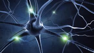
Amyotrophic lateral sclerosis (ALS), also known as motor neurone
disease (MND) or Lou Gehrig's disease, is a neurodegenerative
disease that results in the progressive loss of motor neurons
that control voluntary muscles. ALS is the most common form
of the motor neuron diseases. Early symptoms of ALS include
stiff muscles, muscle twitches, and gradual increasing weakness
and muscle wasting. Limb-onset ALS begins with weakness in
the arms or legs, while bulbar-onset ALS begins with difficulty
speaking or swallowing. Around half of people with ALS develop
at least mild difficulties with thinking and behavior, and
about 15% develop frontotemporal dementia. Motor neuron loss
continues until the ability to eat, speak, move, and finally
the ability to breathe is lost.
Most cases of ALS (about 90% to 95%) have no known cause, and
are known as sporadic ALS. However, both genetic and environmental
factors are believed to be involved. The remaining 5% to 10% of
cases have a genetic cause, often linked to a history of the
disease in the family, and these are known as genetic ALS.
About half of these genetic cases are due to disease-causing
variants in one of two specific genes. The diagnosis is based
on a person's signs and symptoms, with testing conducted to
rule out other potential causes.
There is no known cure for ALS. The goal of treatment is to
slow the disease and improve symptoms.
However, this bibliography specifically searches
PubMed for the idea of treatment in conjunction with ALS to
make it easier to track literature that explores the possibility
of treatment.
Created with PubMed® Query: (( ALS[TIAB] OR "amyotrophic lateral sclerosis"[TIAB] OR "motor neuron disease"[TIAB] ) AND "heat shock"[TIAB] ) NOT pmcbook NOT ispreviousversion
Citations The Papers (from PubMed®)
RevDate: 2019-09-18
CmpDate: 1991-08-01
Ubiquitin and heat shock protein expression in amyotrophic lateral sclerosis.
Neuropathology and applied neurobiology, 17(1):39-45.
The expression of two heat shock proteins, HSP72 and p57, in addition to ubiquitin, has been studied immunocytochemically in nine amyotrophic lateral sclerosis (ALS) cases and 10 age-matched controls. HSP72 and p57 antibodies did not identify the characteristic ubiquitin-immunoreactive inclusions present in anterior horn cells in ALS spinal cord. Antibodies to HSP72, but not to p57 or ubiquitin, strongly labelled structures corresponding to polyglucosan bodies in spinal grey matter. Such immunoreactive profiles were more abundant in ALS cases, although they were also present in control material. They were sometimes identified by haematoxylin and eosin and periodic acid Schiff reaction, but were not labeled by phosphotungstic acid haematoxylin or by antibodies to glial fibrillary acidic protein. Although ubiquitin, HSP72 and p57 are stress-induced proteins, they are expressed differently and might therefore have different significance in neuronal degeneration.
Additional Links: PMID-1647500
Publisher:
PubMed:
Citation:
show bibtex listing
hide bibtex listing
@article {pmid1647500,
year = {1991},
author = {Garofalo, O and Kennedy, PG and Swash, M and Martin, JE and Luthert, P and Anderton, BH and Leigh, PN},
title = {Ubiquitin and heat shock protein expression in amyotrophic lateral sclerosis.},
journal = {Neuropathology and applied neurobiology},
volume = {17},
number = {1},
pages = {39-45},
doi = {10.1111/j.1365-2990.1991.tb00692.x},
pmid = {1647500},
issn = {0305-1846},
support = {//Wellcome Trust/United Kingdom ; },
mesh = {Aged ; Amyotrophic Lateral Sclerosis/immunology/*metabolism/pathology ; Antibodies, Monoclonal ; Brain/pathology ; Female ; Heat-Shock Proteins/*biosynthesis ; Humans ; Immunohistochemistry ; Male ; Middle Aged ; Spinal Cord/metabolism/pathology ; Ubiquitins/*biosynthesis ; },
abstract = {The expression of two heat shock proteins, HSP72 and p57, in addition to ubiquitin, has been studied immunocytochemically in nine amyotrophic lateral sclerosis (ALS) cases and 10 age-matched controls. HSP72 and p57 antibodies did not identify the characteristic ubiquitin-immunoreactive inclusions present in anterior horn cells in ALS spinal cord. Antibodies to HSP72, but not to p57 or ubiquitin, strongly labelled structures corresponding to polyglucosan bodies in spinal grey matter. Such immunoreactive profiles were more abundant in ALS cases, although they were also present in control material. They were sometimes identified by haematoxylin and eosin and periodic acid Schiff reaction, but were not labeled by phosphotungstic acid haematoxylin or by antibodies to glial fibrillary acidic protein. Although ubiquitin, HSP72 and p57 are stress-induced proteins, they are expressed differently and might therefore have different significance in neuronal degeneration.},
}
MeSH Terms:
show MeSH Terms
hide MeSH Terms
Aged
Amyotrophic Lateral Sclerosis/immunology/*metabolism/pathology
Antibodies, Monoclonal
Brain/pathology
Female
Heat-Shock Proteins/*biosynthesis
Humans
Immunohistochemistry
Male
Middle Aged
Spinal Cord/metabolism/pathology
Ubiquitins/*biosynthesis
RevDate: 2020-03-04
CmpDate: 1992-01-23
Purkinje cell toxicity of beta-aminopropionitrile in the rat.
Virchows Archiv. A, Pathological anatomy and histopathology, 419(5):403-408.
Compounds causing neurolathyrism are putative aetiological agents in neurodegenerative disorders including amyotrophic lateral sclerosis. beta-Aminopropionitrile (BAPN) is one such compound. We have administered this lathyrogenic agent at a dose of 1 g/kg by the intraperitoneal route in experiments in adult Sprague-Dawley rats during a period of 10 weeks. The rats developed marked kyphoscoliosis, ataxia with paralysis and muscle wasting of the hind limbs. Vacuolation and loss of Purkinje cells developed, but no anterior horn cell degeneration was noted. Immunohistochemical studies of phosphorylated neurofilaments and the 72 kDa heat shock protein were normal and no intraneuronal ubiquitinated inclusions were seen. High-dose intraperitoneal BAPN in the rat causes Purkinje cell changes, but no other central nervous system abnormalities.
Additional Links: PMID-1750186
PubMed:
Citation:
show bibtex listing
hide bibtex listing
@article {pmid1750186,
year = {1991},
author = {Martin, JE and Sosa-Melgarejo, JA and Swash, M and Mather, K and Leigh, PN and Berry, CL},
title = {Purkinje cell toxicity of beta-aminopropionitrile in the rat.},
journal = {Virchows Archiv. A, Pathological anatomy and histopathology},
volume = {419},
number = {5},
pages = {403-408},
pmid = {1750186},
issn = {0174-7398},
mesh = {Aminopropionitrile/*adverse effects ; Animals ; Female ; Immunohistochemistry ; Injections, Intraperitoneal ; Kyphosis/chemically induced/diagnostic imaging ; Purkinje Cells/*drug effects/metabolism/pathology ; Radiography ; Rats ; Rats, Inbred Strains ; Scoliosis/chemically induced/diagnostic imaging ; },
abstract = {Compounds causing neurolathyrism are putative aetiological agents in neurodegenerative disorders including amyotrophic lateral sclerosis. beta-Aminopropionitrile (BAPN) is one such compound. We have administered this lathyrogenic agent at a dose of 1 g/kg by the intraperitoneal route in experiments in adult Sprague-Dawley rats during a period of 10 weeks. The rats developed marked kyphoscoliosis, ataxia with paralysis and muscle wasting of the hind limbs. Vacuolation and loss of Purkinje cells developed, but no anterior horn cell degeneration was noted. Immunohistochemical studies of phosphorylated neurofilaments and the 72 kDa heat shock protein were normal and no intraneuronal ubiquitinated inclusions were seen. High-dose intraperitoneal BAPN in the rat causes Purkinje cell changes, but no other central nervous system abnormalities.},
}
MeSH Terms:
show MeSH Terms
hide MeSH Terms
Aminopropionitrile/*adverse effects
Animals
Female
Immunohistochemistry
Injections, Intraperitoneal
Kyphosis/chemically induced/diagnostic imaging
Purkinje Cells/*drug effects/metabolism/pathology
Radiography
Rats
Rats, Inbred Strains
Scoliosis/chemically induced/diagnostic imaging
RevDate: 2007-02-22
CmpDate: 1991-07-31
[HSP 70 is associated with abnormal cytoplasmic inclusions characteristic of neurodegenerative diseases].
No to shinkei = Brain and nerve, 43(1):57-60.
Several degenerative diseases of the central nervous system are characterized by the presence of neuronal inclusions. One of these inclusions, neurofibrillary tangles in Alzheimer's disease, has been shown to contain ubiquitin that belongs to a group of proteins known as heat shock proteins. Subsequent studies revealed that ubiquitin is also associated with various neuronal inclusions including Lewy bodies, Pick bodies and hyaline inclusions. Very recently, ubiquitin has been found also to be associated with glial inclusions that are unique to multiple system atrophy. The close association of ubiquitin with varying cellular inclusions, together with its function in the proteolytic process, raised the hypothesis that ubiquitin may be involved in the degradation of abnormal proteins appearing in the damaged neurons and glial cells. In central nervous system, a group of heat shock proteins collectively known as HSP 70 is also present which is constitutive and/or inducible. Since HSP 70 has been suspected to play a crucial role degradation and repair of abnormal intracellular proteins, we hypothesized that HSP 70 may be associated with those inclusions, as the case with ubiquitin. To test this we performed immunohistochemical studies on brain tissues from patients with various neurodegenerative conditions by using specific polyclonal antibody to HSP 70. Brain tissues were obtained at autopsy from each three patients with Alzheimer's disease, Pick's disease, Parkinson's disease, amyotrophic lateral sclerosis (ALS) and multiple system atrophy. Tissues were fixed in buffered formalin and embedded in paraffin. Immunostaining was performed by the standard ABC method using diaminobenzidine as a chromogen. Sections were lightly stained with hematoxylin. Polyclonal antibodies were raised in rabbits against mouse HSP 70.(ABSTRACT TRUNCATED AT 250 WORDS)
Additional Links: PMID-2054224
PubMed:
Citation:
show bibtex listing
hide bibtex listing
@article {pmid2054224,
year = {1991},
author = {Namba, Y and Tomonaga, M and Ohtsuka, K and Oda, M and Ikeda, K},
title = {[HSP 70 is associated with abnormal cytoplasmic inclusions characteristic of neurodegenerative diseases].},
journal = {No to shinkei = Brain and nerve},
volume = {43},
number = {1},
pages = {57-60},
pmid = {2054224},
issn = {0006-8969},
mesh = {Alzheimer Disease/metabolism ; Cytoplasmic Granules/*metabolism ; Dementia/metabolism ; Heat-Shock Proteins/*metabolism ; Humans ; Immunohistochemistry ; *Nerve Degeneration ; Nervous System Diseases/*metabolism/physiopathology ; Parkinson Disease/metabolism ; },
abstract = {Several degenerative diseases of the central nervous system are characterized by the presence of neuronal inclusions. One of these inclusions, neurofibrillary tangles in Alzheimer's disease, has been shown to contain ubiquitin that belongs to a group of proteins known as heat shock proteins. Subsequent studies revealed that ubiquitin is also associated with various neuronal inclusions including Lewy bodies, Pick bodies and hyaline inclusions. Very recently, ubiquitin has been found also to be associated with glial inclusions that are unique to multiple system atrophy. The close association of ubiquitin with varying cellular inclusions, together with its function in the proteolytic process, raised the hypothesis that ubiquitin may be involved in the degradation of abnormal proteins appearing in the damaged neurons and glial cells. In central nervous system, a group of heat shock proteins collectively known as HSP 70 is also present which is constitutive and/or inducible. Since HSP 70 has been suspected to play a crucial role degradation and repair of abnormal intracellular proteins, we hypothesized that HSP 70 may be associated with those inclusions, as the case with ubiquitin. To test this we performed immunohistochemical studies on brain tissues from patients with various neurodegenerative conditions by using specific polyclonal antibody to HSP 70. Brain tissues were obtained at autopsy from each three patients with Alzheimer's disease, Pick's disease, Parkinson's disease, amyotrophic lateral sclerosis (ALS) and multiple system atrophy. Tissues were fixed in buffered formalin and embedded in paraffin. Immunostaining was performed by the standard ABC method using diaminobenzidine as a chromogen. Sections were lightly stained with hematoxylin. Polyclonal antibodies were raised in rabbits against mouse HSP 70.(ABSTRACT TRUNCATED AT 250 WORDS)},
}
MeSH Terms:
show MeSH Terms
hide MeSH Terms
Alzheimer Disease/metabolism
Cytoplasmic Granules/*metabolism
Dementia/metabolism
Heat-Shock Proteins/*metabolism
Humans
Immunohistochemistry
*Nerve Degeneration
Nervous System Diseases/*metabolism/physiopathology
Parkinson Disease/metabolism
RevDate: 2006-11-15
CmpDate: 1995-02-01
Elevated risks for amyotrophic lateral sclerosis and blood disorders in Ashkenazi schizophrenic pedigrees suggest new candidate genes in schizophrenia.
American journal of medical genetics, 54(3):271-278.
Among relatives of Ashkenazi schizophrenic probands the rate of amyotrophic lateral sclerosis was 3/1,000, compared to expected population rates of approximately 2/100,000. Relative risk of bleeding disorders, including hematologic cancers, was increased more than three-fold compared to controls. Co-occurrence of motor neuron disease and blood dyscrasias, accompanied by psychosis, has long been recognized. A virally mediated autoimmune pathogenesis has been proposed. However, the familial co-occurrence of these three disease entities raises the possibility that the disease constellation be considered as a manifestation of a common underlying genetic defect. Such expansion of the spectrum of affectation might enhance the power of both candidate gene and linkage studies. Based on these findings the loci suggested as candidate regions in schizophrenia include a potential hot spot on chromosome 21q21-q22, involving the superoxide dismutase and amyloid precursor protein genes. Alternatively, genes on other chromosomes involved in the expression, transcription, or regulation of these genes, or associated with the illnesses of high frequency in these pedigrees are suggested. Candidates include the choroid plexus transport protein, transthyretin at 18q11.2-q12.1; the t(14;18)(q22;21) characterizing B-cell lymphoma-2, the most common form of hematologic cancer; and the 14q24 locus of early onset Alzheimer's disease, c-Fos, transforming growth factor beta 3, and heat shock protein A2. Expression of hematologic cancers and the suggested candidate genes are known to involve retinoid pathways, and retinoid disregulation has been proposed as a cause of schizophrenia.
Additional Links: PMID-7810588
Publisher:
PubMed:
Citation:
show bibtex listing
hide bibtex listing
@article {pmid7810588,
year = {1994},
author = {Goodman, AB},
title = {Elevated risks for amyotrophic lateral sclerosis and blood disorders in Ashkenazi schizophrenic pedigrees suggest new candidate genes in schizophrenia.},
journal = {American journal of medical genetics},
volume = {54},
number = {3},
pages = {271-278},
doi = {10.1002/ajmg.1320540317},
pmid = {7810588},
issn = {0148-7299},
mesh = {Age Factors ; Amyotrophic Lateral Sclerosis/ethnology/*genetics ; Chromosomes, Human, Pair 14 ; Chromosomes, Human, Pair 18 ; Chromosomes, Human, Pair 21 ; Female ; Hematologic Diseases/ethnology/*genetics ; Humans ; Jews ; Male ; Pedigree ; Risk Factors ; Schizophrenia/ethnology/*genetics ; },
abstract = {Among relatives of Ashkenazi schizophrenic probands the rate of amyotrophic lateral sclerosis was 3/1,000, compared to expected population rates of approximately 2/100,000. Relative risk of bleeding disorders, including hematologic cancers, was increased more than three-fold compared to controls. Co-occurrence of motor neuron disease and blood dyscrasias, accompanied by psychosis, has long been recognized. A virally mediated autoimmune pathogenesis has been proposed. However, the familial co-occurrence of these three disease entities raises the possibility that the disease constellation be considered as a manifestation of a common underlying genetic defect. Such expansion of the spectrum of affectation might enhance the power of both candidate gene and linkage studies. Based on these findings the loci suggested as candidate regions in schizophrenia include a potential hot spot on chromosome 21q21-q22, involving the superoxide dismutase and amyloid precursor protein genes. Alternatively, genes on other chromosomes involved in the expression, transcription, or regulation of these genes, or associated with the illnesses of high frequency in these pedigrees are suggested. Candidates include the choroid plexus transport protein, transthyretin at 18q11.2-q12.1; the t(14;18)(q22;21) characterizing B-cell lymphoma-2, the most common form of hematologic cancer; and the 14q24 locus of early onset Alzheimer's disease, c-Fos, transforming growth factor beta 3, and heat shock protein A2. Expression of hematologic cancers and the suggested candidate genes are known to involve retinoid pathways, and retinoid disregulation has been proposed as a cause of schizophrenia.},
}
MeSH Terms:
show MeSH Terms
hide MeSH Terms
Age Factors
Amyotrophic Lateral Sclerosis/ethnology/*genetics
Chromosomes, Human, Pair 14
Chromosomes, Human, Pair 18
Chromosomes, Human, Pair 21
Female
Hematologic Diseases/ethnology/*genetics
Humans
Jews
Male
Pedigree
Risk Factors
Schizophrenia/ethnology/*genetics
RevDate: 2019-05-14
CmpDate: 1994-06-20
Humoral response to hsp 65 in multiple sclerosis and other neurologic conditions.
Neurology, 44(5):941-946.
The expression of heat shock proteins (hsp) within the target organ is implicated in the pathogenesis of a number of diseases of suspected autoimmune etiology, including MS. To pursue the potential role of a humoral response to the hsp 60/65 kd family in MS, we studied serum and CSF by Western blotting using recombinant Mycobacterium bovis hsp 65 and human hsp 60 as antigens and compared the findings with samples from patients with other neurologic diseases (OND). Analysis of the IgG response in CSF from 18 patients with MS indicated moderate reactivity in 10 cases and no reactivity in eight. In the OND group, reactivity was found in the CSF from one of two patients with Parkinson's disease, four of four Alzheimer's disease patients, and two of two patients with amyotrophic lateral sclerosis. CSF samples from seven of seven patients with subacute sclerosing panencephalitis were negative, as were samples from two normal subjects. There was no reactivity in CSF from two Huntington's disease patients. We conclude that antibodies reactive with hsp 60/65 are present in CSF of some MS patients but are also present in a number of chronic neurodegenerative conditions. The findings indicate that a humoral response to hsp 60/65 in the CSF is not specific for MS.
Additional Links: PMID-8190301
Publisher:
PubMed:
Citation:
show bibtex listing
hide bibtex listing
@article {pmid8190301,
year = {1994},
author = {Gao, YL and Raine, CS and Brosnan, CF},
title = {Humoral response to hsp 65 in multiple sclerosis and other neurologic conditions.},
journal = {Neurology},
volume = {44},
number = {5},
pages = {941-946},
doi = {10.1212/wnl.44.5.941},
pmid = {8190301},
issn = {0028-3878},
support = {NS 07098/NS/NINDS NIH HHS/United States ; NS 08952/NS/NINDS NIH HHS/United States ; NS 11920/NS/NINDS NIH HHS/United States ; },
mesh = {Adult ; Aged ; Antibodies/cerebrospinal fluid ; *Antibody Formation ; *Bacterial Proteins ; Blotting, Western ; Chaperonin 60 ; *Chaperonins ; Female ; Heat-Shock Proteins/*immunology ; Humans ; Male ; Middle Aged ; Multiple Sclerosis/cerebrospinal fluid/*immunology ; Mycobacterium bovis/immunology ; Nervous System Diseases/cerebrospinal fluid/immunology ; },
abstract = {The expression of heat shock proteins (hsp) within the target organ is implicated in the pathogenesis of a number of diseases of suspected autoimmune etiology, including MS. To pursue the potential role of a humoral response to the hsp 60/65 kd family in MS, we studied serum and CSF by Western blotting using recombinant Mycobacterium bovis hsp 65 and human hsp 60 as antigens and compared the findings with samples from patients with other neurologic diseases (OND). Analysis of the IgG response in CSF from 18 patients with MS indicated moderate reactivity in 10 cases and no reactivity in eight. In the OND group, reactivity was found in the CSF from one of two patients with Parkinson's disease, four of four Alzheimer's disease patients, and two of two patients with amyotrophic lateral sclerosis. CSF samples from seven of seven patients with subacute sclerosing panencephalitis were negative, as were samples from two normal subjects. There was no reactivity in CSF from two Huntington's disease patients. We conclude that antibodies reactive with hsp 60/65 are present in CSF of some MS patients but are also present in a number of chronic neurodegenerative conditions. The findings indicate that a humoral response to hsp 60/65 in the CSF is not specific for MS.},
}
MeSH Terms:
show MeSH Terms
hide MeSH Terms
Adult
Aged
Antibodies/cerebrospinal fluid
*Antibody Formation
*Bacterial Proteins
Blotting, Western
Chaperonin 60
*Chaperonins
Female
Heat-Shock Proteins/*immunology
Humans
Male
Middle Aged
Multiple Sclerosis/cerebrospinal fluid/*immunology
Mycobacterium bovis/immunology
Nervous System Diseases/cerebrospinal fluid/immunology
RevDate: 2019-07-24
CmpDate: 1993-12-16
Expression of the human groEL stress-protein homologue in the brain and spinal cord.
Journal of the neurological sciences, 118(2):202-206.
A monoclonal antibody (ML30), previously shown to identify a human mitochondrial protein epitope homologous with the groEL heat-shock protein of bacteria (hsp60), was used in an immunohistochemical survey of the central nervous system in patients dying with no evidence of neurological disease and in tissue from patients dying with various neurological disorders. Staining was performed on frozen tissue sections and on formalin fixed, paraffin embedded tissue. Astrocytes in all areas showed a strong pattern of punctate granular staining, which was increased in astrocytes showing reactive changes. Oligodendrocytes stained lightly in a diffuse granular pattern as did most neurons. Ependymal cells showed apical granular positivity. Expression of the hsp60 epitope recognised by ML30 was not seen in ubiquitinated inclusion bodies in motor neuron disease, neurofibrillary tangles in Alzheimer's disease or Lewy bodies in Parkinson's disease. The epitope recognised by ML30 was stable after formalin fixation and in post mortem tissue up to 96 h after death. Expression of the human groEL stress-protein homologue in brain and spinal cord is consistent with a mitochondrial location and may provide a morphological indicator of the functional or metabolic state of cells, especially glial cells.
Additional Links: PMID-8229070
Publisher:
PubMed:
Citation:
show bibtex listing
hide bibtex listing
@article {pmid8229070,
year = {1993},
author = {Martin, JE and Swash, M and Mather, K and Leigh, PN},
title = {Expression of the human groEL stress-protein homologue in the brain and spinal cord.},
journal = {Journal of the neurological sciences},
volume = {118},
number = {2},
pages = {202-206},
doi = {10.1016/0022-510x(93)90111-b},
pmid = {8229070},
issn = {0022-510X},
support = {//Wellcome Trust/United Kingdom ; },
mesh = {Adolescent ; Adult ; Aged ; Antibodies, Monoclonal ; Astrocytes/metabolism ; Brain/pathology ; Brain Chemistry/*physiology ; Child ; Female ; Heat-Shock Proteins/*biosynthesis/immunology ; Humans ; Immunohistochemistry ; Male ; Middle Aged ; Mitochondria/metabolism ; Nervous System Diseases/pathology ; Neurons/metabolism ; Oligodendroglia/metabolism ; Spinal Cord/*metabolism/pathology ; },
abstract = {A monoclonal antibody (ML30), previously shown to identify a human mitochondrial protein epitope homologous with the groEL heat-shock protein of bacteria (hsp60), was used in an immunohistochemical survey of the central nervous system in patients dying with no evidence of neurological disease and in tissue from patients dying with various neurological disorders. Staining was performed on frozen tissue sections and on formalin fixed, paraffin embedded tissue. Astrocytes in all areas showed a strong pattern of punctate granular staining, which was increased in astrocytes showing reactive changes. Oligodendrocytes stained lightly in a diffuse granular pattern as did most neurons. Ependymal cells showed apical granular positivity. Expression of the hsp60 epitope recognised by ML30 was not seen in ubiquitinated inclusion bodies in motor neuron disease, neurofibrillary tangles in Alzheimer's disease or Lewy bodies in Parkinson's disease. The epitope recognised by ML30 was stable after formalin fixation and in post mortem tissue up to 96 h after death. Expression of the human groEL stress-protein homologue in brain and spinal cord is consistent with a mitochondrial location and may provide a morphological indicator of the functional or metabolic state of cells, especially glial cells.},
}
MeSH Terms:
show MeSH Terms
hide MeSH Terms
Adolescent
Adult
Aged
Antibodies, Monoclonal
Astrocytes/metabolism
Brain/pathology
Brain Chemistry/*physiology
Child
Female
Heat-Shock Proteins/*biosynthesis/immunology
Humans
Immunohistochemistry
Male
Middle Aged
Mitochondria/metabolism
Nervous System Diseases/pathology
Neurons/metabolism
Oligodendroglia/metabolism
Spinal Cord/*metabolism/pathology
RevDate: 2013-04-18
CmpDate: 1996-10-10
Immunohistochemical study of the expression of human groEL-stress protein in human nervous tissue.
The Indian journal of medical research, 103:103-111.
Monoclonal antibody (ML-30) directed against 65 kDa stress protein of mycobacteria, is shown to identify human cellular protein homologous with the groEL heat shock protein in many prokaryotes. Immunohistochemical survey of nervous tissue, both central and peripheral, from patients dying of various inflammatory, degenerative and neoplastic conditions and from experimental animals, using this antibody showed punctate granular staining of the cells to a variable degree. The astrocytes showed strong immunolabelling. The normal neurons and oligodendroglia stained variably, while abnormal neurons were darkly labelled. Ependymal cells showed apical granular positivity. The ubiquitinated inclusion bodies in amyotrophic lateral sclerosis, Alzheimer's disease and Parkinson's disease were not recognised by the ML-30 antibody. In diseased and stressed nervous tissue from experimental animals, the expression of the ML-30 recognisable stress protein was variable. The epitope recognised by ML-30 was found stable in postmortem tissues collected up to 36 h after death and processed for paraffin sectioning, after fixation in formalin for many years. Enhanced expression of the human groEL stress protein homologue in mammalian nervous tissue following various forms of stress may play a role in modulating the extent of tissue damage by autoimmune mechanism because of its high immunogenic nature and constitutive presence in the cells.
Additional Links: PMID-8714148
PubMed:
Citation:
show bibtex listing
hide bibtex listing
@article {pmid8714148,
year = {1996},
author = {Khanna, N and Shankar, SK and Chandramuki, A and Jagannath, C},
title = {Immunohistochemical study of the expression of human groEL-stress protein in human nervous tissue.},
journal = {The Indian journal of medical research},
volume = {103},
number = {},
pages = {103-111},
pmid = {8714148},
issn = {0971-5916},
mesh = {Animals ; Brain Chemistry/*physiology ; Chaperonin 60/*analysis ; Gerbillinae ; Humans ; Immunohistochemistry ; Nerve Tissue Proteins/*analysis ; Rats ; Saimiri ; Spinal Cord/*chemistry ; },
abstract = {Monoclonal antibody (ML-30) directed against 65 kDa stress protein of mycobacteria, is shown to identify human cellular protein homologous with the groEL heat shock protein in many prokaryotes. Immunohistochemical survey of nervous tissue, both central and peripheral, from patients dying of various inflammatory, degenerative and neoplastic conditions and from experimental animals, using this antibody showed punctate granular staining of the cells to a variable degree. The astrocytes showed strong immunolabelling. The normal neurons and oligodendroglia stained variably, while abnormal neurons were darkly labelled. Ependymal cells showed apical granular positivity. The ubiquitinated inclusion bodies in amyotrophic lateral sclerosis, Alzheimer's disease and Parkinson's disease were not recognised by the ML-30 antibody. In diseased and stressed nervous tissue from experimental animals, the expression of the ML-30 recognisable stress protein was variable. The epitope recognised by ML-30 was found stable in postmortem tissues collected up to 36 h after death and processed for paraffin sectioning, after fixation in formalin for many years. Enhanced expression of the human groEL stress protein homologue in mammalian nervous tissue following various forms of stress may play a role in modulating the extent of tissue damage by autoimmune mechanism because of its high immunogenic nature and constitutive presence in the cells.},
}
MeSH Terms:
show MeSH Terms
hide MeSH Terms
Animals
Brain Chemistry/*physiology
Chaperonin 60/*analysis
Gerbillinae
Humans
Immunohistochemistry
Nerve Tissue Proteins/*analysis
Rats
Saimiri
Spinal Cord/*chemistry
RevDate: 2019-08-12
CmpDate: 1997-02-03
Different variants of frontotemporal dementia: a neuropathological and immunohistochemical study.
Acta neuropathologica, 92(2):170-179.
Histological and immunohistochemical findings in 20 cases of frontotemporal dementias-8 cases of dementia of frontal lobe type (DFT), 7 cases of Pick's disease (PD), and 5 cases of motor neuron disease with dementia (MND/D)-are presented. Common features of all three syndromes were: frontotemporal atrophy, involvement of subcortical nuclei, and swollen chromatolytic cells. Ubiquitin (Ub)-positive and tau-negative inclusions in cortical, hippocampal, and motor neurons were found in MND/D and DFT cases, suggesting a common pathogenesis of MND/D and DFT. MND/D showed the same cytoskeletal alterations in motor nuclei as MND without dementia: Bunina bodies and skein-like, Ub-positive inclusions. DFT differed from PD in the preponderance of histopathological changes in upper cortical layers, the sparseness of chromatolytic cells, and the absence of tau-positive Pick bodies (PBs). There were, however, two transitional cases showing Pick-type histology but no PBs, thus linking DFT and PD. PBs expressed chromogranin B and secretoneurin strongly, but chromogranin A only weakly. They were negative for the 70-kDa heat-shock protein, metallothionein, and glutathione-S-transferase.
Additional Links: PMID-8841663
Publisher:
PubMed:
Citation:
show bibtex listing
hide bibtex listing
@article {pmid8841663,
year = {1996},
author = {Bergmann, M and Kuchelmeister, K and Schmid, KW and Kretzschmar, HA and Schröder, R},
title = {Different variants of frontotemporal dementia: a neuropathological and immunohistochemical study.},
journal = {Acta neuropathologica},
volume = {92},
number = {2},
pages = {170-179},
doi = {10.1007/s004010050505},
pmid = {8841663},
issn = {0001-6322},
mesh = {Adult ; Aged ; Atrophy/immunology/pathology ; Dementia/*immunology/*pathology ; Frontal Lobe/*immunology/*pathology ; Humans ; Immunohistochemistry ; Middle Aged ; Motor Neuron Disease/immunology/pathology ; Temporal Lobe/*immunology/*pathology ; },
abstract = {Histological and immunohistochemical findings in 20 cases of frontotemporal dementias-8 cases of dementia of frontal lobe type (DFT), 7 cases of Pick's disease (PD), and 5 cases of motor neuron disease with dementia (MND/D)-are presented. Common features of all three syndromes were: frontotemporal atrophy, involvement of subcortical nuclei, and swollen chromatolytic cells. Ubiquitin (Ub)-positive and tau-negative inclusions in cortical, hippocampal, and motor neurons were found in MND/D and DFT cases, suggesting a common pathogenesis of MND/D and DFT. MND/D showed the same cytoskeletal alterations in motor nuclei as MND without dementia: Bunina bodies and skein-like, Ub-positive inclusions. DFT differed from PD in the preponderance of histopathological changes in upper cortical layers, the sparseness of chromatolytic cells, and the absence of tau-positive Pick bodies (PBs). There were, however, two transitional cases showing Pick-type histology but no PBs, thus linking DFT and PD. PBs expressed chromogranin B and secretoneurin strongly, but chromogranin A only weakly. They were negative for the 70-kDa heat-shock protein, metallothionein, and glutathione-S-transferase.},
}
MeSH Terms:
show MeSH Terms
hide MeSH Terms
Adult
Aged
Atrophy/immunology/pathology
Dementia/*immunology/*pathology
Frontal Lobe/*immunology/*pathology
Humans
Immunohistochemistry
Middle Aged
Motor Neuron Disease/immunology/pathology
Temporal Lobe/*immunology/*pathology
RevDate: 2018-11-13
CmpDate: 1997-08-26
Pathological characterization of astrocytic hyaline inclusions in familial amyotrophic lateral sclerosis.
The American journal of pathology, 151(2):611-620.
To clarify the pathological characteristics of astrocytic hyaline inclusions (Ast-HIs) in patients with familial amyotrophic lateral sclerosis (FALS) with neuronal Lewy-body-like hyaline inclusions (LBHIs), eight autopsies on members of four different families, including two long-term surviving patients with clinical courses of over 10 years, were analyzed. Ast-HIs were found only in the two long-term surviving patients who belonged to different families and to different races. Ast-HIs were ultrastructurally composed of 15- to 25-nm granule-coated fibrils that had immunoreactivities to superoxide dismutase 1 (SOD1) and ubiquitin. Approximately 50% of the Ast-HIs expressed alpha B-crystallin, metallothionein, glutamine synthetase, and tubulin (alpha and beta) at various intensities. Some Ast-HIs reacted with antibodies to tau protein, S-100 protein, and heat shock protein 27. The Ast-HIs were not stained for glial fibrillary acidic protein. Our results suggest a cooperative role of superoxide dismutase 1, ubiquitin, and cytoskeletal proteins in the formation of granule-coated fibrils (namely, Ast-HIs) and provide evidence that Ast-HIs are formed in certain long-surviving familial amyotrophic lateral sclerosis patients with neuronal Lewy-body-like hyaline inclusions.
Additional Links: PMID-9273821
PubMed:
Citation:
show bibtex listing
hide bibtex listing
@article {pmid9273821,
year = {1997},
author = {Kato, S and Hayashi, H and Nakashima, K and Nanba, E and Kato, M and Hirano, A and Nakano, I and Asayama, K and Ohama, E},
title = {Pathological characterization of astrocytic hyaline inclusions in familial amyotrophic lateral sclerosis.},
journal = {The American journal of pathology},
volume = {151},
number = {2},
pages = {611-620},
pmid = {9273821},
issn = {0002-9440},
mesh = {Adult ; Aged ; Amyotrophic Lateral Sclerosis/genetics/*pathology ; Astrocytes/*pathology/ultrastructure ; Brain/pathology/ultrastructure ; Female ; Humans ; Hyalin/*ultrastructure ; Immunohistochemistry ; Lewy Bodies/*ultrastructure ; Male ; Microscopy, Electron ; Middle Aged ; Spinal Cord/pathology/ultrastructure ; },
abstract = {To clarify the pathological characteristics of astrocytic hyaline inclusions (Ast-HIs) in patients with familial amyotrophic lateral sclerosis (FALS) with neuronal Lewy-body-like hyaline inclusions (LBHIs), eight autopsies on members of four different families, including two long-term surviving patients with clinical courses of over 10 years, were analyzed. Ast-HIs were found only in the two long-term surviving patients who belonged to different families and to different races. Ast-HIs were ultrastructurally composed of 15- to 25-nm granule-coated fibrils that had immunoreactivities to superoxide dismutase 1 (SOD1) and ubiquitin. Approximately 50% of the Ast-HIs expressed alpha B-crystallin, metallothionein, glutamine synthetase, and tubulin (alpha and beta) at various intensities. Some Ast-HIs reacted with antibodies to tau protein, S-100 protein, and heat shock protein 27. The Ast-HIs were not stained for glial fibrillary acidic protein. Our results suggest a cooperative role of superoxide dismutase 1, ubiquitin, and cytoskeletal proteins in the formation of granule-coated fibrils (namely, Ast-HIs) and provide evidence that Ast-HIs are formed in certain long-surviving familial amyotrophic lateral sclerosis patients with neuronal Lewy-body-like hyaline inclusions.},
}
MeSH Terms:
show MeSH Terms
hide MeSH Terms
Adult
Aged
Amyotrophic Lateral Sclerosis/genetics/*pathology
Astrocytes/*pathology/ultrastructure
Brain/pathology/ultrastructure
Female
Humans
Hyalin/*ultrastructure
Immunohistochemistry
Lewy Bodies/*ultrastructure
Male
Microscopy, Electron
Middle Aged
Spinal Cord/pathology/ultrastructure
RevDate: 2021-02-17
CmpDate: 1998-03-26
Role of sphingolipid-mediated cell death in neurodegenerative diseases.
Journal of lipid research, 39(1):1-16.
The metazoan nervous system gives rise intradevelopmentally to many more neurons than ultimately survive in the adult. Such excess cells are eliminated through programmed cell death or apoptosis. As is true for cells of other lineages, neuronal survival is sustained by an array of growth factors, such that withdrawal of neurotrophic support results in apoptotic cell death. Apoptosis is therefore believed to represent a beneficial process essential to normal development of central and peripheral nervous system (CNS and PNS) structures. Although the initiation of neuronal apoptosis in response to numerous extracellular agents has been widely reported, the regulatory mechanisms underlying this mode of cell death remain incompletely understood. In recent years, the contribution of lipid-dependent signaling systems, such as the sphingomyelin pathway, to regulation of cell survival has received considerable attention, leading to the identification of lethal functions for the lipid effectors ceramide and sphingosine in both normal and pathophysiological conditions. Moreover, the apoptotic capacities of several cytotoxic receptor systems (e.g., CD120a, CD95) and many environmental stresses (e.g., ionizing radiation, heat-shock, oxidative stress) are now known to derive from the activation of multiple signaling cascades by ceramide or, under some circumstances, by sphingosine. Inappropriate initiation of apoptosis has been proposed to underlie the progressive neuronal attrition associated with various neurodegenerative diseases such as Alzheimer's disease (AD), Parkinson's disease (PD), amyotrophic lateral sclerosis (ALS), and other neurological disorders that are characterized by the gradual loss of specific populations of neurons. In such pathophysiological states, neuronal cell death can result in specific disorders of movement and diverse impairments of CNS and PNS function. In some autoimmune neurological diseases such as Guillain-Barré syndrome, demyelinating polyneuropathy, and motoneuron disease, persistent immunological attack of microvascular endothelial cells by glycolipid-directed autoantibodies may lead to extensive cellular damages, resulting in increased permeability across brain-nerve barrier (BNB) and/or blood-brain barrier (BBB).
Additional Links: PMID-9469581
PubMed:
Citation:
show bibtex listing
hide bibtex listing
@article {pmid9469581,
year = {1998},
author = {Ariga, T and Jarvis, WD and Yu, RK},
title = {Role of sphingolipid-mediated cell death in neurodegenerative diseases.},
journal = {Journal of lipid research},
volume = {39},
number = {1},
pages = {1-16},
pmid = {9469581},
issn = {0022-2275},
support = {CA-09380/CA/NCI NIH HHS/United States ; NS-11853/NS/NINDS NIH HHS/United States ; NS-26994/NS/NINDS NIH HHS/United States ; },
mesh = {Animals ; *Apoptosis ; Gangliosides/physiology ; Glycolipids/physiology ; Humans ; Lipids/physiology ; Neurodegenerative Diseases/*pathology ; Second Messenger Systems ; Sphingolipids/*physiology ; },
abstract = {The metazoan nervous system gives rise intradevelopmentally to many more neurons than ultimately survive in the adult. Such excess cells are eliminated through programmed cell death or apoptosis. As is true for cells of other lineages, neuronal survival is sustained by an array of growth factors, such that withdrawal of neurotrophic support results in apoptotic cell death. Apoptosis is therefore believed to represent a beneficial process essential to normal development of central and peripheral nervous system (CNS and PNS) structures. Although the initiation of neuronal apoptosis in response to numerous extracellular agents has been widely reported, the regulatory mechanisms underlying this mode of cell death remain incompletely understood. In recent years, the contribution of lipid-dependent signaling systems, such as the sphingomyelin pathway, to regulation of cell survival has received considerable attention, leading to the identification of lethal functions for the lipid effectors ceramide and sphingosine in both normal and pathophysiological conditions. Moreover, the apoptotic capacities of several cytotoxic receptor systems (e.g., CD120a, CD95) and many environmental stresses (e.g., ionizing radiation, heat-shock, oxidative stress) are now known to derive from the activation of multiple signaling cascades by ceramide or, under some circumstances, by sphingosine. Inappropriate initiation of apoptosis has been proposed to underlie the progressive neuronal attrition associated with various neurodegenerative diseases such as Alzheimer's disease (AD), Parkinson's disease (PD), amyotrophic lateral sclerosis (ALS), and other neurological disorders that are characterized by the gradual loss of specific populations of neurons. In such pathophysiological states, neuronal cell death can result in specific disorders of movement and diverse impairments of CNS and PNS function. In some autoimmune neurological diseases such as Guillain-Barré syndrome, demyelinating polyneuropathy, and motoneuron disease, persistent immunological attack of microvascular endothelial cells by glycolipid-directed autoantibodies may lead to extensive cellular damages, resulting in increased permeability across brain-nerve barrier (BNB) and/or blood-brain barrier (BBB).},
}
MeSH Terms:
show MeSH Terms
hide MeSH Terms
Animals
*Apoptosis
Gangliosides/physiology
Glycolipids/physiology
Humans
Lipids/physiology
Neurodegenerative Diseases/*pathology
Second Messenger Systems
Sphingolipids/*physiology
RevDate: 2019-06-30
CmpDate: 1999-02-11
Up-regulation of protein chaperones preserves viability of cells expressing toxic Cu/Zn-superoxide dismutase mutants associated with amyotrophic lateral sclerosis.
Journal of neurochemistry, 72(2):693-699.
Mutations in the Cu/Zn-superoxide dismutase (SOD-1) gene underlie some familial cases of amyotrophic lateral sclerosis, a neurodegenerative disorder characterized by loss of cortical, brainstem, and spinal motor neurons. We present evidence that SOD-1 mutants alter the activity of molecular chaperones that aid in proper protein folding and targeting of abnormal proteins for degradation. In a cultured cell line (NIH 3T3), resistance to mutant SOD-1 toxicity correlated with increased overall chaperoning activity (measured by the ability of cytosolic extracts to prevent heat denaturation of catalase) as well as with up-regulation of individual chaperones/stress proteins. In transgenic mice expressing human SOD-1 with the G93A mutation, chaperoning activity was decreased in lumbar spinal cord but increased or unchanged in clinically unaffected tissues. Increasing the level of the stress-inducible chaperone 70-kDa heat shock protein by gene transfer reduced formation of mutant SOD-containing proteinaceous aggregates in cultured primary motor neurons expressing G93A SOD-1 and prolonged their survival. We propose that insufficiency of molecular chaperones may be directly involved in loss of motor neurons in this disease.
Additional Links: PMID-9930742
Publisher:
PubMed:
Citation:
show bibtex listing
hide bibtex listing
@article {pmid9930742,
year = {1999},
author = {Bruening, W and Roy, J and Giasson, B and Figlewicz, DA and Mushynski, WE and Durham, HD},
title = {Up-regulation of protein chaperones preserves viability of cells expressing toxic Cu/Zn-superoxide dismutase mutants associated with amyotrophic lateral sclerosis.},
journal = {Journal of neurochemistry},
volume = {72},
number = {2},
pages = {693-699},
doi = {10.1046/j.1471-4159.1999.0720693.x},
pmid = {9930742},
issn = {0022-3042},
support = {NIHRO1/HR/NHLBI NIH HHS/United States ; },
mesh = {3T3 Cells ; Amyotrophic Lateral Sclerosis/*metabolism ; Animals ; Cell Survival/physiology ; Chaperonins/*metabolism ; Gene Expression Regulation, Enzymologic ; Gene Transfer Techniques ; HSP70 Heat-Shock Proteins/genetics/metabolism ; Heat-Shock Response/physiology ; Humans ; Mice ; Mice, Transgenic ; Motor Neurons/cytology/enzymology ; Mutation/physiology ; Neuroprotective Agents/metabolism ; Spinal Cord/cytology ; Superoxide Dismutase/*genetics/metabolism ; Transfection ; Up-Regulation/physiology ; },
abstract = {Mutations in the Cu/Zn-superoxide dismutase (SOD-1) gene underlie some familial cases of amyotrophic lateral sclerosis, a neurodegenerative disorder characterized by loss of cortical, brainstem, and spinal motor neurons. We present evidence that SOD-1 mutants alter the activity of molecular chaperones that aid in proper protein folding and targeting of abnormal proteins for degradation. In a cultured cell line (NIH 3T3), resistance to mutant SOD-1 toxicity correlated with increased overall chaperoning activity (measured by the ability of cytosolic extracts to prevent heat denaturation of catalase) as well as with up-regulation of individual chaperones/stress proteins. In transgenic mice expressing human SOD-1 with the G93A mutation, chaperoning activity was decreased in lumbar spinal cord but increased or unchanged in clinically unaffected tissues. Increasing the level of the stress-inducible chaperone 70-kDa heat shock protein by gene transfer reduced formation of mutant SOD-containing proteinaceous aggregates in cultured primary motor neurons expressing G93A SOD-1 and prolonged their survival. We propose that insufficiency of molecular chaperones may be directly involved in loss of motor neurons in this disease.},
}
MeSH Terms:
show MeSH Terms
hide MeSH Terms
3T3 Cells
Amyotrophic Lateral Sclerosis/*metabolism
Animals
Cell Survival/physiology
Chaperonins/*metabolism
Gene Expression Regulation, Enzymologic
Gene Transfer Techniques
HSP70 Heat-Shock Proteins/genetics/metabolism
Heat-Shock Response/physiology
Humans
Mice
Mice, Transgenic
Motor Neurons/cytology/enzymology
Mutation/physiology
Neuroprotective Agents/metabolism
Spinal Cord/cytology
Superoxide Dismutase/*genetics/metabolism
Transfection
Up-Regulation/physiology
RevDate: 2021-02-09
CmpDate: 2001-05-31
Mutant Cu/Zn-superoxide dismutase proteins have altered solubility and interact with heat shock/stress proteins in models of amyotrophic lateral sclerosis.
The Journal of biological chemistry, 276(16):12791-12796.
Mutations in the Cu/Zn-superoxide dismutase (SOD-1) gene are responsible for a familial form of amyotrophic lateral sclerosis. In humans and experimental models, death of motor neurons is preceded by formation of cytoplasmic aggregates containing mutant SOD-1 protein. In our previous studies, heat shock protein 70 (HSP70) prolonged viability of cultured motor neurons expressing mutant human SOD-1 and reduced formation of aggregates. In this paper, we report that mutant SOD-1 proteins have altered solubility in cells relative to wild-type SOD-1 and can form a direct association with HSP70 and other stress proteins. Whereas wild-type human and endogenous mouse SOD-1 were detergent-soluble, a portion of mutant SOD-1 was detergent-insoluble in protein extracts of NIH3T3 transfected with SOD-1 gene constructs, spinal cord cultures established from G93A SOD-1 transgenic mouse embryos, and lumbar spinal cord from adult G93A transgenic mice. A direct association of HSP70, HSP40, and alphaB-crystallin with mutant SOD-1 (G93A or G41S), but not wild-type or endogenous mouse SOD-1, was demonstrated by coimmunoprecipitation. Mutant SOD-1.HSP70 complexes were predominantly in the detergent-insoluble fraction. However, only a small percentage of total cellular mutant SOD-1 was detergent-insoluble, suggesting that mutation-induced alteration of protein conformation may not in itself be sufficient for direct interaction with heat shock proteins.
Additional Links: PMID-11278741
Publisher:
PubMed:
Citation:
show bibtex listing
hide bibtex listing
@article {pmid11278741,
year = {2001},
author = {Shinder, GA and Lacourse, MC and Minotti, S and Durham, HD},
title = {Mutant Cu/Zn-superoxide dismutase proteins have altered solubility and interact with heat shock/stress proteins in models of amyotrophic lateral sclerosis.},
journal = {The Journal of biological chemistry},
volume = {276},
number = {16},
pages = {12791-12796},
doi = {10.1074/jbc.M010759200},
pmid = {11278741},
issn = {0021-9258},
mesh = {3T3 Cells ; Amino Acid Substitution ; Animals ; Disease Models, Animal ; Heat-Shock Proteins/chemistry/*metabolism ; Humans ; Isoenzymes/chemistry/genetics/metabolism ; Mice ; Mice, Transgenic ; Motor Neuron Disease/*enzymology/*genetics ; Mutagenesis, Site-Directed ; Recombinant Proteins/chemistry/metabolism ; Solubility ; Superoxide Dismutase/*chemistry/genetics/*metabolism ; Transfection ; },
abstract = {Mutations in the Cu/Zn-superoxide dismutase (SOD-1) gene are responsible for a familial form of amyotrophic lateral sclerosis. In humans and experimental models, death of motor neurons is preceded by formation of cytoplasmic aggregates containing mutant SOD-1 protein. In our previous studies, heat shock protein 70 (HSP70) prolonged viability of cultured motor neurons expressing mutant human SOD-1 and reduced formation of aggregates. In this paper, we report that mutant SOD-1 proteins have altered solubility in cells relative to wild-type SOD-1 and can form a direct association with HSP70 and other stress proteins. Whereas wild-type human and endogenous mouse SOD-1 were detergent-soluble, a portion of mutant SOD-1 was detergent-insoluble in protein extracts of NIH3T3 transfected with SOD-1 gene constructs, spinal cord cultures established from G93A SOD-1 transgenic mouse embryos, and lumbar spinal cord from adult G93A transgenic mice. A direct association of HSP70, HSP40, and alphaB-crystallin with mutant SOD-1 (G93A or G41S), but not wild-type or endogenous mouse SOD-1, was demonstrated by coimmunoprecipitation. Mutant SOD-1.HSP70 complexes were predominantly in the detergent-insoluble fraction. However, only a small percentage of total cellular mutant SOD-1 was detergent-insoluble, suggesting that mutation-induced alteration of protein conformation may not in itself be sufficient for direct interaction with heat shock proteins.},
}
MeSH Terms:
show MeSH Terms
hide MeSH Terms
3T3 Cells
Amino Acid Substitution
Animals
Disease Models, Animal
Heat-Shock Proteins/chemistry/*metabolism
Humans
Isoenzymes/chemistry/genetics/metabolism
Mice
Mice, Transgenic
Motor Neuron Disease/*enzymology/*genetics
Mutagenesis, Site-Directed
Recombinant Proteins/chemistry/metabolism
Solubility
Superoxide Dismutase/*chemistry/genetics/*metabolism
Transfection
RevDate: 2023-03-20
CmpDate: 2001-10-25
Heat shock protein 60 in corpora amylacea.
Pathology oncology research : POR, 7(2):140-144.
Heat shock protein 60 representation in the corpora amylacea of the brain was investigated in five different neurological diseases. In the cases with cerebral infarct, amyotrophic lateral sclerosis, multiple sclerosis, acute disseminated encephalomyelitis and primary tumors of the nervous system the corpora amylacea showed similar appearance with strong HSP-60 positivity in all investigated disorders at the predilection sites. In the inflammatory diseases, besides corpora amylacea, several cellular elements exhibited HSP-60 immunostaining too. In these cases, the widespread HSP-60 immunoreactivity associated with relative moderate corpora amylacea production as compared to other diseases. From this contradiction we concluded the corpora amylacea participate in the cellular stress reaction but stress protein synthesis certainly is not the primary event in corpora amylacea formation. In the development of the corpora amylacea the incipient process is most probably degenerative in nature, which later on is accompanied by stress protein synthesis and slow growing of these round structures designated for a protective role in the brain. However, the role of the stress protein synthesis in the corpora amylacea formation and growth was not unequivocally answered in this study. It is necessary to perform further comparative investigations of the stress protein representation and corpora amylacea formation in different diseases which may help in discovering useful pathogenetic data and the biological role of this degenerative structure.
Additional Links: PMID-11458278
PubMed:
Citation:
show bibtex listing
hide bibtex listing
@article {pmid11458278,
year = {2001},
author = {Gáti, I and Leel-Ossy, L},
title = {Heat shock protein 60 in corpora amylacea.},
journal = {Pathology oncology research : POR},
volume = {7},
number = {2},
pages = {140-144},
pmid = {11458278},
issn = {1219-4956},
mesh = {Astrocytes/chemistry ; Biomarkers ; Brain/*pathology ; *Brain Chemistry ; Brain Diseases/*metabolism/pathology ; Brain Neoplasms/chemistry/pathology ; Cerebral Infarction/metabolism/pathology ; Chaperonin 60/*analysis ; Encephalomyelitis, Acute Disseminated/metabolism/pathology ; Glioblastoma/chemistry/pathology ; Humans ; Inclusion Bodies/*chemistry ; Motor Neuron Disease/metabolism/pathology ; Multiple Sclerosis/metabolism/pathology ; Polysaccharides/analysis ; },
abstract = {Heat shock protein 60 representation in the corpora amylacea of the brain was investigated in five different neurological diseases. In the cases with cerebral infarct, amyotrophic lateral sclerosis, multiple sclerosis, acute disseminated encephalomyelitis and primary tumors of the nervous system the corpora amylacea showed similar appearance with strong HSP-60 positivity in all investigated disorders at the predilection sites. In the inflammatory diseases, besides corpora amylacea, several cellular elements exhibited HSP-60 immunostaining too. In these cases, the widespread HSP-60 immunoreactivity associated with relative moderate corpora amylacea production as compared to other diseases. From this contradiction we concluded the corpora amylacea participate in the cellular stress reaction but stress protein synthesis certainly is not the primary event in corpora amylacea formation. In the development of the corpora amylacea the incipient process is most probably degenerative in nature, which later on is accompanied by stress protein synthesis and slow growing of these round structures designated for a protective role in the brain. However, the role of the stress protein synthesis in the corpora amylacea formation and growth was not unequivocally answered in this study. It is necessary to perform further comparative investigations of the stress protein representation and corpora amylacea formation in different diseases which may help in discovering useful pathogenetic data and the biological role of this degenerative structure.},
}
MeSH Terms:
show MeSH Terms
hide MeSH Terms
Astrocytes/chemistry
Biomarkers
Brain/*pathology
*Brain Chemistry
Brain Diseases/*metabolism/pathology
Brain Neoplasms/chemistry/pathology
Cerebral Infarction/metabolism/pathology
Chaperonin 60/*analysis
Encephalomyelitis, Acute Disseminated/metabolism/pathology
Glioblastoma/chemistry/pathology
Humans
Inclusion Bodies/*chemistry
Motor Neuron Disease/metabolism/pathology
Multiple Sclerosis/metabolism/pathology
Polysaccharides/analysis
RevDate: 2022-04-08
CmpDate: 2001-12-21
Oxidative stress and signal transduction in Saccharomyces cerevisiae: insights into ageing, apoptosis and diseases.
Molecular aspects of medicine, 22(4-5):217-246.
In yeast, as in higher eukaryotes, reactive oxygen species are produced as normal by-products of cellular metabolism. Under physiological conditions, the cell defence mechanisms are able to avoid molecular damages. This balance is disturbed when yeast cells are exposed to diverse environmental stress conditions, such as the presence of oxidants, heat shock, ethanol and metal ions. The increased production of reactive oxygen species is sensed by the cell, leading to the induction of defence mechanisms - the oxidative stress response. The present review discusses the mechanisms by which reactive oxygen species are sensed and the signalling pathways that are coupled with changes in genomic expression programs. Yeast has been used as an eukaryotic cell system to characterise the molecular mechanisms underlying the oxidative stress response. Furthermore, yeast has been utilised to elucidate the role of oxidative stress in ageing, apoptosis, and diseases, such as familial amyotrophic lateral sclerosis and Friedreich's ataxia.
Additional Links: PMID-11679167
Publisher:
PubMed:
Citation:
show bibtex listing
hide bibtex listing
@article {pmid11679167,
year = {2001},
author = {Costa, V and Moradas-Ferreira, P},
title = {Oxidative stress and signal transduction in Saccharomyces cerevisiae: insights into ageing, apoptosis and diseases.},
journal = {Molecular aspects of medicine},
volume = {22},
number = {4-5},
pages = {217-246},
doi = {10.1016/s0098-2997(01)00012-7},
pmid = {11679167},
issn = {0098-2997},
mesh = {Aging/*metabolism ; Animals ; *Apoptosis ; Disease ; Humans ; *Oxidative Stress ; Saccharomyces cerevisiae/*metabolism/physiology ; *Signal Transduction ; },
abstract = {In yeast, as in higher eukaryotes, reactive oxygen species are produced as normal by-products of cellular metabolism. Under physiological conditions, the cell defence mechanisms are able to avoid molecular damages. This balance is disturbed when yeast cells are exposed to diverse environmental stress conditions, such as the presence of oxidants, heat shock, ethanol and metal ions. The increased production of reactive oxygen species is sensed by the cell, leading to the induction of defence mechanisms - the oxidative stress response. The present review discusses the mechanisms by which reactive oxygen species are sensed and the signalling pathways that are coupled with changes in genomic expression programs. Yeast has been used as an eukaryotic cell system to characterise the molecular mechanisms underlying the oxidative stress response. Furthermore, yeast has been utilised to elucidate the role of oxidative stress in ageing, apoptosis, and diseases, such as familial amyotrophic lateral sclerosis and Friedreich's ataxia.},
}
MeSH Terms:
show MeSH Terms
hide MeSH Terms
Aging/*metabolism
Animals
*Apoptosis
Disease
Humans
*Oxidative Stress
Saccharomyces cerevisiae/*metabolism/physiology
*Signal Transduction
RevDate: 2018-11-13
CmpDate: 2002-07-01
15-Deoxy-Delta(12,14)-prostaglandin J(2): the endogenous electrophile that induces neuronal apoptosis.
Proceedings of the National Academy of Sciences of the United States of America, 99(11):7367-7372.
Prostaglandin D(2) (PGD(2)), a major cyclooxygenase product in a variety of tissues and cells, readily undergoes dehydration to yield the bioactive cyclopentenone-type PGs of the J(2)-series, such as 15-deoxy-Delta(12,14)-PGJ(2) (15d-PGJ(2)). The observation that the level of 15d-PGJ(2) increased in the tissue cells from patients with sporadic amyotrophic lateral sclerosis suggested that the formation of 15d-PGJ(2) may be closely associated with neuronal cell death during chronic inflammatory processes. In vitro experiments using SH-SY5Y human neuroblastoma cells revealed that 15d-PGJ(2) induced apoptotic cell death. An oligonucleotide microarray analysis demonstrated that, in addition to the heat shock-responsive and redox-responsive genes, the p53-responsive genes, such as gadd45, cyclin G1, and cathepsin D, were significantly up-regulated in the cells treated with 15d-PGJ(2). Indeed, the 15d-PGJ(2) induced accumulation and phosphorylation of p53, which was accompanied by a preferential redistribution of the p53 protein in the nuclei of the cells and by a time-dependent increase in p53 DNA binding activity, suggesting that p53 accumulated in response to the treatment with 15d-PGJ(2) was functional. The 15d-PGJ(2)-induced accumulation of p53 resulted in the activation of a death-inducing caspase cascade mediated by Fas and the Fas ligand.
Additional Links: PMID-12032289
PubMed:
Citation:
show bibtex listing
hide bibtex listing
@article {pmid12032289,
year = {2002},
author = {Kondo, M and Shibata, T and Kumagai, T and Osawa, T and Shibata, N and Kobayashi, M and Sasaki, S and Iwata, M and Noguchi, N and Uchida, K},
title = {15-Deoxy-Delta(12,14)-prostaglandin J(2): the endogenous electrophile that induces neuronal apoptosis.},
journal = {Proceedings of the National Academy of Sciences of the United States of America},
volume = {99},
number = {11},
pages = {7367-7372},
pmid = {12032289},
issn = {0027-8424},
mesh = {Adult ; Aged ; Apoptosis/*drug effects ; Female ; Flow Cytometry ; Gene Expression Regulation/drug effects ; Genes, p53 ; Humans ; Immunologic Factors/*pharmacology ; Male ; Middle Aged ; Motor Neuron Disease/pathology ; Motor Neurons/drug effects/pathology/physiology ; Neuroblastoma ; Neurons/cytology/*physiology ; Oligonucleotide Array Sequence Analysis ; Prostaglandin D2/*analogs & derivatives/analysis/*pharmacology ; Tumor Cells, Cultured ; Tumor Suppressor Protein p53/genetics ; },
abstract = {Prostaglandin D(2) (PGD(2)), a major cyclooxygenase product in a variety of tissues and cells, readily undergoes dehydration to yield the bioactive cyclopentenone-type PGs of the J(2)-series, such as 15-deoxy-Delta(12,14)-PGJ(2) (15d-PGJ(2)). The observation that the level of 15d-PGJ(2) increased in the tissue cells from patients with sporadic amyotrophic lateral sclerosis suggested that the formation of 15d-PGJ(2) may be closely associated with neuronal cell death during chronic inflammatory processes. In vitro experiments using SH-SY5Y human neuroblastoma cells revealed that 15d-PGJ(2) induced apoptotic cell death. An oligonucleotide microarray analysis demonstrated that, in addition to the heat shock-responsive and redox-responsive genes, the p53-responsive genes, such as gadd45, cyclin G1, and cathepsin D, were significantly up-regulated in the cells treated with 15d-PGJ(2). Indeed, the 15d-PGJ(2) induced accumulation and phosphorylation of p53, which was accompanied by a preferential redistribution of the p53 protein in the nuclei of the cells and by a time-dependent increase in p53 DNA binding activity, suggesting that p53 accumulated in response to the treatment with 15d-PGJ(2) was functional. The 15d-PGJ(2)-induced accumulation of p53 resulted in the activation of a death-inducing caspase cascade mediated by Fas and the Fas ligand.},
}
MeSH Terms:
show MeSH Terms
hide MeSH Terms
Adult
Aged
Apoptosis/*drug effects
Female
Flow Cytometry
Gene Expression Regulation/drug effects
Genes, p53
Humans
Immunologic Factors/*pharmacology
Male
Middle Aged
Motor Neuron Disease/pathology
Motor Neurons/drug effects/pathology/physiology
Neuroblastoma
Neurons/cytology/*physiology
Oligonucleotide Array Sequence Analysis
Prostaglandin D2/*analogs & derivatives/analysis/*pharmacology
Tumor Cells, Cultured
Tumor Suppressor Protein p53/genetics
RevDate: 2023-01-31
CmpDate: 2002-07-19
Amyotrophic lateral sclerosis: a proposed mechanism.
Proceedings of the National Academy of Sciences of the United States of America, 99(13):9010-9014.
Missense mutations in Cu,Zn-superoxide dismutase (SOD1) account for approximately 20% of familial amyotrophic lateral sclerosis (FALS) through some, as yet undefined, toxic gain of function that leads to gradual death of motor neurons. Mitochondrial swelling and vacuolization are early signs of incipient motor neuron death in FALS. We previously reported that SOD1 exists in the intermembrane space of mitochondria. Herein, we demonstrate that the entry of SOD1 into mitochondria depends on demetallation and that heat shock proteins (Hsp70, Hsp27, or Hsp25) block the uptake of the FALS-associated mutant SOD1 (G37R, G41D, or G93A), while having no effect on wild-type SOD1. The binding of mutant SOD1 to Hsps in the extract of neuroblastoma cells leads to formation of sedimentable aggregates. Many antiapoptotic effects of Hsps have been reported. We now propose that this binding of Hsps to mutant forms of a protein abundant in motor neurons, such as SOD1, makes Hsps unavailable for their antiapoptotic functions and leads ultimately to motor neuron death. It also appears that the Hsp-SOD1 complex recruits other proteins present in the neuroblastoma cell and presumably in motor neurons to form sedimentable aggregates.
Additional Links: PMID-12060716
PubMed:
Citation:
show bibtex listing
hide bibtex listing
@article {pmid12060716,
year = {2002},
author = {Okado-Matsumoto, A and Fridovich, I},
title = {Amyotrophic lateral sclerosis: a proposed mechanism.},
journal = {Proceedings of the National Academy of Sciences of the United States of America},
volume = {99},
number = {13},
pages = {9010-9014},
pmid = {12060716},
issn = {0027-8424},
support = {R01 DK059868/DK/NIDDK NIH HHS/United States ; 1R01DK59868-01/DK/NIDDK NIH HHS/United States ; },
mesh = {Amyotrophic Lateral Sclerosis/enzymology/*physiopathology ; Animals ; Humans ; Mice ; Mitochondria, Liver/enzymology ; Mitochondrial Swelling ; Mutation ; Superoxide Dismutase/genetics/metabolism ; },
abstract = {Missense mutations in Cu,Zn-superoxide dismutase (SOD1) account for approximately 20% of familial amyotrophic lateral sclerosis (FALS) through some, as yet undefined, toxic gain of function that leads to gradual death of motor neurons. Mitochondrial swelling and vacuolization are early signs of incipient motor neuron death in FALS. We previously reported that SOD1 exists in the intermembrane space of mitochondria. Herein, we demonstrate that the entry of SOD1 into mitochondria depends on demetallation and that heat shock proteins (Hsp70, Hsp27, or Hsp25) block the uptake of the FALS-associated mutant SOD1 (G37R, G41D, or G93A), while having no effect on wild-type SOD1. The binding of mutant SOD1 to Hsps in the extract of neuroblastoma cells leads to formation of sedimentable aggregates. Many antiapoptotic effects of Hsps have been reported. We now propose that this binding of Hsps to mutant forms of a protein abundant in motor neurons, such as SOD1, makes Hsps unavailable for their antiapoptotic functions and leads ultimately to motor neuron death. It also appears that the Hsp-SOD1 complex recruits other proteins present in the neuroblastoma cell and presumably in motor neurons to form sedimentable aggregates.},
}
MeSH Terms:
show MeSH Terms
hide MeSH Terms
Amyotrophic Lateral Sclerosis/enzymology/*physiopathology
Animals
Humans
Mice
Mitochondria, Liver/enzymology
Mitochondrial Swelling
Mutation
Superoxide Dismutase/genetics/metabolism
RevDate: 2016-11-24
CmpDate: 2002-09-09
Effect of geranylgeranylaceton on cellular damage induced by proteasome inhibition in cultured spinal neurons.
Journal of neuroscience research, 69(3):373-381.
We investigated the effect of two proteasome inhibitors, lactacystin and epoxomicin, on cultured spinal cord neurons. The incubation of spinal neurons with proteasome inhibitors for 24 hr induced neurotoxicity in a dose-dependent manner. We found motor neurons to be more vulnerable to proteasome-induced neurotoxicity than nonmotor neurons. The staining of cell bodies in treated motor neurons was markedly disrupted and showed characteristic granular patterns. Proteasome-induced neurotoxicity is accompanied by apoptotic nuclear changes, posttranslational modification of the cellular proteins, generation of intracellular free radicals, reduction in the amount of reduced glutathione, and mitochondrial dysfunction. Neurotoxicity was reduced by the administration of low concentrations (1-100 nM) of geranylgeranylacetone (GGA), which is widely used as an antiulcer drug, although higher concentrations of this drug produced neurotoxicity in spinal cord neurons. GGA was found to induce the expression of heat shock protein 70 as well as thioredoxin, which may partly contribute to the protective effect of GGA. These data suggest that the inhibition of proteasome may play a role in the mechanism of neurodegenerative diseases of the spinal cord, such as amyotrophic lateral sclerosis, and that the use of GGA may be effective in the treatment of these conditions.
Additional Links: PMID-12125078
Publisher:
PubMed:
Citation:
show bibtex listing
hide bibtex listing
@article {pmid12125078,
year = {2002},
author = {Kikuchi, S and Shinpo, K and Takeuchi, M and Tsuji, S and Yabe, I and Niino, M and Tashiro, K},
title = {Effect of geranylgeranylaceton on cellular damage induced by proteasome inhibition in cultured spinal neurons.},
journal = {Journal of neuroscience research},
volume = {69},
number = {3},
pages = {373-381},
doi = {10.1002/jnr.10298},
pmid = {12125078},
issn = {0360-4012},
mesh = {Acetylcysteine/adverse effects/*analogs & derivatives ; Animals ; Cell Survival/drug effects ; Cells, Cultured ; Cysteine Endopeptidases/*metabolism ; Cysteine Proteinase Inhibitors/adverse effects ; Diterpenes/*pharmacology ; Dose-Response Relationship, Drug ; Glutathione/drug effects/metabolism ; HSP70 Heat-Shock Proteins/metabolism ; Immunoblotting ; Immunohistochemistry ; Mitochondria/drug effects/metabolism ; Multienzyme Complexes/*antagonists & inhibitors/*metabolism ; Neurons/drug effects/*metabolism ; Neuroprotective Agents/*pharmacology ; Oligopeptides/adverse effects ; Proteasome Endopeptidase Complex ; Rats ; Rats, Sprague-Dawley ; Reactive Oxygen Species/metabolism ; Spinal Cord/cytology/drug effects/*metabolism ; Thioredoxins/metabolism ; Time Factors ; },
abstract = {We investigated the effect of two proteasome inhibitors, lactacystin and epoxomicin, on cultured spinal cord neurons. The incubation of spinal neurons with proteasome inhibitors for 24 hr induced neurotoxicity in a dose-dependent manner. We found motor neurons to be more vulnerable to proteasome-induced neurotoxicity than nonmotor neurons. The staining of cell bodies in treated motor neurons was markedly disrupted and showed characteristic granular patterns. Proteasome-induced neurotoxicity is accompanied by apoptotic nuclear changes, posttranslational modification of the cellular proteins, generation of intracellular free radicals, reduction in the amount of reduced glutathione, and mitochondrial dysfunction. Neurotoxicity was reduced by the administration of low concentrations (1-100 nM) of geranylgeranylacetone (GGA), which is widely used as an antiulcer drug, although higher concentrations of this drug produced neurotoxicity in spinal cord neurons. GGA was found to induce the expression of heat shock protein 70 as well as thioredoxin, which may partly contribute to the protective effect of GGA. These data suggest that the inhibition of proteasome may play a role in the mechanism of neurodegenerative diseases of the spinal cord, such as amyotrophic lateral sclerosis, and that the use of GGA may be effective in the treatment of these conditions.},
}
MeSH Terms:
show MeSH Terms
hide MeSH Terms
Acetylcysteine/adverse effects/*analogs & derivatives
Animals
Cell Survival/drug effects
Cells, Cultured
Cysteine Endopeptidases/*metabolism
Cysteine Proteinase Inhibitors/adverse effects
Diterpenes/*pharmacology
Dose-Response Relationship, Drug
Glutathione/drug effects/metabolism
HSP70 Heat-Shock Proteins/metabolism
Immunoblotting
Immunohistochemistry
Mitochondria/drug effects/metabolism
Multienzyme Complexes/*antagonists & inhibitors/*metabolism
Neurons/drug effects/*metabolism
Neuroprotective Agents/*pharmacology
Oligopeptides/adverse effects
Proteasome Endopeptidase Complex
Rats
Rats, Sprague-Dawley
Reactive Oxygen Species/metabolism
Spinal Cord/cytology/drug effects/*metabolism
Thioredoxins/metabolism
Time Factors
RevDate: 2019-06-14
CmpDate: 2002-11-27
Hsp70 and Hsp40 improve neurite outgrowth and suppress intracytoplasmic aggregate formation in cultured neuronal cells expressing mutant SOD1.
Brain research, 949(1-2):11-22.
Mutations of the superoxide dismutase 1 (SOD1) gene cause familial amyotrophic lateral sclerosis (FALS). Intracytoplasmic aggregate formation consisting of mutant SOD1 is the histological hallmark of FALS. Since a previous report revealed that Hsp70 reduced aggregate formation and cell death in a cell model of FALS, here we examined the combined effects of Hsp70 and its cofactor, Hsp40, on a cell model of FALS. The combination of Hsp70 and Hsp40 reduced intracytoplasmic aggregates and markedly improved neurite outgrowth. They also prevented cell death to a relatively lesser extent. Neurite outgrowth was recognized almost exclusively in the cells without intracytoplasmic aggregates. Hsp70 and Hsp40 were upregulated in cells expressing mutant SOD1, and were colocalized with intracytoplasmic aggregates of mutant SOD1. These findings suggest that heat shock proteins (HSPs) promote neurite outgrowth by suppressing intracytoplasmic aggregate formation and restoring cellular dysfunctions. This is the first demonstration that overexpression of HSPs improved neurite outgrowth as it suppressed intracytoplasmic aggregate formation and cell death in a cultured neuronal cell model of FALS. These findings may provide a basis for the utilization of HSPs in developing a treatment for FALS.
Additional Links: PMID-12213295
Publisher:
PubMed:
Citation:
show bibtex listing
hide bibtex listing
@article {pmid12213295,
year = {2002},
author = {Takeuchi, H and Kobayashi, Y and Yoshihara, T and Niwa, J and Doyu, M and Ohtsuka, K and Sobue, G},
title = {Hsp70 and Hsp40 improve neurite outgrowth and suppress intracytoplasmic aggregate formation in cultured neuronal cells expressing mutant SOD1.},
journal = {Brain research},
volume = {949},
number = {1-2},
pages = {11-22},
doi = {10.1016/s0006-8993(02)02568-4},
pmid = {12213295},
issn = {0006-8993},
mesh = {Amyotrophic Lateral Sclerosis/metabolism ; Baculoviridae ; Blotting, Western ; Cell Culture Techniques ; Cell Death/genetics ; Cytoplasm/*metabolism/ultrastructure ; Fluorescent Antibody Technique ; Gene Expression Regulation, Enzymologic ; HSP40 Heat-Shock Proteins ; HSP70 Heat-Shock Proteins/biosynthesis/*metabolism ; Heat-Shock Proteins/biosynthesis/*metabolism ; Humans ; Microscopy, Confocal ; *Mutation ; *Neurites/metabolism ; Superoxide Dismutase/*genetics ; Superoxide Dismutase-1 ; Transfection ; Up-Regulation ; },
abstract = {Mutations of the superoxide dismutase 1 (SOD1) gene cause familial amyotrophic lateral sclerosis (FALS). Intracytoplasmic aggregate formation consisting of mutant SOD1 is the histological hallmark of FALS. Since a previous report revealed that Hsp70 reduced aggregate formation and cell death in a cell model of FALS, here we examined the combined effects of Hsp70 and its cofactor, Hsp40, on a cell model of FALS. The combination of Hsp70 and Hsp40 reduced intracytoplasmic aggregates and markedly improved neurite outgrowth. They also prevented cell death to a relatively lesser extent. Neurite outgrowth was recognized almost exclusively in the cells without intracytoplasmic aggregates. Hsp70 and Hsp40 were upregulated in cells expressing mutant SOD1, and were colocalized with intracytoplasmic aggregates of mutant SOD1. These findings suggest that heat shock proteins (HSPs) promote neurite outgrowth by suppressing intracytoplasmic aggregate formation and restoring cellular dysfunctions. This is the first demonstration that overexpression of HSPs improved neurite outgrowth as it suppressed intracytoplasmic aggregate formation and cell death in a cultured neuronal cell model of FALS. These findings may provide a basis for the utilization of HSPs in developing a treatment for FALS.},
}
MeSH Terms:
show MeSH Terms
hide MeSH Terms
Amyotrophic Lateral Sclerosis/metabolism
Baculoviridae
Blotting, Western
Cell Culture Techniques
Cell Death/genetics
Cytoplasm/*metabolism/ultrastructure
Fluorescent Antibody Technique
Gene Expression Regulation, Enzymologic
HSP40 Heat-Shock Proteins
HSP70 Heat-Shock Proteins/biosynthesis/*metabolism
Heat-Shock Proteins/biosynthesis/*metabolism
Humans
Microscopy, Confocal
*Mutation
*Neurites/metabolism
Superoxide Dismutase/*genetics
Superoxide Dismutase-1
Transfection
Up-Regulation
RevDate: 2020-12-08
CmpDate: 2002-12-09
Upregulation of HSP27 in a transgenic model of ALS.
Journal of neuropathology and experimental neurology, 61(11):968-974.
Mutations of the SOD1 gene underlie 1 form of familial amyotrophic lateral sclerosis (ALS). Their pathogenic mechanism remains uncertain, but is thought to involve oxidative stress and abnormal protein aggregation, 2 processes known to induce heat shock proteins (HSPs). We studied the expression of 3 HSPs (alphaB-crystallin, HSP27, and HSP70) in transgenic mice overexpressing human mutant (G93A and G37R) SOD1, using a combination of immunohistochemistry and immunoblotting. Quantitative Western blot analysis demonstrated alphaB-crystallin and HSP27 to be upregulated in the spinal cord of mutant SOD1 mice compared to mice overexpressing wild-type SOD1. HSP70 levels were normal. Immunocytochemical studies of the ventral horn of the spinal cord demonstrated HSP27 to be localized in the nucleus of neurons and glial cells in presymptomatic and early symptomatic animals, where it often was punctate in pattern. In the later stages of the disease, HSP27 was predominantly present in the cytoplasm of reactive glial cells. The early nuclear localization was confirmed by Western blot analysis of spinal cord nuclear and cytoplasmic fractions. In contrast to HSP27, alphaB-crystallin was localized exclusively in the cytoplasm of reactive glial cells.
Additional Links: PMID-12430713
Publisher:
PubMed:
Citation:
show bibtex listing
hide bibtex listing
@article {pmid12430713,
year = {2002},
author = {Vleminckx, V and Van Damme, P and Goffin, K and Delye, H and Van Den Bosch, L and Robberecht, W},
title = {Upregulation of HSP27 in a transgenic model of ALS.},
journal = {Journal of neuropathology and experimental neurology},
volume = {61},
number = {11},
pages = {968-974},
doi = {10.1093/jnen/61.11.968},
pmid = {12430713},
issn = {0022-3069},
mesh = {Amyotrophic Lateral Sclerosis/genetics/*pathology/*physiopathology ; Animals ; Blotting, Western ; Disease Models, Animal ; HSP70 Heat-Shock Proteins/metabolism ; *Heat-Shock Proteins ; Immunohistochemistry ; Mice ; Mice, Transgenic ; Molecular Chaperones ; Mutation/genetics ; Neoplasm Proteins/*analysis ; Neuroglia/pathology ; Neurons/pathology ; Oxidative Stress/physiology ; Spinal Cord/*pathology/physiopathology ; Superoxide Dismutase/genetics ; Superoxide Dismutase-1 ; Up-Regulation/*physiology ; alpha-Crystallin B Chain/metabolism ; },
abstract = {Mutations of the SOD1 gene underlie 1 form of familial amyotrophic lateral sclerosis (ALS). Their pathogenic mechanism remains uncertain, but is thought to involve oxidative stress and abnormal protein aggregation, 2 processes known to induce heat shock proteins (HSPs). We studied the expression of 3 HSPs (alphaB-crystallin, HSP27, and HSP70) in transgenic mice overexpressing human mutant (G93A and G37R) SOD1, using a combination of immunohistochemistry and immunoblotting. Quantitative Western blot analysis demonstrated alphaB-crystallin and HSP27 to be upregulated in the spinal cord of mutant SOD1 mice compared to mice overexpressing wild-type SOD1. HSP70 levels were normal. Immunocytochemical studies of the ventral horn of the spinal cord demonstrated HSP27 to be localized in the nucleus of neurons and glial cells in presymptomatic and early symptomatic animals, where it often was punctate in pattern. In the later stages of the disease, HSP27 was predominantly present in the cytoplasm of reactive glial cells. The early nuclear localization was confirmed by Western blot analysis of spinal cord nuclear and cytoplasmic fractions. In contrast to HSP27, alphaB-crystallin was localized exclusively in the cytoplasm of reactive glial cells.},
}
MeSH Terms:
show MeSH Terms
hide MeSH Terms
Amyotrophic Lateral Sclerosis/genetics/*pathology/*physiopathology
Animals
Blotting, Western
Disease Models, Animal
HSP70 Heat-Shock Proteins/metabolism
*Heat-Shock Proteins
Immunohistochemistry
Mice
Mice, Transgenic
Molecular Chaperones
Mutation/genetics
Neoplasm Proteins/*analysis
Neuroglia/pathology
Neurons/pathology
Oxidative Stress/physiology
Spinal Cord/*pathology/physiopathology
Superoxide Dismutase/genetics
Superoxide Dismutase-1
Up-Regulation/*physiology
alpha-Crystallin B Chain/metabolism
RevDate: 2019-11-20
Geographical and seasonal correlation of multiple sclerosis to sporadic schizophrenia.
International journal of health geographics, 1(1):5.
BACKGROUND: Clusters by season and locality reveal a striking epidemiological overlap between sporadic schizophrenia and multiple sclerosis (MS). As the birth excesses of those individuals who later in life develop schizophrenia mirror the seasonal distribution of Ixodid ticks, a meta analysis has been performed between all neuropsychiatric birth excesses including MS and the epidemiology of spirochaetal infectious diseases. RESULTS: The prevalence of MS and schizophrenic birth excesses entirely spares the tropical belt where human treponematoses are endemic, whereas in more temperate climates infection rates of Borrelia garinii in ticks collected from seabirds match the global geographic distribution of MS. If the seasonal fluctuations of Lyme borreliosis in Europe are taken into account, the birth excesses of MS and those of schizophrenia are nine months apart, reflecting the activity of Ixodes ricinus at the time of embryonic implantation and birth. In America, this nine months' shift between MS and schizophrenic births is also reflected by the periodicity of Borrelia burgdorferi transmitting Ixodes pacificus ticks along the West Coast and the periodicity of Ixodes scapularis along the East Coast. With respect to Ixodid tick activity, amongst the neuropsychiatric birth excesses only amyotrophic lateral sclerosis (ALS) shows a similar seasonal trend. CONCLUSION: It cannot be excluded at present that maternal infection by Borrelia burgdorferi poses a risk to the unborn. The seasonal and geographical overlap between schizophrenia, MS and neuroborreliosis rather emphasises a causal relation that derives from exposure to a flagellar virulence factor at conception and delivery. It is hoped that the pathogenic correlation of spirochaetal virulence to temperature and heat shock proteins (HSP) might encourage a new direction of research in molecular epidemiology.
Additional Links: PMID-12537588
PubMed:
Citation:
show bibtex listing
hide bibtex listing
@article {pmid12537588,
year = {2002},
author = {Fritzsche, M},
title = {Geographical and seasonal correlation of multiple sclerosis to sporadic schizophrenia.},
journal = {International journal of health geographics},
volume = {1},
number = {1},
pages = {5},
pmid = {12537588},
issn = {1476-072X},
abstract = {BACKGROUND: Clusters by season and locality reveal a striking epidemiological overlap between sporadic schizophrenia and multiple sclerosis (MS). As the birth excesses of those individuals who later in life develop schizophrenia mirror the seasonal distribution of Ixodid ticks, a meta analysis has been performed between all neuropsychiatric birth excesses including MS and the epidemiology of spirochaetal infectious diseases. RESULTS: The prevalence of MS and schizophrenic birth excesses entirely spares the tropical belt where human treponematoses are endemic, whereas in more temperate climates infection rates of Borrelia garinii in ticks collected from seabirds match the global geographic distribution of MS. If the seasonal fluctuations of Lyme borreliosis in Europe are taken into account, the birth excesses of MS and those of schizophrenia are nine months apart, reflecting the activity of Ixodes ricinus at the time of embryonic implantation and birth. In America, this nine months' shift between MS and schizophrenic births is also reflected by the periodicity of Borrelia burgdorferi transmitting Ixodes pacificus ticks along the West Coast and the periodicity of Ixodes scapularis along the East Coast. With respect to Ixodid tick activity, amongst the neuropsychiatric birth excesses only amyotrophic lateral sclerosis (ALS) shows a similar seasonal trend. CONCLUSION: It cannot be excluded at present that maternal infection by Borrelia burgdorferi poses a risk to the unborn. The seasonal and geographical overlap between schizophrenia, MS and neuroborreliosis rather emphasises a causal relation that derives from exposure to a flagellar virulence factor at conception and delivery. It is hoped that the pathogenic correlation of spirochaetal virulence to temperature and heat shock proteins (HSP) might encourage a new direction of research in molecular epidemiology.},
}
RevDate: 2021-12-09
CmpDate: 2003-04-17
Heat shock protein 70 chaperone overexpression ameliorates phenotypes of the spinal and bulbar muscular atrophy transgenic mouse model by reducing nuclear-localized mutant androgen receptor protein.
The Journal of neuroscience : the official journal of the Society for Neuroscience, 23(6):2203-2211.
Spinal and bulbar muscular atrophy (SBMA) is an inherited motor neuron disease caused by the expansion of the polyglutamine (polyQ) tract within the androgen receptor (AR). The nuclear inclusions consisting of the mutant AR protein are characteristic and combine with many components of ubiquitin-proteasome and molecular chaperone pathways, raising the possibility that misfolding and altered degradation of mutant AR may be involved in the pathogenesis. We have reported that the overexpression of heat shock protein (HSP) chaperones reduces mutant AR aggregation and cell death in a neuronal cell model (Kobayashi et al., 2000). To determine whether increasing the expression level of chaperone improves the phenotype in a mouse model, we cross-bred SBMA transgenic mice with mice overexpressing the inducible form of human HSP70. We demonstrated that high expression of HSP70 markedly ameliorated the motor function of the SBMA model mice. In double-transgenic mice, the nuclear-localized mutant AR protein, particularly that of the large complex form, was significantly reduced. Monomeric mutant AR was also reduced in amount by HSP70 overexpression, suggesting the enhanced degradation of mutant AR. These findings suggest that HSP70 overexpression ameliorates SBMA phenotypes in mice by reducing nuclear-localized mutant AR, probably caused by enhanced mutant AR degradation. Our study may provide the basis for the development of an HSP70-related therapy for SBMA and other polyQ diseases.
Additional Links: PMID-12657679
PubMed:
Citation:
show bibtex listing
hide bibtex listing
@article {pmid12657679,
year = {2003},
author = {Adachi, H and Katsuno, M and Minamiyama, M and Sang, C and Pagoulatos, G and Angelidis, C and Kusakabe, M and Yoshiki, A and Kobayashi, Y and Doyu, M and Sobue, G},
title = {Heat shock protein 70 chaperone overexpression ameliorates phenotypes of the spinal and bulbar muscular atrophy transgenic mouse model by reducing nuclear-localized mutant androgen receptor protein.},
journal = {The Journal of neuroscience : the official journal of the Society for Neuroscience},
volume = {23},
number = {6},
pages = {2203-2211},
pmid = {12657679},
issn = {1529-2401},
mesh = {Animals ; Blotting, Western ; Cell Nucleus/*metabolism/pathology ; Crosses, Genetic ; Disease Models, Animal ; Disease Progression ; Gene Expression ; HSP70 Heat-Shock Proteins/*biosynthesis/genetics ; Humans ; Immunohistochemistry ; Macromolecular Substances ; Male ; Mice ; Mice, Transgenic ; Molecular Chaperones/*biosynthesis/genetics ; Motor Activity/genetics ; Muscular Atrophy, Spinal/genetics/pathology/*physiopathology ; Mutation ; Phenotype ; Receptors, Androgen/genetics/*metabolism ; Trinucleotide Repeat Expansion/genetics ; },
abstract = {Spinal and bulbar muscular atrophy (SBMA) is an inherited motor neuron disease caused by the expansion of the polyglutamine (polyQ) tract within the androgen receptor (AR). The nuclear inclusions consisting of the mutant AR protein are characteristic and combine with many components of ubiquitin-proteasome and molecular chaperone pathways, raising the possibility that misfolding and altered degradation of mutant AR may be involved in the pathogenesis. We have reported that the overexpression of heat shock protein (HSP) chaperones reduces mutant AR aggregation and cell death in a neuronal cell model (Kobayashi et al., 2000). To determine whether increasing the expression level of chaperone improves the phenotype in a mouse model, we cross-bred SBMA transgenic mice with mice overexpressing the inducible form of human HSP70. We demonstrated that high expression of HSP70 markedly ameliorated the motor function of the SBMA model mice. In double-transgenic mice, the nuclear-localized mutant AR protein, particularly that of the large complex form, was significantly reduced. Monomeric mutant AR was also reduced in amount by HSP70 overexpression, suggesting the enhanced degradation of mutant AR. These findings suggest that HSP70 overexpression ameliorates SBMA phenotypes in mice by reducing nuclear-localized mutant AR, probably caused by enhanced mutant AR degradation. Our study may provide the basis for the development of an HSP70-related therapy for SBMA and other polyQ diseases.},
}
MeSH Terms:
show MeSH Terms
hide MeSH Terms
Animals
Blotting, Western
Cell Nucleus/*metabolism/pathology
Crosses, Genetic
Disease Models, Animal
Disease Progression
Gene Expression
HSP70 Heat-Shock Proteins/*biosynthesis/genetics
Humans
Immunohistochemistry
Macromolecular Substances
Male
Mice
Mice, Transgenic
Molecular Chaperones/*biosynthesis/genetics
Motor Activity/genetics
Muscular Atrophy, Spinal/genetics/pathology/*physiopathology
Mutation
Phenotype
Receptors, Androgen/genetics/*metabolism
Trinucleotide Repeat Expansion/genetics
RevDate: 2020-02-25
CmpDate: 2003-08-14
High threshold for induction of the stress response in motor neurons is associated with failure to activate HSF1.
The Journal of neuroscience : the official journal of the Society for Neuroscience, 23(13):5789-5798.
Heat shock protein 70 (Hsp70) protects cultured motor neurons from the toxic effects of mutations in Cu/Zn-superoxide dismutase (SOD-1), which is responsible for a familial form of the disease, amyotrophic lateral sclerosis (ALS). Here, the endogenous heat shock response of motor neurons was investigated to determine whether a high threshold for activating this protective mechanism contributes to their vulnerability to stresses associated with ALS. When heat shocked, cultured motor neurons failed to express Hsp70 or transactivate a green fluorescent protein reporter gene driven by the Hsp70 promoter, although Hsp70 was induced in glial cells. No increase in Hsp70 occurred in motor neurons after exposure to excitotoxic glutamate or expression of mutant SOD-1 with a glycine--> alanine substitution at residue 93 (G93A), nor was Hsp70 increased in spinal cords of G93A SOD-1 transgenic mice or sporadic or familial ALS patients. In contrast, strong Hsp70 induction occurred in motor neurons with expression of a constitutively active form of heat shock transcription factor (HSF)-1 or when proteasome activity was sufficiently inhibited to induce accumulation of an alternative transcription factor HSF2. These results indicate that the high threshold for induction of the stress response in motor neurons stems from an impaired ability to activate the main heat shock-stress sensor, HSF1.
Additional Links: PMID-12843283
PubMed:
Citation:
show bibtex listing
hide bibtex listing
@article {pmid12843283,
year = {2003},
author = {Batulan, Z and Shinder, GA and Minotti, S and He, BP and Doroudchi, MM and Nalbantoglu, J and Strong, MJ and Durham, HD},
title = {High threshold for induction of the stress response in motor neurons is associated with failure to activate HSF1.},
journal = {The Journal of neuroscience : the official journal of the Society for Neuroscience},
volume = {23},
number = {13},
pages = {5789-5798},
pmid = {12843283},
issn = {1529-2401},
mesh = {Amyotrophic Lateral Sclerosis/genetics/*metabolism/pathology ; Animals ; Cells, Cultured ; Cysteine Endopeptidases/metabolism ; DNA-Binding Proteins/*metabolism ; Disease Models, Animal ; Enzyme Inhibitors/pharmacology ; *Gene Expression Regulation ; Genes, Reporter ; Glutamic Acid/toxicity ; HSP70 Heat-Shock Proteins/genetics/metabolism ; Heat Shock Transcription Factors ; Heat-Shock Proteins/metabolism ; *Heat-Shock Response/physiology ; Hot Temperature ; Humans ; In Vitro Techniques ; Mice ; Mice, Transgenic ; Motor Neurons/drug effects/*metabolism/pathology ; Multienzyme Complexes/antagonists & inhibitors/metabolism ; Promoter Regions, Genetic ; Proteasome Endopeptidase Complex ; Spinal Cord/cytology/metabolism/pathology ; Superoxide Dismutase/biosynthesis/genetics ; Transcription Factors/metabolism ; },
abstract = {Heat shock protein 70 (Hsp70) protects cultured motor neurons from the toxic effects of mutations in Cu/Zn-superoxide dismutase (SOD-1), which is responsible for a familial form of the disease, amyotrophic lateral sclerosis (ALS). Here, the endogenous heat shock response of motor neurons was investigated to determine whether a high threshold for activating this protective mechanism contributes to their vulnerability to stresses associated with ALS. When heat shocked, cultured motor neurons failed to express Hsp70 or transactivate a green fluorescent protein reporter gene driven by the Hsp70 promoter, although Hsp70 was induced in glial cells. No increase in Hsp70 occurred in motor neurons after exposure to excitotoxic glutamate or expression of mutant SOD-1 with a glycine--> alanine substitution at residue 93 (G93A), nor was Hsp70 increased in spinal cords of G93A SOD-1 transgenic mice or sporadic or familial ALS patients. In contrast, strong Hsp70 induction occurred in motor neurons with expression of a constitutively active form of heat shock transcription factor (HSF)-1 or when proteasome activity was sufficiently inhibited to induce accumulation of an alternative transcription factor HSF2. These results indicate that the high threshold for induction of the stress response in motor neurons stems from an impaired ability to activate the main heat shock-stress sensor, HSF1.},
}
MeSH Terms:
show MeSH Terms
hide MeSH Terms
Amyotrophic Lateral Sclerosis/genetics/*metabolism/pathology
Animals
Cells, Cultured
Cysteine Endopeptidases/metabolism
DNA-Binding Proteins/*metabolism
Disease Models, Animal
Enzyme Inhibitors/pharmacology
*Gene Expression Regulation
Genes, Reporter
Glutamic Acid/toxicity
HSP70 Heat-Shock Proteins/genetics/metabolism
Heat Shock Transcription Factors
Heat-Shock Proteins/metabolism
*Heat-Shock Response/physiology
Hot Temperature
Humans
In Vitro Techniques
Mice
Mice, Transgenic
Motor Neurons/drug effects/*metabolism/pathology
Multienzyme Complexes/antagonists & inhibitors/metabolism
Promoter Regions, Genetic
Proteasome Endopeptidase Complex
Spinal Cord/cytology/metabolism/pathology
Superoxide Dismutase/biosynthesis/genetics
Transcription Factors/metabolism
RevDate: 2019-09-22
CmpDate: 2004-01-16
Protein aggregation in motor neurone disorders.
Neuropathology and applied neurobiology, 29(6):529-545.
Toxicity associated with abnormal protein folding and protein aggregation are major hypotheses for neurodegeneration. This article comparatively reviews the experimental and human tissue-based evidence for the involvement of such mechanisms in neuronal death associated with the motor system disorders of X-linked spinobulbar muscular atrophy (SBMA; Kennedy's disease) and amyotrophic lateral sclerosis (ALS), especially disease related to mutations in the superoxide dismutase (SOD1) gene. Evidence from transgenic mouse, Drosophila and cell culture models of SBMA, in common with other trinucleotide repeat expansion disorders, show protein aggregation of the mutated androgen receptor, and intraneuronal accumulation of aggregated protein, to be obligate mechanisms. Strong experimental data link these phenomena with downstream biochemical events involving gene transcription pathways (CREB-binding protein) and interactions with protein chaperone systems. Manipulations of these pathways are already established in experimental systems of trinucleotide repeat disorders as potential beneficial targets for therapeutic activity. In contrast, the evidence for the role of protein aggregation in models of SOD1-linked familial ALS is less clear-cut. Several classes of intraneuronal inclusion body have been described, some of which are invariably present. However, the lack of understanding of the biochemical basis of the most frequent inclusion in sporadic ALS, the ubiquitinated inclusion, has hampered research. The toxicity associated with expression of mutant SOD1 has been intensively studied however. Abnormal protein aggregation and folding is the only one of the four major hypotheses for the mechanism of neuronal degeneration in this disorder currently under investigation (the others comprise oxidative stress, axonal transport and cytoskeletal dysfunctions, and glutamatergic excitotoxicity). Whilst hyaline inclusions, which are strongly immunoreactive to SOD1, are linked to degeneration in SOD1 mutant mouse models, the evidence from human tissue is less consistent and convincing. A role for mutant SOD1 aggregation in the mitochondrial dysfunction associated with ALS, and in potentially toxic interactions with heat shock proteins, both leading to apoptosis, are supported by some experimental data. Direct in vitro data on mutant SOD1 show evidence for spontaneous oligomerization, but the role of such oligomers remains to be elucidated, and therapeutic strategies are less well developed for this familial variant of ALS.
Additional Links: PMID-14636160
Publisher:
PubMed:
Citation:
show bibtex listing
hide bibtex listing
@article {pmid14636160,
year = {2003},
author = {Wood, JD and Beaujeux, TP and Shaw, PJ},
title = {Protein aggregation in motor neurone disorders.},
journal = {Neuropathology and applied neurobiology},
volume = {29},
number = {6},
pages = {529-545},
doi = {10.1046/j.0305-1846.2003.00518.x},
pmid = {14636160},
issn = {0305-1846},
mesh = {Animals ; Disease Models, Animal ; Humans ; Inclusion Bodies/*pathology ; Motor Neuron Disease/genetics/*pathology ; Mutation ; Protein Conformation ; Proteins/chemistry/*metabolism ; Superoxide Dismutase/genetics ; },
abstract = {Toxicity associated with abnormal protein folding and protein aggregation are major hypotheses for neurodegeneration. This article comparatively reviews the experimental and human tissue-based evidence for the involvement of such mechanisms in neuronal death associated with the motor system disorders of X-linked spinobulbar muscular atrophy (SBMA; Kennedy's disease) and amyotrophic lateral sclerosis (ALS), especially disease related to mutations in the superoxide dismutase (SOD1) gene. Evidence from transgenic mouse, Drosophila and cell culture models of SBMA, in common with other trinucleotide repeat expansion disorders, show protein aggregation of the mutated androgen receptor, and intraneuronal accumulation of aggregated protein, to be obligate mechanisms. Strong experimental data link these phenomena with downstream biochemical events involving gene transcription pathways (CREB-binding protein) and interactions with protein chaperone systems. Manipulations of these pathways are already established in experimental systems of trinucleotide repeat disorders as potential beneficial targets for therapeutic activity. In contrast, the evidence for the role of protein aggregation in models of SOD1-linked familial ALS is less clear-cut. Several classes of intraneuronal inclusion body have been described, some of which are invariably present. However, the lack of understanding of the biochemical basis of the most frequent inclusion in sporadic ALS, the ubiquitinated inclusion, has hampered research. The toxicity associated with expression of mutant SOD1 has been intensively studied however. Abnormal protein aggregation and folding is the only one of the four major hypotheses for the mechanism of neuronal degeneration in this disorder currently under investigation (the others comprise oxidative stress, axonal transport and cytoskeletal dysfunctions, and glutamatergic excitotoxicity). Whilst hyaline inclusions, which are strongly immunoreactive to SOD1, are linked to degeneration in SOD1 mutant mouse models, the evidence from human tissue is less consistent and convincing. A role for mutant SOD1 aggregation in the mitochondrial dysfunction associated with ALS, and in potentially toxic interactions with heat shock proteins, both leading to apoptosis, are supported by some experimental data. Direct in vitro data on mutant SOD1 show evidence for spontaneous oligomerization, but the role of such oligomers remains to be elucidated, and therapeutic strategies are less well developed for this familial variant of ALS.},
}
MeSH Terms:
show MeSH Terms
hide MeSH Terms
Animals
Disease Models, Animal
Humans
Inclusion Bodies/*pathology
Motor Neuron Disease/genetics/*pathology
Mutation
Protein Conformation
Proteins/chemistry/*metabolism
Superoxide Dismutase/genetics
RevDate: 2012-11-15
CmpDate: 2004-06-08
Treatment with arimoclomol, a coinducer of heat shock proteins, delays disease progression in ALS mice.
Nature medicine, 10(4):402-405.
Amyotrophic lateral sclerosis (ALS) is a fatal neurodegenerative condition in which motoneurons of the spinal cord and motor cortex die, resulting in progressive paralysis. This condition has no cure and results in eventual death, usually within 1-5 years of diagnosis. Although the specific etiology of ALS is unknown, 20% of familial cases of the disease carry mutations in the gene encoding Cu/Zn superoxide dismutase-1 (SOD1). Transgenic mice overexpressing human mutant SOD1 have a phenotype and pathology that are very similar to that seen in human ALS patients. Here we show that treatment with arimoclomol, a coinducer of heat shock proteins (HSPs), significantly delays disease progression in mice expressing a SOD1 mutant in which glycine is substituted with alanine at position 93 (SOD1(G93A)). Arimoclomol-treated SOD1(G93A) mice show marked improvement in hind limb muscle function and motoneuron survival in the later stages of the disease, resulting in a 22% increase in lifespan. Pharmacological activation of the heat shock response may therefore be a successful therapeutic approach to treating ALS, and possibly other neurodegenerative diseases.
Additional Links: PMID-15034571
Publisher:
PubMed:
Citation:
show bibtex listing
hide bibtex listing
@article {pmid15034571,
year = {2004},
author = {Kieran, D and Kalmar, B and Dick, JR and Riddoch-Contreras, J and Burnstock, G and Greensmith, L},
title = {Treatment with arimoclomol, a coinducer of heat shock proteins, delays disease progression in ALS mice.},
journal = {Nature medicine},
volume = {10},
number = {4},
pages = {402-405},
doi = {10.1038/nm1021},
pmid = {15034571},
issn = {1078-8956},
mesh = {Amyotrophic Lateral Sclerosis/*drug therapy/enzymology/pathology/physiopathology ; Animals ; Disease Progression ; Heat-Shock Proteins/*biosynthesis ; Humans ; Hydroxylamines/pharmacology/*therapeutic use ; Mice ; Mice, Transgenic ; Motor Neurons/pathology ; Mutation ; Superoxide Dismutase/genetics ; },
abstract = {Amyotrophic lateral sclerosis (ALS) is a fatal neurodegenerative condition in which motoneurons of the spinal cord and motor cortex die, resulting in progressive paralysis. This condition has no cure and results in eventual death, usually within 1-5 years of diagnosis. Although the specific etiology of ALS is unknown, 20% of familial cases of the disease carry mutations in the gene encoding Cu/Zn superoxide dismutase-1 (SOD1). Transgenic mice overexpressing human mutant SOD1 have a phenotype and pathology that are very similar to that seen in human ALS patients. Here we show that treatment with arimoclomol, a coinducer of heat shock proteins (HSPs), significantly delays disease progression in mice expressing a SOD1 mutant in which glycine is substituted with alanine at position 93 (SOD1(G93A)). Arimoclomol-treated SOD1(G93A) mice show marked improvement in hind limb muscle function and motoneuron survival in the later stages of the disease, resulting in a 22% increase in lifespan. Pharmacological activation of the heat shock response may therefore be a successful therapeutic approach to treating ALS, and possibly other neurodegenerative diseases.},
}
MeSH Terms:
show MeSH Terms
hide MeSH Terms
Amyotrophic Lateral Sclerosis/*drug therapy/enzymology/pathology/physiopathology
Animals
Disease Progression
Heat-Shock Proteins/*biosynthesis
Humans
Hydroxylamines/pharmacology/*therapeutic use
Mice
Mice, Transgenic
Motor Neurons/pathology
Mutation
Superoxide Dismutase/genetics
RevDate: 2018-11-13
CmpDate: 2004-12-08
Spinal and bulbar muscular atrophy: ligand-dependent pathogenesis and therapeutic perspectives.
Journal of molecular medicine (Berlin, Germany), 82(5):298-307.
Spinal and bulbar muscular atrophy (SBMA) is a late-onset motor neuron disease characterized by proximal muscle atrophy, weakness, contraction fasciculations, and bulbar involvement. SBMA exclusively affects males, while females are usually asymptomatic. The molecular basis of SBMA is the expansion of a trinucleotide CAG repeat, which encodes the polyglutamine (polyQ) tract in the first exon of the androgen receptor (AR) gene. The histopathological hallmark is the presence of nuclear inclusions containing mutant truncated ARs with expanded polyQ tracts in the residual motor neurons in the brainstem and spinal cord, as well as in some other visceral organs. The AR ligand, testosterone, accelerates AR dissociation from heat shock proteins and thus its nuclear translocation. Ligand-dependent nuclear accumulation of mutant ARs has been implicated in the pathogenesis of SBMA. Transgenic mice carrying the full-length human AR gene with an expanded polyQ tract demonstrate neuromuscular phenotypes, which are profound in males. Their SBMA-like phenotypes are rescued by castration, and aggravated by testosterone administration. Leuprorelin, an LHRH agonist that reduces testosterone release from the testis, inhibits nuclear accumulation of mutant ARs, resulting in the rescue of motor dysfunction in the male transgenic mice. However, flutamide, an androgen antagonist promoting nuclear translocation of the AR, yielded no therapeutic effect. The degradation and cleavage of the AR protein are also influenced by the ligand, contributing to the pathogenesis. Testosterone thus appears to be the key molecule in the pathogenesis of SBMA, as well as main therapeutic target of this disease.
Additional Links: PMID-15133611
PubMed:
Citation:
show bibtex listing
hide bibtex listing
@article {pmid15133611,
year = {2004},
author = {Katsuno, M and Adachi, H and Tanaka, F and Sobue, G},
title = {Spinal and bulbar muscular atrophy: ligand-dependent pathogenesis and therapeutic perspectives.},
journal = {Journal of molecular medicine (Berlin, Germany)},
volume = {82},
number = {5},
pages = {298-307},
pmid = {15133611},
issn = {0946-2716},
mesh = {Animals ; Disease Models, Animal ; Humans ; Leuprolide/therapeutic use ; Ligands ; Mice ; Muscle, Skeletal/pathology ; Muscular Atrophy, Spinal/*drug therapy/etiology/*genetics ; Mutation ; Peptides/genetics ; Receptors, Androgen/*genetics/*metabolism ; Spinal Cord/pathology ; },
abstract = {Spinal and bulbar muscular atrophy (SBMA) is a late-onset motor neuron disease characterized by proximal muscle atrophy, weakness, contraction fasciculations, and bulbar involvement. SBMA exclusively affects males, while females are usually asymptomatic. The molecular basis of SBMA is the expansion of a trinucleotide CAG repeat, which encodes the polyglutamine (polyQ) tract in the first exon of the androgen receptor (AR) gene. The histopathological hallmark is the presence of nuclear inclusions containing mutant truncated ARs with expanded polyQ tracts in the residual motor neurons in the brainstem and spinal cord, as well as in some other visceral organs. The AR ligand, testosterone, accelerates AR dissociation from heat shock proteins and thus its nuclear translocation. Ligand-dependent nuclear accumulation of mutant ARs has been implicated in the pathogenesis of SBMA. Transgenic mice carrying the full-length human AR gene with an expanded polyQ tract demonstrate neuromuscular phenotypes, which are profound in males. Their SBMA-like phenotypes are rescued by castration, and aggravated by testosterone administration. Leuprorelin, an LHRH agonist that reduces testosterone release from the testis, inhibits nuclear accumulation of mutant ARs, resulting in the rescue of motor dysfunction in the male transgenic mice. However, flutamide, an androgen antagonist promoting nuclear translocation of the AR, yielded no therapeutic effect. The degradation and cleavage of the AR protein are also influenced by the ligand, contributing to the pathogenesis. Testosterone thus appears to be the key molecule in the pathogenesis of SBMA, as well as main therapeutic target of this disease.},
}
MeSH Terms:
show MeSH Terms
hide MeSH Terms
Animals
Disease Models, Animal
Humans
Leuprolide/therapeutic use
Ligands
Mice
Muscle, Skeletal/pathology
Muscular Atrophy, Spinal/*drug therapy/etiology/*genetics
Mutation
Peptides/genetics
Receptors, Androgen/*genetics/*metabolism
Spinal Cord/pathology
RevDate: 2016-11-24
CmpDate: 2004-08-06
CHIP promotes proteasomal degradation of familial ALS-linked mutant SOD1 by ubiquitinating Hsp/Hsc70.
Journal of neurochemistry, 90(1):231-244.
Over 100 mutants in superoxide dismutase 1 (SOD1) are reported in familial amyotrophic lateral sclerosis (ALS). However, the precise mechanism by which they are degraded through a ubiquitin-proteasomal pathway (UPP) remains unclear. Here, we report that heat-shock protein (Hsp) or heat-shock cognate (Hsc)70, and the carboxyl terminus of the Hsc70-interacting protein (CHIP), are involved in proteasomal degradation of mutant SOD1. Only mutant SOD1 interacted with Hsp/Hsc70 in vivo, and in vitro experiments revealed that Hsp/Hsc70 preferentially interacted with apo-SOD1 or dithiothreitol (DTT)-treated holo-SOD1, compared with metallated or oxidized forms. CHIP, a binding partner of Hsp/Hsc70, interacted only with mutant SOD1 and promoted its degradation. Both Hsp70 and CHIP promoted polyubiquitination of mutant SOD1-associated molecules, but not of mutant SOD1, indicating that mutant SOD1 is not a substrate of CHIP. Moreover, mutant SOD1-associated Hsp/Hsc70, a known substrate of CHIP, was polyubiquitinated in vivo, and polyubiquitinated Hsc70 by CHIP interacted with the S5a subunit of the 26S proteasome in vitro. Furthermore, CHIP was predominantly expressed in spinal neurons, and ubiquitinated inclusions in the spinal motor neurons of hSOD1(G93A) transgenic mice were CHIP-immunoreactive. Taken together, we propose a novel pathway in which ubiquitinated Hsp/Hsc70 might deliver mutant SOD1 to, and facilitate its degradation, at the proteasome.
Additional Links: PMID-15198682
Publisher:
PubMed:
Citation:
show bibtex listing
hide bibtex listing
@article {pmid15198682,
year = {2004},
author = {Urushitani, M and Kurisu, J and Tateno, M and Hatakeyama, S and Nakayama, K and Kato, S and Takahashi, R},
title = {CHIP promotes proteasomal degradation of familial ALS-linked mutant SOD1 by ubiquitinating Hsp/Hsc70.},
journal = {Journal of neurochemistry},
volume = {90},
number = {1},
pages = {231-244},
doi = {10.1111/j.1471-4159.2004.02486.x},
pmid = {15198682},
issn = {0022-3042},
mesh = {Amyotrophic Lateral Sclerosis/enzymology/genetics/*metabolism ; Animals ; Cells, Cultured ; Disease Models, Animal ; HSC70 Heat-Shock Proteins ; HSP70 Heat-Shock Proteins/genetics/*metabolism ; Humans ; Inclusion Bodies/metabolism ; Macromolecular Substances ; Mice ; Mice, Transgenic ; Motor Neurons/metabolism ; Mutation ; Peptide Hydrolases/*metabolism ; *Proteasome Endopeptidase Complex ; Protein Subunits/metabolism ; Superoxide Dismutase/genetics/*metabolism ; Transfection ; Ubiquitin/*metabolism ; Ubiquitin-Protein Ligases/genetics/metabolism/*physiology ; },
abstract = {Over 100 mutants in superoxide dismutase 1 (SOD1) are reported in familial amyotrophic lateral sclerosis (ALS). However, the precise mechanism by which they are degraded through a ubiquitin-proteasomal pathway (UPP) remains unclear. Here, we report that heat-shock protein (Hsp) or heat-shock cognate (Hsc)70, and the carboxyl terminus of the Hsc70-interacting protein (CHIP), are involved in proteasomal degradation of mutant SOD1. Only mutant SOD1 interacted with Hsp/Hsc70 in vivo, and in vitro experiments revealed that Hsp/Hsc70 preferentially interacted with apo-SOD1 or dithiothreitol (DTT)-treated holo-SOD1, compared with metallated or oxidized forms. CHIP, a binding partner of Hsp/Hsc70, interacted only with mutant SOD1 and promoted its degradation. Both Hsp70 and CHIP promoted polyubiquitination of mutant SOD1-associated molecules, but not of mutant SOD1, indicating that mutant SOD1 is not a substrate of CHIP. Moreover, mutant SOD1-associated Hsp/Hsc70, a known substrate of CHIP, was polyubiquitinated in vivo, and polyubiquitinated Hsc70 by CHIP interacted with the S5a subunit of the 26S proteasome in vitro. Furthermore, CHIP was predominantly expressed in spinal neurons, and ubiquitinated inclusions in the spinal motor neurons of hSOD1(G93A) transgenic mice were CHIP-immunoreactive. Taken together, we propose a novel pathway in which ubiquitinated Hsp/Hsc70 might deliver mutant SOD1 to, and facilitate its degradation, at the proteasome.},
}
MeSH Terms:
show MeSH Terms
hide MeSH Terms
Amyotrophic Lateral Sclerosis/enzymology/genetics/*metabolism
Animals
Cells, Cultured
Disease Models, Animal
HSC70 Heat-Shock Proteins
HSP70 Heat-Shock Proteins/genetics/*metabolism
Humans
Inclusion Bodies/metabolism
Macromolecular Substances
Mice
Mice, Transgenic
Motor Neurons/metabolism
Mutation
Peptide Hydrolases/*metabolism
*Proteasome Endopeptidase Complex
Protein Subunits/metabolism
Superoxide Dismutase/genetics/*metabolism
Transfection
Ubiquitin/*metabolism
Ubiquitin-Protein Ligases/genetics/metabolism/*physiology
RevDate: 2020-12-08
CmpDate: 2004-09-07
Decrease of Hsp25 protein expression precedes degeneration of motoneurons in ALS-SOD1 mice.
The European journal of neuroscience, 20(1):14-28.
We have investigated the expression of Hsp25, a heat shock protein constitutively expressed in motoneurons, in amyotrophic lateral sclerosis (ALS) mice that express G93A mutant SOD1 (G93A mice). Immunocytochemistry and Western blotting showed that a decrease of Hsp25 protein expression occurred in motoneurons of G93A mice prior to the onset of motoneuron death and muscle weakness. This decrease in Hsp25 expression also preceded the appearance of SOD1 aggregates as identified by cellulose acetate filtration and Western blot analysis. In contrast to Hsp25 protein levels, Hsp25 mRNA as determined by in situ hybridization and RT-PCR, remained unchanged. This suggests that the decrease in Hsp25 protein levels occurs post-transcriptionally. In view of the cytoprotective properties of Hsp25 and the temporal relationship between decreased Hsp25 expression and the onset of motoneuron death, it is feasible that reduced Hsp25 concentration contributes to the degeneration of motoneurons in G93A mice. These data are consistent with the idea that mutant SOD1 may reduce the availability of the protein quality control machinery in motoneurons.
Additional Links: PMID-15245475
Publisher:
PubMed:
Citation:
show bibtex listing
hide bibtex listing
@article {pmid15245475,
year = {2004},
author = {Maatkamp, A and Vlug, A and Haasdijk, E and Troost, D and French, PJ and Jaarsma, D},
title = {Decrease of Hsp25 protein expression precedes degeneration of motoneurons in ALS-SOD1 mice.},
journal = {The European journal of neuroscience},
volume = {20},
number = {1},
pages = {14-28},
doi = {10.1111/j.1460-9568.2004.03430.x},
pmid = {15245475},
issn = {0953-816X},
mesh = {Activating Transcription Factor 3 ; Adult ; Age Factors ; Aged ; Amyotrophic Lateral Sclerosis/genetics/*metabolism ; Animals ; Animals, Newborn ; Blotting, Western/methods ; Case-Control Studies ; Choline O-Acetyltransferase/metabolism ; Disease Models, Animal ; Female ; Gene Expression ; Gene Expression Regulation, Developmental ; HSP27 Heat-Shock Proteins ; Heat-Shock Proteins/genetics/*metabolism ; Humans ; Immunohistochemistry/methods/statistics & numerical data ; In Situ Hybridization/methods ; Male ; Mice ; Mice, Transgenic ; Middle Aged ; Molecular Chaperones ; Motor Neurons/*metabolism ; Neoplasm Proteins/genetics/*metabolism ; Nerve Degeneration/genetics/*metabolism ; Postmortem Changes ; RNA, Messenger/metabolism ; Reverse Transcriptase Polymerase Chain Reaction/methods ; Spinal Cord/cytology/metabolism ; Superoxide Dismutase/genetics/metabolism ; Superoxide Dismutase-1 ; Transcription Factors/metabolism ; Ubiquitin/metabolism ; },
abstract = {We have investigated the expression of Hsp25, a heat shock protein constitutively expressed in motoneurons, in amyotrophic lateral sclerosis (ALS) mice that express G93A mutant SOD1 (G93A mice). Immunocytochemistry and Western blotting showed that a decrease of Hsp25 protein expression occurred in motoneurons of G93A mice prior to the onset of motoneuron death and muscle weakness. This decrease in Hsp25 expression also preceded the appearance of SOD1 aggregates as identified by cellulose acetate filtration and Western blot analysis. In contrast to Hsp25 protein levels, Hsp25 mRNA as determined by in situ hybridization and RT-PCR, remained unchanged. This suggests that the decrease in Hsp25 protein levels occurs post-transcriptionally. In view of the cytoprotective properties of Hsp25 and the temporal relationship between decreased Hsp25 expression and the onset of motoneuron death, it is feasible that reduced Hsp25 concentration contributes to the degeneration of motoneurons in G93A mice. These data are consistent with the idea that mutant SOD1 may reduce the availability of the protein quality control machinery in motoneurons.},
}
MeSH Terms:
show MeSH Terms
hide MeSH Terms
Activating Transcription Factor 3
Adult
Age Factors
Aged
Amyotrophic Lateral Sclerosis/genetics/*metabolism
Animals
Animals, Newborn
Blotting, Western/methods
Case-Control Studies
Choline O-Acetyltransferase/metabolism
Disease Models, Animal
Female
Gene Expression
Gene Expression Regulation, Developmental
HSP27 Heat-Shock Proteins
Heat-Shock Proteins/genetics/*metabolism
Humans
Immunohistochemistry/methods/statistics & numerical data
In Situ Hybridization/methods
Male
Mice
Mice, Transgenic
Middle Aged
Molecular Chaperones
Motor Neurons/*metabolism
Neoplasm Proteins/genetics/*metabolism
Nerve Degeneration/genetics/*metabolism
Postmortem Changes
RNA, Messenger/metabolism
Reverse Transcriptase Polymerase Chain Reaction/methods
Spinal Cord/cytology/metabolism
Superoxide Dismutase/genetics/metabolism
Superoxide Dismutase-1
Transcription Factors/metabolism
Ubiquitin/metabolism
RevDate: 2022-03-30
CmpDate: 2005-03-07
Nitric oxide and cellular stress response in brain aging and neurodegenerative disorders: the role of vitagenes.
In vivo (Athens, Greece), 18(3):245-267.
Nitric oxide and other reactive nitrogen species appear to play crucial roles in the brain such as neuromodulation, neurotransmission and synaptic plasticity, but are also involved in pathological processes such as neurodegeneration and neuroinflammation. Acute and chronic inflammation result in increased nitrogen monoxide formation and nitrosative stress. It is now well documented that NO and its toxic metabolite, peroxynitrite, can inhibit components of the mitochondrial respiratory chain leading to cellular energy deficiency and, eventually, to cell death. Within the brain, the susceptibility of different brain cell types to NO and peroxynitrite exposure may be dependent on factors such as the intracellular reduced glutathione and cellular stress resistance signal pathways. Thus neurons, in contrast to astrocytes, appear particularly vulnerable to the effect of nitrosative stress. Evidence is now available to support this scenario for neurological disorders such as Alzheimer's disease, amyotrophic lateral sclerosis, Parkinson's disease, multiple sclerosis and Huntington's disease, but also in the brain damage following ischemia and reperfusion, Down's syndrome and mitochondrial encephalopathies. To survive different types of injuries, brain cells have evolved integrated responses, the so-called longevity assurance processes, composed of several genes termed vitagenes and including, among others, members of the HSP system, such as HSP70 and HSP32, to detect and control diverse forms of stress. In particular, HSP32, also known as heme oxygenase-1 (HO-1), has received considerable attention, as it has been recently demonstrated that HO-1 induction, by generating the vasoactive molecule carbon monoxide and the potent antioxidant bilirubin, could represent a protective system potentially active against brain oxidative injury. Increasing evidence suggests that the HO-1 gene is redox-regulated and its expression appears closely related to conditions of oxidative and nitrosative stress. An amount of experimental evidence indicates that increased rate of free radical generation and decreased efficiency of the reparative/degradative mechanisms, such as proteolysis, are factors that primarily contribute to age-related elevation in the level of oxidative stress and brain damage. Given the broad cytoprotective properties of the heat shock response there is now strong interest in discovering and developing pharmacological agents capable of inducing such a response. These findings have led to new perspectives in medicine and pharmacology, as molecules inducing this defense mechanism appear to be possible candidates for novel, cytoprotective strategies. Particularly, manipulation of endogenous cellular defense mechanisms such as the heat shock response, through nutritional antioxidants or pharmacological compounds, represents an innovative approach to therapeutic intervention in diseases causing tissue damage, such as neurodegeneration. Consistent with this notion, maintenance or recovery of the activity of vitagenes may possibly delay the aging process and decrease the occurrence of age-related diseases with resulting prolongation of a healthy life span.
Additional Links: PMID-15341181
PubMed:
Citation:
show bibtex listing
hide bibtex listing
@article {pmid15341181,
year = {2004},
author = {Calabrese, V and Boyd-Kimball, D and Scapagnini, G and Butterfield, DA},
title = {Nitric oxide and cellular stress response in brain aging and neurodegenerative disorders: the role of vitagenes.},
journal = {In vivo (Athens, Greece)},
volume = {18},
number = {3},
pages = {245-267},
pmid = {15341181},
issn = {0258-851X},
mesh = {Aging/*physiology ; Animals ; Brain/*growth & development ; Cell Death ; Cell Survival ; Humans ; Neurodegenerative Diseases/genetics/*physiopathology ; Nitric Oxide/*physiology ; Oxidative Stress/*physiology ; },
abstract = {Nitric oxide and other reactive nitrogen species appear to play crucial roles in the brain such as neuromodulation, neurotransmission and synaptic plasticity, but are also involved in pathological processes such as neurodegeneration and neuroinflammation. Acute and chronic inflammation result in increased nitrogen monoxide formation and nitrosative stress. It is now well documented that NO and its toxic metabolite, peroxynitrite, can inhibit components of the mitochondrial respiratory chain leading to cellular energy deficiency and, eventually, to cell death. Within the brain, the susceptibility of different brain cell types to NO and peroxynitrite exposure may be dependent on factors such as the intracellular reduced glutathione and cellular stress resistance signal pathways. Thus neurons, in contrast to astrocytes, appear particularly vulnerable to the effect of nitrosative stress. Evidence is now available to support this scenario for neurological disorders such as Alzheimer's disease, amyotrophic lateral sclerosis, Parkinson's disease, multiple sclerosis and Huntington's disease, but also in the brain damage following ischemia and reperfusion, Down's syndrome and mitochondrial encephalopathies. To survive different types of injuries, brain cells have evolved integrated responses, the so-called longevity assurance processes, composed of several genes termed vitagenes and including, among others, members of the HSP system, such as HSP70 and HSP32, to detect and control diverse forms of stress. In particular, HSP32, also known as heme oxygenase-1 (HO-1), has received considerable attention, as it has been recently demonstrated that HO-1 induction, by generating the vasoactive molecule carbon monoxide and the potent antioxidant bilirubin, could represent a protective system potentially active against brain oxidative injury. Increasing evidence suggests that the HO-1 gene is redox-regulated and its expression appears closely related to conditions of oxidative and nitrosative stress. An amount of experimental evidence indicates that increased rate of free radical generation and decreased efficiency of the reparative/degradative mechanisms, such as proteolysis, are factors that primarily contribute to age-related elevation in the level of oxidative stress and brain damage. Given the broad cytoprotective properties of the heat shock response there is now strong interest in discovering and developing pharmacological agents capable of inducing such a response. These findings have led to new perspectives in medicine and pharmacology, as molecules inducing this defense mechanism appear to be possible candidates for novel, cytoprotective strategies. Particularly, manipulation of endogenous cellular defense mechanisms such as the heat shock response, through nutritional antioxidants or pharmacological compounds, represents an innovative approach to therapeutic intervention in diseases causing tissue damage, such as neurodegeneration. Consistent with this notion, maintenance or recovery of the activity of vitagenes may possibly delay the aging process and decrease the occurrence of age-related diseases with resulting prolongation of a healthy life span.},
}
MeSH Terms:
show MeSH Terms
hide MeSH Terms
Aging/*physiology
Animals
Brain/*growth & development
Cell Death
Cell Survival
Humans
Neurodegenerative Diseases/genetics/*physiopathology
Nitric Oxide/*physiology
Oxidative Stress/*physiology
RevDate: 2006-11-15
CmpDate: 2004-11-23
Prevalence of anti-heat shock protein antibodies in cerebrospinal fluids of patients with Guillain-Barré syndrome.
Journal of neuroimmunology, 156(1-2):204-209.
We examined antibodies against 10 heat shock proteins (HSPs) in cerebrospinal fluids (CSF) and sera from patients with Guillain-Barré syndrome (GBS). Significantly higher IgG antibody titers against HSP27, HSP60, HSP70 and HSP90 family, including mycobacterial HSP65 and Escherichia coli GroEL, were found in CSF from GBS patients as compared with motor neuron disease. Serum IgG antibodies against each HSP showed no difference between GBS patients and normal controls. GBS seems to be induced by reactive autoimmune responses frequently triggered by infections. The CSF antibodies against HSPs may modify the immune responses and/or cell-protective functions of HSPs in the pathophysiology of GBS.
Additional Links: PMID-15465612
Publisher:
PubMed:
Citation:
show bibtex listing
hide bibtex listing
@article {pmid15465612,
year = {2004},
author = {Yonekura, K and Yokota, S and Tanaka, S and Kubota, H and Fujii, N and Matsumoto, H and Chiba, S},
title = {Prevalence of anti-heat shock protein antibodies in cerebrospinal fluids of patients with Guillain-Barré syndrome.},
journal = {Journal of neuroimmunology},
volume = {156},
number = {1-2},
pages = {204-209},
doi = {10.1016/j.jneuroim.2004.07.017},
pmid = {15465612},
issn = {0165-5728},
mesh = {Adult ; Autoantibodies/*cerebrospinal fluid ; Female ; Guillain-Barre Syndrome/*cerebrospinal fluid/epidemiology/*immunology ; Heat-Shock Proteins/*cerebrospinal fluid/*immunology ; Humans ; Immunoglobulin G/cerebrospinal fluid ; Immunoglobulin M/cerebrospinal fluid ; Male ; Middle Aged ; Seroepidemiologic Studies ; },
abstract = {We examined antibodies against 10 heat shock proteins (HSPs) in cerebrospinal fluids (CSF) and sera from patients with Guillain-Barré syndrome (GBS). Significantly higher IgG antibody titers against HSP27, HSP60, HSP70 and HSP90 family, including mycobacterial HSP65 and Escherichia coli GroEL, were found in CSF from GBS patients as compared with motor neuron disease. Serum IgG antibodies against each HSP showed no difference between GBS patients and normal controls. GBS seems to be induced by reactive autoimmune responses frequently triggered by infections. The CSF antibodies against HSPs may modify the immune responses and/or cell-protective functions of HSPs in the pathophysiology of GBS.},
}
MeSH Terms:
show MeSH Terms
hide MeSH Terms
Adult
Autoantibodies/*cerebrospinal fluid
Female
Guillain-Barre Syndrome/*cerebrospinal fluid/epidemiology/*immunology
Heat-Shock Proteins/*cerebrospinal fluid/*immunology
Humans
Immunoglobulin G/cerebrospinal fluid
Immunoglobulin M/cerebrospinal fluid
Male
Middle Aged
Seroepidemiologic Studies
RevDate: 2023-11-03
CmpDate: 2004-12-17
Dysregulation of stathmin, a microtubule-destabilizing protein, and up-regulation of Hsp25, Hsp27, and the antioxidant peroxiredoxin 6 in a mouse model of familial amyotrophic lateral sclerosis.
The American journal of pathology, 165(5):1701-1718.
Gain-of-function mutations of the Cu/Zn superoxide dismutase (SOD1) gene cause dominantly inherited familial amyotrophic lateral sclerosis. The identification of differentially regulated proteins in spinal cords of paralyzed mice expressing SOD1(G93A) may contribute to understanding mechanisms of toxicity by mutant SOD1. Protein profiling showed dysregulation of Stathmin with a marked decrease of its most acidic and phosphorylated isoform, and up-regulation of heat shock proteins 25 and 27, peroxiredoxin 6, phosphatidylinositol transfer protein-alpha, apolipoprotein E, and ferritin heavy chain. Stathmin accumulated in the cytoplasm of 30% of spinal cord motor neurons with fragmented Golgi apparatus. Overexpression of Stathmin in HeLa cells was associated with collapse of microtubule networks and Golgi fragmentation. These results, together with the decrease of one Stathmin isoform, suggest a role of the protein in Golgi fragmentation. Mutant SOD1 co-precipitated and co-localized with Hsp25 in neurons and astrocytes. Mutant SOD1 may thus deprive cells of the anti-apoptotic and other protective activities of Hsp25. Astrocytes contained peroxiredoxin 6, a unique nonredundant antioxidant. The up-regulation of peroxiredoxin 6 probably constitutes a defense to oxidative stress induced by SOD1(G93A). Direct effects of SOD1(G93A) or sequential reactions triggered by the mutant may cause the protein changes.
Additional Links: PMID-15509539
PubMed:
Citation:
show bibtex listing
hide bibtex listing
@article {pmid15509539,
year = {2004},
author = {Strey, CW and Spellman, D and Stieber, A and Gonatas, JO and Wang, X and Lambris, JD and Gonatas, NK},
title = {Dysregulation of stathmin, a microtubule-destabilizing protein, and up-regulation of Hsp25, Hsp27, and the antioxidant peroxiredoxin 6 in a mouse model of familial amyotrophic lateral sclerosis.},
journal = {The American journal of pathology},
volume = {165},
number = {5},
pages = {1701-1718},
pmid = {15509539},
issn = {0002-9440},
support = {NS 36732/NS/NINDS NIH HHS/United States ; },
mesh = {Amyotrophic Lateral Sclerosis/*genetics ; Animals ; Antioxidants/*pharmacology ; Astrocytes/metabolism ; Blotting, Western ; Electrophoresis, Gel, Two-Dimensional ; *Gene Expression Regulation ; Golgi Apparatus/metabolism ; HSP27 Heat-Shock Proteins ; HeLa Cells ; Heat-Shock Proteins/*biosynthesis ; Humans ; Immunohistochemistry ; Immunoprecipitation ; Mass Spectrometry ; Mice ; Mice, Transgenic ; Microscopy, Fluorescence ; Microtubule Proteins/*biosynthesis/genetics/metabolism ; Microtubules/metabolism ; Models, Biological ; Molecular Chaperones ; Motor Neurons/metabolism ; Mutation ; Neoplasm Proteins/*biosynthesis ; Neurodegenerative Diseases/metabolism ; Neurons/metabolism ; Parkinson Disease/metabolism ; Peroxidases/*biosynthesis ; Peroxiredoxin VI ; Peroxiredoxins ; Phosphoproteins/*biosynthesis/genetics/metabolism ; Phosphorylation ; Plasmids/metabolism ; Protein Isoforms ; Protein Structure, Tertiary ; Spinal Cord/metabolism ; Stathmin ; Transfection ; *Up-Regulation ; },
abstract = {Gain-of-function mutations of the Cu/Zn superoxide dismutase (SOD1) gene cause dominantly inherited familial amyotrophic lateral sclerosis. The identification of differentially regulated proteins in spinal cords of paralyzed mice expressing SOD1(G93A) may contribute to understanding mechanisms of toxicity by mutant SOD1. Protein profiling showed dysregulation of Stathmin with a marked decrease of its most acidic and phosphorylated isoform, and up-regulation of heat shock proteins 25 and 27, peroxiredoxin 6, phosphatidylinositol transfer protein-alpha, apolipoprotein E, and ferritin heavy chain. Stathmin accumulated in the cytoplasm of 30% of spinal cord motor neurons with fragmented Golgi apparatus. Overexpression of Stathmin in HeLa cells was associated with collapse of microtubule networks and Golgi fragmentation. These results, together with the decrease of one Stathmin isoform, suggest a role of the protein in Golgi fragmentation. Mutant SOD1 co-precipitated and co-localized with Hsp25 in neurons and astrocytes. Mutant SOD1 may thus deprive cells of the anti-apoptotic and other protective activities of Hsp25. Astrocytes contained peroxiredoxin 6, a unique nonredundant antioxidant. The up-regulation of peroxiredoxin 6 probably constitutes a defense to oxidative stress induced by SOD1(G93A). Direct effects of SOD1(G93A) or sequential reactions triggered by the mutant may cause the protein changes.},
}
MeSH Terms:
show MeSH Terms
hide MeSH Terms
Amyotrophic Lateral Sclerosis/*genetics
Animals
Antioxidants/*pharmacology
Astrocytes/metabolism
Blotting, Western
Electrophoresis, Gel, Two-Dimensional
*Gene Expression Regulation
Golgi Apparatus/metabolism
HSP27 Heat-Shock Proteins
HeLa Cells
Heat-Shock Proteins/*biosynthesis
Humans
Immunohistochemistry
Immunoprecipitation
Mass Spectrometry
Mice
Mice, Transgenic
Microscopy, Fluorescence
Microtubule Proteins/*biosynthesis/genetics/metabolism
Microtubules/metabolism
Models, Biological
Molecular Chaperones
Motor Neurons/metabolism
Mutation
Neoplasm Proteins/*biosynthesis
Neurodegenerative Diseases/metabolism
Neurons/metabolism
Parkinson Disease/metabolism
Peroxidases/*biosynthesis
Peroxiredoxin VI
Peroxiredoxins
Phosphoproteins/*biosynthesis/genetics/metabolism
Phosphorylation
Plasmids/metabolism
Protein Isoforms
Protein Structure, Tertiary
Spinal Cord/metabolism
Stathmin
Transfection
*Up-Regulation
RevDate: 2025-05-29
CmpDate: 2005-06-23
Protein nitration in a mouse model of familial amyotrophic lateral sclerosis: possible multifunctional role in the pathogenesis.
The Journal of biological chemistry, 280(16):16295-16304.
Multiple mechanisms have been proposed to contribute to amyotrophic lateral sclerosis (ALS) pathogenesis, including oxidative stress. Early evidence of a role for oxidative damage was based on the finding, in patients and murine models, of high levels of markers, such as free nitrotyrosine (NT). However, no comprehensive study on the protein targets of nitration in ALS has been reported. We found an increased level of NT immunoreactivity in spinal cord protein extracts of a transgenic mouse model of familial ALS (FALS) at a presymptomatic stage of the disease compared with age-matched controls. NT immunoreactivity is increased in the soluble fraction of spinal cord homogenates and is found as a punctate staining in motor neuron perikarya of presymptomatic FALS mice. Using a proteome-based strategy, we identified proteins nitrated in vivo, under physiological or pathological conditions, and compared their level of specific nitration. alpha- and gamma-enolase, ATP synthase beta chain, and heat shock cognate 71-kDa protein and actin were overnitrated in presymptomatic FALS mice. We identified by matrix-assisted laser desorption/ionization mass spectrometry 16 sites of nitration in proteins oxidized in vivo. In particular, alpha-enolase nitration at Tyr(43), target also of phosphorylation, brings additional evidence on the possible interference of nitration with phosphorylation. In conclusion, we propose that protein nitration may have a role in ALS pathogenesis, acting directly by inhibiting the function of specific proteins and indirectly interfering with protein degradation pathways and phosphorylation cascades.
Additional Links: PMID-15699043
Publisher:
PubMed:
Citation:
show bibtex listing
hide bibtex listing
@article {pmid15699043,
year = {2005},
author = {Casoni, F and Basso, M and Massignan, T and Gianazza, E and Cheroni, C and Salmona, M and Bendotti, C and Bonetto, V},
title = {Protein nitration in a mouse model of familial amyotrophic lateral sclerosis: possible multifunctional role in the pathogenesis.},
journal = {The Journal of biological chemistry},
volume = {280},
number = {16},
pages = {16295-16304},
doi = {10.1074/jbc.M413111200},
pmid = {15699043},
issn = {0021-9258},
support = {TCP01010/TI_/Telethon/Italy ; },
mesh = {Amino Acid Substitution ; Amyotrophic Lateral Sclerosis/etiology/*metabolism ; Animals ; Disease Models, Animal ; Mice ; Mice, Transgenic ; Mutation ; Nitrates/*metabolism ; Proteins/*metabolism ; Spectrometry, Mass, Matrix-Assisted Laser Desorption-Ionization ; Spinal Cord/metabolism ; Superoxide Dismutase/*genetics/metabolism ; Superoxide Dismutase-1 ; },
abstract = {Multiple mechanisms have been proposed to contribute to amyotrophic lateral sclerosis (ALS) pathogenesis, including oxidative stress. Early evidence of a role for oxidative damage was based on the finding, in patients and murine models, of high levels of markers, such as free nitrotyrosine (NT). However, no comprehensive study on the protein targets of nitration in ALS has been reported. We found an increased level of NT immunoreactivity in spinal cord protein extracts of a transgenic mouse model of familial ALS (FALS) at a presymptomatic stage of the disease compared with age-matched controls. NT immunoreactivity is increased in the soluble fraction of spinal cord homogenates and is found as a punctate staining in motor neuron perikarya of presymptomatic FALS mice. Using a proteome-based strategy, we identified proteins nitrated in vivo, under physiological or pathological conditions, and compared their level of specific nitration. alpha- and gamma-enolase, ATP synthase beta chain, and heat shock cognate 71-kDa protein and actin were overnitrated in presymptomatic FALS mice. We identified by matrix-assisted laser desorption/ionization mass spectrometry 16 sites of nitration in proteins oxidized in vivo. In particular, alpha-enolase nitration at Tyr(43), target also of phosphorylation, brings additional evidence on the possible interference of nitration with phosphorylation. In conclusion, we propose that protein nitration may have a role in ALS pathogenesis, acting directly by inhibiting the function of specific proteins and indirectly interfering with protein degradation pathways and phosphorylation cascades.},
}
MeSH Terms:
show MeSH Terms
hide MeSH Terms
Amino Acid Substitution
Amyotrophic Lateral Sclerosis/etiology/*metabolism
Animals
Disease Models, Animal
Mice
Mice, Transgenic
Mutation
Nitrates/*metabolism
Proteins/*metabolism
Spectrometry, Mass, Matrix-Assisted Laser Desorption-Ionization
Spinal Cord/metabolism
Superoxide Dismutase/*genetics/metabolism
Superoxide Dismutase-1
RevDate: 2016-11-24
CmpDate: 2005-09-26
Proteomic analysis of 4-hydroxy-2-nonenal-modified proteins in G93A-SOD1 transgenic mice--a model of familial amyotrophic lateral sclerosis.
Free radical biology & medicine, 38(7):960-968.
Amyotrophic lateral sclerosis (ALS) is an age-related, fatal motor neuron degenerative disease occurring both sporadically (sALS) and heritably (fALS), with inherited cases accounting for approximately 10% of diagnoses. Although multiple mechanisms likely contribute to the pathogenesis of motor neuron injury in ALS, recent advances suggest that oxidative stress may play a significant role in the amplification, and possibly the initiation, of the disease. Lipid peroxidation is one of the several outcomes of oxidative stress. Since the central nervous system (CNS) is enriched with polyunsaturated fatty acids, it is particularly vulnerable to membrane-associated oxidative stress. Peroxidation of cellular membrane lipids or circulating lipoprotein molecules generates highly reactive aldehydes, among which is 4-hydroxy-2-nonenal (HNE). HNE levels are increased in spinal cord motor neurons of ALS patients, indicating that lipid peroxidation is associated with the motor neuron degeneration in ALS. In the present study, we used a parallel proteomic approach to identify HNE-modified proteins in the spinal cord tissue of a model of fALS, G93A-SOD1 transgenic mice, in comparison to the nontransgenic mice. We found three significantly HNE-modified proteins in the spinal cord of G93A-SOD1 transgenic mice: dihydropyrimidinase-related protein 2 (DRP-2), heat-shock protein 70 (Hsp70), and possibly alpha-enolase. These results support the role of oxidative stress as a major mechanism in the pathogenesis of ALS. Structural alteration and activity decline of functional proteins may consistently contribute to the neurodegeneration process in ALS.
Additional Links: PMID-15749392
Publisher:
PubMed:
Citation:
show bibtex listing
hide bibtex listing
@article {pmid15749392,
year = {2005},
author = {Perluigi, M and Fai Poon, H and Hensley, K and Pierce, WM and Klein, JB and Calabrese, V and De Marco, C and Butterfield, DA},
title = {Proteomic analysis of 4-hydroxy-2-nonenal-modified proteins in G93A-SOD1 transgenic mice--a model of familial amyotrophic lateral sclerosis.},
journal = {Free radical biology & medicine},
volume = {38},
number = {7},
pages = {960-968},
doi = {10.1016/j.freeradbiomed.2004.12.021},
pmid = {15749392},
issn = {0891-5849},
support = {AG-05119/AG/NIA NIH HHS/United States ; AG-10836/AG/NIA NIH HHS/United States ; NS044154/NS/NINDS NIH HHS/United States ; },
mesh = {Aldehydes/*analysis ; Amyotrophic Lateral Sclerosis/genetics/*metabolism ; Animals ; Disease Models, Animal ; HSP70 Heat-Shock Proteins/*analysis/metabolism ; Intercellular Signaling Peptides and Proteins ; Lipid Peroxidation ; Mice ; Mice, Transgenic ; Nerve Tissue Proteins/*analysis/metabolism ; Oxidative Stress ; Phosphopyruvate Hydratase/*analysis/metabolism ; Proteomics ; Spinal Cord/*chemistry/metabolism ; Superoxide Dismutase/genetics ; },
abstract = {Amyotrophic lateral sclerosis (ALS) is an age-related, fatal motor neuron degenerative disease occurring both sporadically (sALS) and heritably (fALS), with inherited cases accounting for approximately 10% of diagnoses. Although multiple mechanisms likely contribute to the pathogenesis of motor neuron injury in ALS, recent advances suggest that oxidative stress may play a significant role in the amplification, and possibly the initiation, of the disease. Lipid peroxidation is one of the several outcomes of oxidative stress. Since the central nervous system (CNS) is enriched with polyunsaturated fatty acids, it is particularly vulnerable to membrane-associated oxidative stress. Peroxidation of cellular membrane lipids or circulating lipoprotein molecules generates highly reactive aldehydes, among which is 4-hydroxy-2-nonenal (HNE). HNE levels are increased in spinal cord motor neurons of ALS patients, indicating that lipid peroxidation is associated with the motor neuron degeneration in ALS. In the present study, we used a parallel proteomic approach to identify HNE-modified proteins in the spinal cord tissue of a model of fALS, G93A-SOD1 transgenic mice, in comparison to the nontransgenic mice. We found three significantly HNE-modified proteins in the spinal cord of G93A-SOD1 transgenic mice: dihydropyrimidinase-related protein 2 (DRP-2), heat-shock protein 70 (Hsp70), and possibly alpha-enolase. These results support the role of oxidative stress as a major mechanism in the pathogenesis of ALS. Structural alteration and activity decline of functional proteins may consistently contribute to the neurodegeneration process in ALS.},
}
MeSH Terms:
show MeSH Terms
hide MeSH Terms
Aldehydes/*analysis
Amyotrophic Lateral Sclerosis/genetics/*metabolism
Animals
Disease Models, Animal
HSP70 Heat-Shock Proteins/*analysis/metabolism
Intercellular Signaling Peptides and Proteins
Lipid Peroxidation
Mice
Mice, Transgenic
Nerve Tissue Proteins/*analysis/metabolism
Oxidative Stress
Phosphopyruvate Hydratase/*analysis/metabolism
Proteomics
Spinal Cord/*chemistry/metabolism
Superoxide Dismutase/genetics
RevDate: 2022-03-09
CmpDate: 2005-07-05
Hsp27 and Hsp70 administered in combination have a potent protective effect against FALS-associated SOD1-mutant-induced cell death in mammalian neuronal cells.
Brain research. Molecular brain research, 134(2):256-274.
Amyotrophic lateral sclerosis (ALS) is an adult-onset degenerative disorder characterised by the death of motor neurons in the cortex, brainstem, and spinal cord; resulting in progressive muscle weakness, atrophy, and death from respiratory paralysis, usually within 3-5 years of symptom onset. Approximately 10% of ALS cases are familial (FALS). Mutations in superoxide dismutase-1 (SOD1) cause approximately 20% of FALS cases and there is overwhelming evidence that a toxic gain of function is the cause of the disease. We have previously shown that FALS-associated SOD1 disease mutants enhanced neuronal death in response to a wide range of stimuli tested whereas wt-SOD1 protected against all insults. We demonstrate for the first time that over-expression of either heat shock protein Hsp27 or Hsp70 has a protective effect against SOD1 disease associated mutant-induced cell death. However, over-expression of Hsp27 and Hsp70 together has a greater potent anti-apoptotic effect, than when expressed singly, against the damaging effects of mutant SOD1. Our results indicate that FALS-associated SOD1 disease mutants possess enhanced death-inducing properties and lead to increased apoptosis which can be prevented by either the use of specific caspase inhibitors or Hsp27 and/or Hsp70 over-expression. This potent protective effect of Hsp27 and Hsp70 against the FALS-associated SOD1 disease mutants may be of potential therapeutic importance.
Additional Links: PMID-15836922
Publisher:
PubMed:
Citation:
show bibtex listing
hide bibtex listing
@article {pmid15836922,
year = {2005},
author = {Patel, YJ and Payne Smith, MD and de Belleroche, J and Latchman, DS},
title = {Hsp27 and Hsp70 administered in combination have a potent protective effect against FALS-associated SOD1-mutant-induced cell death in mammalian neuronal cells.},
journal = {Brain research. Molecular brain research},
volume = {134},
number = {2},
pages = {256-274},
doi = {10.1016/j.molbrainres.2004.10.028},
pmid = {15836922},
issn = {0169-328X},
mesh = {Amino Acid Chloromethyl Ketones/pharmacology ; Amyotrophic Lateral Sclerosis/genetics/pathology/physiopathology/*prevention & control ; Analysis of Variance ; Animals ; Animals, Newborn ; Blotting, Western/methods ; Cell Death/drug effects/genetics ; Cells, Cultured ; Cricetinae ; Culture Media, Serum-Free/pharmacology ; Disease Models, Animal ; Drug Combinations ; Drug Interactions ; Enzyme Inhibitors/pharmacology ; Ganglia, Spinal/*cytology ; Genetic Vectors/physiology ; Green Fluorescent Proteins/metabolism ; HIV-1/physiology ; HSP27 Heat-Shock Proteins ; HSP70 Heat-Shock Proteins/*administration & dosage ; Heat-Shock Proteins/*administration & dosage ; In Situ Nick-End Labeling/methods ; Mutagenesis/physiology ; Mutation ; Neoplasm Proteins/*administration & dosage ; Neurons/*drug effects ; Neuroprotective Agents/pharmacology ; Rats ; Staurosporine/pharmacology ; Superoxide Dismutase/genetics ; Superoxide Dismutase-1 ; Time Factors ; Transfection/methods ; },
abstract = {Amyotrophic lateral sclerosis (ALS) is an adult-onset degenerative disorder characterised by the death of motor neurons in the cortex, brainstem, and spinal cord; resulting in progressive muscle weakness, atrophy, and death from respiratory paralysis, usually within 3-5 years of symptom onset. Approximately 10% of ALS cases are familial (FALS). Mutations in superoxide dismutase-1 (SOD1) cause approximately 20% of FALS cases and there is overwhelming evidence that a toxic gain of function is the cause of the disease. We have previously shown that FALS-associated SOD1 disease mutants enhanced neuronal death in response to a wide range of stimuli tested whereas wt-SOD1 protected against all insults. We demonstrate for the first time that over-expression of either heat shock protein Hsp27 or Hsp70 has a protective effect against SOD1 disease associated mutant-induced cell death. However, over-expression of Hsp27 and Hsp70 together has a greater potent anti-apoptotic effect, than when expressed singly, against the damaging effects of mutant SOD1. Our results indicate that FALS-associated SOD1 disease mutants possess enhanced death-inducing properties and lead to increased apoptosis which can be prevented by either the use of specific caspase inhibitors or Hsp27 and/or Hsp70 over-expression. This potent protective effect of Hsp27 and Hsp70 against the FALS-associated SOD1 disease mutants may be of potential therapeutic importance.},
}
MeSH Terms:
show MeSH Terms
hide MeSH Terms
Amino Acid Chloromethyl Ketones/pharmacology
Amyotrophic Lateral Sclerosis/genetics/pathology/physiopathology/*prevention & control
Analysis of Variance
Animals
Animals, Newborn
Blotting, Western/methods
Cell Death/drug effects/genetics
Cells, Cultured
Cricetinae
Culture Media, Serum-Free/pharmacology
Disease Models, Animal
Drug Combinations
Drug Interactions
Enzyme Inhibitors/pharmacology
Ganglia, Spinal/*cytology
Genetic Vectors/physiology
Green Fluorescent Proteins/metabolism
HIV-1/physiology
HSP27 Heat-Shock Proteins
HSP70 Heat-Shock Proteins/*administration & dosage
Heat-Shock Proteins/*administration & dosage
In Situ Nick-End Labeling/methods
Mutagenesis/physiology
Mutation
Neoplasm Proteins/*administration & dosage
Neurons/*drug effects
Neuroprotective Agents/pharmacology
Rats
Staurosporine/pharmacology
Superoxide Dismutase/genetics
Superoxide Dismutase-1
Time Factors
Transfection/methods
RevDate: 2016-11-24
CmpDate: 2005-07-05
Elevation of the Hsp70 chaperone does not effect toxicity in mouse models of familial amyotrophic lateral sclerosis.
Journal of neurochemistry, 93(4):875-882.
Mutations in copper/zinc superoxide dismutase (SOD1) account for 10-20% of a familial form of amyotrophic lateral sclerosis (ALS). A common feature of SOD1 mutants is abnormal aggregation of the aberrant SOD1 in neurons and glia. We now report that in ALS transgenic mouse models the constitutively expressed heat shock protein 70 (Hsp70) is mislocalized into aggregates together with mutant SOD1 and ubiquitin. Forcing increased synthesis of Hsp70 ameliorates both aggregate formation and toxicity in primary motor neurons in culture. However, chronic increase in an inducible form of Hsp70 to about 10-fold its normal level is shown here not to affect disease course or pathology developed in mice from accumulation of any of three familial ALS causing SOD1 mutants with different underlying biochemical characteristics. Therefore, increasing Hsp70 to a level that is protective in mouse models of acute ischemic insult and selected neurodegenerative disorders is not sufficient to ameliorate mutant SOD1-mediated toxicity.
Additional Links: PMID-15857390
Publisher:
PubMed:
Citation:
show bibtex listing
hide bibtex listing
@article {pmid15857390,
year = {2005},
author = {Liu, J and Shinobu, LA and Ward, CM and Young, D and Cleveland, DW},
title = {Elevation of the Hsp70 chaperone does not effect toxicity in mouse models of familial amyotrophic lateral sclerosis.},
journal = {Journal of neurochemistry},
volume = {93},
number = {4},
pages = {875-882},
doi = {10.1111/j.1471-4159.2005.03054.x},
pmid = {15857390},
issn = {0022-3042},
support = {R37 NS027036/NS/NINDS NIH HHS/United States ; NS27036/NS/NINDS NIH HHS/United States ; },
mesh = {Age Factors ; Age of Onset ; *Amyotrophic Lateral Sclerosis/genetics/metabolism/physiopathology ; Animals ; Behavior, Animal/physiology ; Blotting, Western/methods ; Body Weight/genetics ; Brain/anatomy & histology/metabolism ; Disease Models, Animal ; Gene Expression Regulation/*physiology ; HSP40 Heat-Shock Proteins ; HSP70 Heat-Shock Proteins/genetics/*metabolism ; HSP90 Heat-Shock Proteins/genetics/metabolism ; Heat-Shock Proteins/genetics/metabolism ; Immunohistochemistry/methods ; Mice ; Mice, Inbred C57BL ; Mice, Transgenic ; Motor Neurons/metabolism ; Psychomotor Performance/physiology ; Spinal Cord/cytology ; Superoxide Dismutase/genetics ; Superoxide Dismutase-1 ; },
abstract = {Mutations in copper/zinc superoxide dismutase (SOD1) account for 10-20% of a familial form of amyotrophic lateral sclerosis (ALS). A common feature of SOD1 mutants is abnormal aggregation of the aberrant SOD1 in neurons and glia. We now report that in ALS transgenic mouse models the constitutively expressed heat shock protein 70 (Hsp70) is mislocalized into aggregates together with mutant SOD1 and ubiquitin. Forcing increased synthesis of Hsp70 ameliorates both aggregate formation and toxicity in primary motor neurons in culture. However, chronic increase in an inducible form of Hsp70 to about 10-fold its normal level is shown here not to affect disease course or pathology developed in mice from accumulation of any of three familial ALS causing SOD1 mutants with different underlying biochemical characteristics. Therefore, increasing Hsp70 to a level that is protective in mouse models of acute ischemic insult and selected neurodegenerative disorders is not sufficient to ameliorate mutant SOD1-mediated toxicity.},
}
MeSH Terms:
show MeSH Terms
hide MeSH Terms
Age Factors
Age of Onset
*Amyotrophic Lateral Sclerosis/genetics/metabolism/physiopathology
Animals
Behavior, Animal/physiology
Blotting, Western/methods
Body Weight/genetics
Brain/anatomy & histology/metabolism
Disease Models, Animal
Gene Expression Regulation/*physiology
HSP40 Heat-Shock Proteins
HSP70 Heat-Shock Proteins/genetics/*metabolism
HSP90 Heat-Shock Proteins/genetics/metabolism
Heat-Shock Proteins/genetics/metabolism
Immunohistochemistry/methods
Mice
Mice, Inbred C57BL
Mice, Transgenic
Motor Neurons/metabolism
Psychomotor Performance/physiology
Spinal Cord/cytology
Superoxide Dismutase/genetics
Superoxide Dismutase-1
RevDate: 2016-11-24
CmpDate: 2005-07-18
Mutant SOD1 alters the motor neuronal transcriptome: implications for familial ALS.
Brain : a journal of neurology, 128(Pt 7):1686-1706.
Familial amyotrophic lateral sclerosis (FALS) is caused, in 20% of cases, by mutations in the Cu/Zn superoxide dismutase gene (SOD1). Although motor neuron injury occurs through a toxic gain of function, the precise mechanism(s) remains unclear. Using an established NSC34 cellular model for SOD1-associated FALS, we investigated the effects of mutant SOD1 specifically in cells modelling the vulnerable cell population, the motor neurons, without contamination from non-neuronal cells present in CNS. Using gene expression profiling, 268 transcripts were differentially expressed in the presence of mutant human G93A SOD1. Of these, 197 were decreased, demonstrating that the presence of mutant SOD1 leads to a marked degree of transcriptional repression. Amongst these were a group of antioxidant response element (ARE) genes encoding phase II detoxifying enzymes and antioxidant response proteins (so-called 'programmed cell life' genes), the expression of which is regulated by the transcription factor NRF2. We provide evidence that dysregulation of Nrf2 and the ARE, coupled with reduced pentose phosphate pathway activity and decreased generation of NADPH, represent significant and hitherto unrecognized components of the toxic gain of function of mutant SOD1. Other genes of interest significantly altered in the presence of mutant SOD1 include several previously implicated in neurodegeneration, as well as genes involved in protein degradation, the immune response, cell death/survival and the heat shock response. Preliminary studies on isolated motor neurons from SOD1-associated motor neuron disease cases suggest key genes are also differently expressed in the human disease.
Additional Links: PMID-15872021
Publisher:
PubMed:
Citation:
show bibtex listing
hide bibtex listing
@article {pmid15872021,
year = {2005},
author = {Kirby, J and Halligan, E and Baptista, MJ and Allen, S and Heath, PR and Holden, H and Barber, SC and Loynes, CA and Wood-Allum, CA and Lunec, J and Shaw, PJ},
title = {Mutant SOD1 alters the motor neuronal transcriptome: implications for familial ALS.},
journal = {Brain : a journal of neurology},
volume = {128},
number = {Pt 7},
pages = {1686-1706},
doi = {10.1093/brain/awh503},
pmid = {15872021},
issn = {1460-2156},
mesh = {Amyotrophic Lateral Sclerosis/*genetics/metabolism/pathology ; Antioxidants ; Apoptosis/genetics ; Cell Line ; Cell Survival/genetics ; DNA-Binding Proteins/*genetics ; Female ; Gene Expression Profiling ; *Gene Expression Regulation ; Humans ; Male ; Middle Aged ; Motor Neurons/*metabolism/pathology ; *Mutation ; NF-E2-Related Factor 2 ; Nerve Degeneration/genetics ; Oligonucleotide Array Sequence Analysis ; Response Elements ; Superoxide Dismutase/*genetics/metabolism ; Superoxide Dismutase-1 ; Trans-Activators/*genetics ; },
abstract = {Familial amyotrophic lateral sclerosis (FALS) is caused, in 20% of cases, by mutations in the Cu/Zn superoxide dismutase gene (SOD1). Although motor neuron injury occurs through a toxic gain of function, the precise mechanism(s) remains unclear. Using an established NSC34 cellular model for SOD1-associated FALS, we investigated the effects of mutant SOD1 specifically in cells modelling the vulnerable cell population, the motor neurons, without contamination from non-neuronal cells present in CNS. Using gene expression profiling, 268 transcripts were differentially expressed in the presence of mutant human G93A SOD1. Of these, 197 were decreased, demonstrating that the presence of mutant SOD1 leads to a marked degree of transcriptional repression. Amongst these were a group of antioxidant response element (ARE) genes encoding phase II detoxifying enzymes and antioxidant response proteins (so-called 'programmed cell life' genes), the expression of which is regulated by the transcription factor NRF2. We provide evidence that dysregulation of Nrf2 and the ARE, coupled with reduced pentose phosphate pathway activity and decreased generation of NADPH, represent significant and hitherto unrecognized components of the toxic gain of function of mutant SOD1. Other genes of interest significantly altered in the presence of mutant SOD1 include several previously implicated in neurodegeneration, as well as genes involved in protein degradation, the immune response, cell death/survival and the heat shock response. Preliminary studies on isolated motor neurons from SOD1-associated motor neuron disease cases suggest key genes are also differently expressed in the human disease.},
}
MeSH Terms:
show MeSH Terms
hide MeSH Terms
Amyotrophic Lateral Sclerosis/*genetics/metabolism/pathology
Antioxidants
Apoptosis/genetics
Cell Line
Cell Survival/genetics
DNA-Binding Proteins/*genetics
Female
Gene Expression Profiling
*Gene Expression Regulation
Humans
Male
Middle Aged
Motor Neurons/*metabolism/pathology
*Mutation
NF-E2-Related Factor 2
Nerve Degeneration/genetics
Oligonucleotide Array Sequence Analysis
Response Elements
Superoxide Dismutase/*genetics/metabolism
Superoxide Dismutase-1
Trans-Activators/*genetics
RevDate: 2021-01-05
CmpDate: 2005-08-22
Oxidative stress, mitochondrial dysfunction and cellular stress response in Friedreich's ataxia.
Journal of the neurological sciences, 233(1-2):145-162.
There is significant evidence that the pathogenesis of several neurodegenerative diseases, including Parkinson's disease, Alzheimer's disease, Friedreich's ataxia (FRDA), multiple sclerosis and amyotrophic lateral sclerosis, may involve the generation of reactive oxygen species (ROS) and/or reactive nitrogen species (RNS) associated with mitochondrial dysfunction. The mitochondrial genome may play an essential role in the pathogenesis of these diseases, and evidence for mitochondria being a site of damage in neurodegenerative disorders is based in part on observed decreases in the respiratory chain complex activities in Parkinson's, Alzheimer's, and Huntington's disease. Such defects in respiratory complex activities, possibly associated with oxidant/antioxidant imbalance, are thought to underlie defects in energy metabolism and induce cellular degeneration. The precise sequence of events in FRDA pathogenesis is uncertain. The impaired intramitochondrial metabolism with increased free iron levels and a defective mitochondrial respiratory chain, associated with increased free radical generation and oxidative damage, may be considered possible mechanisms that compromise cell viability. Recent evidence suggests that frataxin might detoxify ROS via activation of glutathione peroxidase and elevation of thiols, and in addition, that decreased expression of frataxin protein is associated with FRDA. Many approaches have been undertaken to understand FRDA, but the heterogeneity of the etiologic factors makes it difficult to define the clinically most important factor determining the onset and progression of the disease. However, increasing evidence indicates that factors such as oxidative stress and disturbed protein metabolism and their interaction in a vicious cycle are central to FRDA pathogenesis. Brains of FRDA patients undergo many changes, such as disruption of protein synthesis and degradation, classically associated with the heat shock response, which is one form of stress response. Heat shock proteins are proteins serving as molecular chaperones involved in the protection of cells from various forms of stress. In the central nervous system, heat shock protein (HSP) synthesis is induced not only after hyperthermia, but also following alterations in the intracellular redox environment. The major neurodegenerative diseases, Alzheimer's disease (AD), Parkinson's disease (PD), amyotrophic lateral sclerosis (ALS), multiple sclerosis (MS), Huntington's disease (HD) and FRDA are all associated with the presence of abnormal proteins. Among the various HSPs, HSP32, also known as heme oxygenase I (HO-1), has received considerable attention, as it has been recently demonstrated that HO-1 induction, by generating the vasoactive molecule carbon monoxide and the potent antioxidant bilirubin, could represent a protective system potentially active against brain oxidative injury. Given the broad cytoprotective properties of the heat shock response there is now strong interest in discovering and developing pharmacological agents capable of inducing the heat shock response. This may open up new perspectives in medicine, as molecules inducing this defense mechanism appear to be possible candidates for novel cytoprotective strategies. In particular, manipulation of endogenous cellular defense mechanisms, such as the heat shock response, through nutritional antioxidants, pharmacological compounds or gene transduction, may represent an innovative approach to therapeutic intervention in diseases causing tissue damage, such as neurodegeneration.
Additional Links: PMID-15896810
Publisher:
PubMed:
Citation:
show bibtex listing
hide bibtex listing
@article {pmid15896810,
year = {2005},
author = {Calabrese, V and Lodi, R and Tonon, C and D'Agata, V and Sapienza, M and Scapagnini, G and Mangiameli, A and Pennisi, G and Stella, AM and Butterfield, DA},
title = {Oxidative stress, mitochondrial dysfunction and cellular stress response in Friedreich's ataxia.},
journal = {Journal of the neurological sciences},
volume = {233},
number = {1-2},
pages = {145-162},
doi = {10.1016/j.jns.2005.03.012},
pmid = {15896810},
issn = {0022-510X},
support = {//Wellcome Trust/United Kingdom ; },
mesh = {Aging/physiology ; Animals ; Antioxidants/therapeutic use ; DNA, Mitochondrial/genetics/metabolism ; Energy Metabolism/physiology ; Friedreich Ataxia/genetics/metabolism/*physiopathology ; Heat-Shock Proteins/metabolism ; Humans ; Mitochondrial Diseases/genetics/metabolism/*physiopathology ; Neurodegenerative Diseases/drug therapy/genetics/metabolism/*physiopathology ; Oxidative Stress/*physiology ; Reactive Nitrogen Species/metabolism ; Reactive Oxygen Species/metabolism ; },
abstract = {There is significant evidence that the pathogenesis of several neurodegenerative diseases, including Parkinson's disease, Alzheimer's disease, Friedreich's ataxia (FRDA), multiple sclerosis and amyotrophic lateral sclerosis, may involve the generation of reactive oxygen species (ROS) and/or reactive nitrogen species (RNS) associated with mitochondrial dysfunction. The mitochondrial genome may play an essential role in the pathogenesis of these diseases, and evidence for mitochondria being a site of damage in neurodegenerative disorders is based in part on observed decreases in the respiratory chain complex activities in Parkinson's, Alzheimer's, and Huntington's disease. Such defects in respiratory complex activities, possibly associated with oxidant/antioxidant imbalance, are thought to underlie defects in energy metabolism and induce cellular degeneration. The precise sequence of events in FRDA pathogenesis is uncertain. The impaired intramitochondrial metabolism with increased free iron levels and a defective mitochondrial respiratory chain, associated with increased free radical generation and oxidative damage, may be considered possible mechanisms that compromise cell viability. Recent evidence suggests that frataxin might detoxify ROS via activation of glutathione peroxidase and elevation of thiols, and in addition, that decreased expression of frataxin protein is associated with FRDA. Many approaches have been undertaken to understand FRDA, but the heterogeneity of the etiologic factors makes it difficult to define the clinically most important factor determining the onset and progression of the disease. However, increasing evidence indicates that factors such as oxidative stress and disturbed protein metabolism and their interaction in a vicious cycle are central to FRDA pathogenesis. Brains of FRDA patients undergo many changes, such as disruption of protein synthesis and degradation, classically associated with the heat shock response, which is one form of stress response. Heat shock proteins are proteins serving as molecular chaperones involved in the protection of cells from various forms of stress. In the central nervous system, heat shock protein (HSP) synthesis is induced not only after hyperthermia, but also following alterations in the intracellular redox environment. The major neurodegenerative diseases, Alzheimer's disease (AD), Parkinson's disease (PD), amyotrophic lateral sclerosis (ALS), multiple sclerosis (MS), Huntington's disease (HD) and FRDA are all associated with the presence of abnormal proteins. Among the various HSPs, HSP32, also known as heme oxygenase I (HO-1), has received considerable attention, as it has been recently demonstrated that HO-1 induction, by generating the vasoactive molecule carbon monoxide and the potent antioxidant bilirubin, could represent a protective system potentially active against brain oxidative injury. Given the broad cytoprotective properties of the heat shock response there is now strong interest in discovering and developing pharmacological agents capable of inducing the heat shock response. This may open up new perspectives in medicine, as molecules inducing this defense mechanism appear to be possible candidates for novel cytoprotective strategies. In particular, manipulation of endogenous cellular defense mechanisms, such as the heat shock response, through nutritional antioxidants, pharmacological compounds or gene transduction, may represent an innovative approach to therapeutic intervention in diseases causing tissue damage, such as neurodegeneration.},
}
MeSH Terms:
show MeSH Terms
hide MeSH Terms
Aging/physiology
Animals
Antioxidants/therapeutic use
DNA, Mitochondrial/genetics/metabolism
Energy Metabolism/physiology
Friedreich Ataxia/genetics/metabolism/*physiopathology
Heat-Shock Proteins/metabolism
Humans
Mitochondrial Diseases/genetics/metabolism/*physiopathology
Neurodegenerative Diseases/drug therapy/genetics/metabolism/*physiopathology
Oxidative Stress/*physiology
Reactive Nitrogen Species/metabolism
Reactive Oxygen Species/metabolism
RevDate: 2006-11-15
CmpDate: 2006-08-09
Temporal profiles of neuronal degeneration, glial proliferation, and cell death in hNFL(+/+) and NFL(-/-) mice.
Glia, 52(1):59-69.
Neurofilament (NF) aggregate formation within motor neurons is a pathological hallmark of both the sporadic and familial forms of amyotrophic lateral sclerosis (ALS). The relationship between aggregate formation and both microglial and astrocytic proliferation, as well as additional neuropathological features of ALS, is unknown. To examine this, we have used transgenic mice that develop NF aggregates, through either a lack of the low-molecular-weight NF subunit [NFL (-/-)] or the overexpression of human NFL [hNFL (+/+)]. Transgenic and wild-type C57bl/6 mice were examined from 1 month to 18 months of age, and the temporal pattern of motor neuron degeneration, microglial and astrocytic proliferation, and heat shock protein-70 (HSP-70) expression characterized. We observed three overlapping phases in both transgenic mice, including transient aggregate formation, reactive microgliosis, and progressive motor neuron loss. However, only NFL (-/-) mice demonstrated significant astrogliosis and HSP-70 upregulation in both motor neurons and astrocytes. These in vivo models suggest that the development of NF aggregates in motor neurons leads to motor neuron death, but that the interaction between the degenerating motor neurons and the adjacent non-neuronal cells may differ significantly depending on the etiology of the NF aggregate itself.
Additional Links: PMID-15920739
Publisher:
PubMed:
Citation:
show bibtex listing
hide bibtex listing
@article {pmid15920739,
year = {2005},
author = {McLean, JR and Sanelli, TR and Leystra-Lantz, C and He, BP and Strong, MJ},
title = {Temporal profiles of neuronal degeneration, glial proliferation, and cell death in hNFL(+/+) and NFL(-/-) mice.},
journal = {Glia},
volume = {52},
number = {1},
pages = {59-69},
doi = {10.1002/glia.20218},
pmid = {15920739},
issn = {0894-1491},
mesh = {Amyotrophic Lateral Sclerosis/genetics/metabolism/*physiopathology ; Animals ; Astrocytes/metabolism/pathology ; Caspase 3 ; Caspases/metabolism ; Cell Death/physiology ; Cell Proliferation ; Disease Models, Animal ; Gliosis/genetics/metabolism/*physiopathology ; HSP70 Heat-Shock Proteins/genetics/metabolism ; Inclusion Bodies/genetics/metabolism/pathology ; Mice ; Mice, Inbred C57BL ; Mice, Knockout ; Mice, Transgenic ; Microglia/metabolism/pathology ; Motor Neurons/metabolism/pathology ; Nerve Degeneration/genetics/metabolism/*physiopathology ; Neurofilament Proteins/*genetics ; Time Factors ; },
abstract = {Neurofilament (NF) aggregate formation within motor neurons is a pathological hallmark of both the sporadic and familial forms of amyotrophic lateral sclerosis (ALS). The relationship between aggregate formation and both microglial and astrocytic proliferation, as well as additional neuropathological features of ALS, is unknown. To examine this, we have used transgenic mice that develop NF aggregates, through either a lack of the low-molecular-weight NF subunit [NFL (-/-)] or the overexpression of human NFL [hNFL (+/+)]. Transgenic and wild-type C57bl/6 mice were examined from 1 month to 18 months of age, and the temporal pattern of motor neuron degeneration, microglial and astrocytic proliferation, and heat shock protein-70 (HSP-70) expression characterized. We observed three overlapping phases in both transgenic mice, including transient aggregate formation, reactive microgliosis, and progressive motor neuron loss. However, only NFL (-/-) mice demonstrated significant astrogliosis and HSP-70 upregulation in both motor neurons and astrocytes. These in vivo models suggest that the development of NF aggregates in motor neurons leads to motor neuron death, but that the interaction between the degenerating motor neurons and the adjacent non-neuronal cells may differ significantly depending on the etiology of the NF aggregate itself.},
}
MeSH Terms:
show MeSH Terms
hide MeSH Terms
Amyotrophic Lateral Sclerosis/genetics/metabolism/*physiopathology
Animals
Astrocytes/metabolism/pathology
Caspase 3
Caspases/metabolism
Cell Death/physiology
Cell Proliferation
Disease Models, Animal
Gliosis/genetics/metabolism/*physiopathology
HSP70 Heat-Shock Proteins/genetics/metabolism
Inclusion Bodies/genetics/metabolism/pathology
Mice
Mice, Inbred C57BL
Mice, Knockout
Mice, Transgenic
Microglia/metabolism/pathology
Motor Neurons/metabolism/pathology
Nerve Degeneration/genetics/metabolism/*physiopathology
Neurofilament Proteins/*genetics
Time Factors
RevDate: 2026-01-28
CmpDate: 2005-10-25
Phosphatidylinositol 3-kinase activator reduces motor neuronal cell death induced by G93A or A4V mutant SOD1 gene.
Toxicology, 213(1-2):45-55.
The primary pathogenic mechanism of amyotrophic lateral sclerosis (ALS) remains largely unclear. We recently observed that motoneuron cell death mediated by G93A or A4V mutant SOD1, causing familial ALS, was related with decrease of survival signals, such as phosphatidylinositol 3-kinase (PI3-K) and Akt, which play a pivotal role in neuronal survival. Using a G93A or A4V mutant SOD1 transfected VSC4.1 motoneuron cells (G93A or A4V cells, respectively), we presently investigated whether PI3-K activator could reduce mutant SOD1-mediated motoneuron cell death. To investigate the effect of PI3-K activator on viability of G93A and A4V cells, these cells were treated with 10, 50 or 100ng/ml PI3-K activator for 24h and viability and intracellular signals, including Akt, glycogen synthase kinase-3 (GSK-3), heat shock transcription factor-1 (HSTF-1), cytosolic cytochrome c, caspase-3 and poly(ADP-ribose) polymerase (PARP), were compared with those without treatment (control). Compared with non-treated control G93A or A4V cells, the PI3-K activator treatment increased their viability by enhancing the survival signals, including pAkt, pGSK-3, and by inhibiting the death signals, including caspase-3 activation and PARP cleavage. These results suggest that PI3-K activator protects G93A or A4V cells from mutant SOD1-mediated motoneuron cell death by both activating survival signals and inactivating death signals.
Additional Links: PMID-15996807
Publisher:
PubMed:
Citation:
show bibtex listing
hide bibtex listing
@article {pmid15996807,
year = {2005},
author = {Koh, SH and Roh, H and Lee, SM and Kim, HJ and Kim, M and Lee, KW and Kim, HT and Kim, J and Kim, SH},
title = {Phosphatidylinositol 3-kinase activator reduces motor neuronal cell death induced by G93A or A4V mutant SOD1 gene.},
journal = {Toxicology},
volume = {213},
number = {1-2},
pages = {45-55},
doi = {10.1016/j.tox.2005.05.009},
pmid = {15996807},
issn = {0300-483X},
mesh = {Amyotrophic Lateral Sclerosis/enzymology ; Apoptosis/*physiology ; Blotting, Western ; Caspase 3 ; Caspases/metabolism ; Cell Survival/drug effects ; Chromones/pharmacology ; Enzyme Activation/drug effects ; Glycogen Synthase Kinase 3/metabolism ; Glycogen Synthase Kinase 3 beta ; Humans ; Morpholines/pharmacology ; Motor Neurons/cytology/drug effects/*enzymology ; Phosphatidylinositol 3-Kinases/*metabolism ; Phosphoinositide-3 Kinase Inhibitors ; Point Mutation ; Poly(ADP-ribose) Polymerases/metabolism ; Protein Kinase Inhibitors/pharmacology ; Protein Serine-Threonine Kinases/metabolism ; Proto-Oncogene Proteins/metabolism ; Proto-Oncogene Proteins c-akt ; Signal Transduction ; Superoxide Dismutase/genetics/*metabolism ; },
abstract = {The primary pathogenic mechanism of amyotrophic lateral sclerosis (ALS) remains largely unclear. We recently observed that motoneuron cell death mediated by G93A or A4V mutant SOD1, causing familial ALS, was related with decrease of survival signals, such as phosphatidylinositol 3-kinase (PI3-K) and Akt, which play a pivotal role in neuronal survival. Using a G93A or A4V mutant SOD1 transfected VSC4.1 motoneuron cells (G93A or A4V cells, respectively), we presently investigated whether PI3-K activator could reduce mutant SOD1-mediated motoneuron cell death. To investigate the effect of PI3-K activator on viability of G93A and A4V cells, these cells were treated with 10, 50 or 100ng/ml PI3-K activator for 24h and viability and intracellular signals, including Akt, glycogen synthase kinase-3 (GSK-3), heat shock transcription factor-1 (HSTF-1), cytosolic cytochrome c, caspase-3 and poly(ADP-ribose) polymerase (PARP), were compared with those without treatment (control). Compared with non-treated control G93A or A4V cells, the PI3-K activator treatment increased their viability by enhancing the survival signals, including pAkt, pGSK-3, and by inhibiting the death signals, including caspase-3 activation and PARP cleavage. These results suggest that PI3-K activator protects G93A or A4V cells from mutant SOD1-mediated motoneuron cell death by both activating survival signals and inactivating death signals.},
}
MeSH Terms:
show MeSH Terms
hide MeSH Terms
Amyotrophic Lateral Sclerosis/enzymology
Apoptosis/*physiology
Blotting, Western
Caspase 3
Caspases/metabolism
Cell Survival/drug effects
Chromones/pharmacology
Enzyme Activation/drug effects
Glycogen Synthase Kinase 3/metabolism
Glycogen Synthase Kinase 3 beta
Humans
Morpholines/pharmacology
Motor Neurons/cytology/drug effects/*enzymology
Phosphatidylinositol 3-Kinases/*metabolism
Phosphoinositide-3 Kinase Inhibitors
Point Mutation
Poly(ADP-ribose) Polymerases/metabolism
Protein Kinase Inhibitors/pharmacology
Protein Serine-Threonine Kinases/metabolism
Proto-Oncogene Proteins/metabolism
Proto-Oncogene Proteins c-akt
Signal Transduction
Superoxide Dismutase/genetics/*metabolism
RevDate: 2016-11-24
CmpDate: 2005-10-20
Somatodendritic accumulation of misfolded SOD1-L126Z in motor neurons mediates degeneration: alphaB-crystallin modulates aggregation.
Human molecular genetics, 14(16):2335-2347.
Mice expressing variants of superoxide dismutase-1 (SOD1) encoding C-terminal truncation mutations linked to familial amyotrophic lateral sclerosis (FALS) have begun to define the role of misfolding and aggregation in the pathogenesis of disease. Here, we examine transgenic mice expressing SOD1-L126Z (Z = stop-truncation of last 28 amino acids), finding that detergent-insoluble mutant protein specifically accumulates in somatodendritic compartments. Soluble forms of the SOD1-L126Z were virtually undetectable in spinal cord at any age and the levels of accumulated protein directly correlated with disease symptoms. Neither soluble nor insoluble forms of SOD1-L126Z were transported to distal axons. In vitro, small heat shock protein (Hsp) alphaB-crystallin suppressed the in vitro aggregation of SOD1-L126Z. In vivo, alphaB-crystallin immunoreactivity was most abundant in oligodendrocytes and up-regulated in astrocytes of symptomatic mice; neither of these cell-types accumulated mutant SOD1 immunoreactivity. These results suggest that damage to motor neuron cell bodies and dendrites within the spinal cord can be sufficient to induce motor neuron disease and that the activities of chaperones may modulate the cellular specificity of mutant SOD1 accumulation.
Additional Links: PMID-16000321
Publisher:
PubMed:
Citation:
show bibtex listing
hide bibtex listing
@article {pmid16000321,
year = {2005},
author = {Wang, J and Xu, G and Li, H and Gonzales, V and Fromholt, D and Karch, C and Copeland, NG and Jenkins, NA and Borchelt, DR},
title = {Somatodendritic accumulation of misfolded SOD1-L126Z in motor neurons mediates degeneration: alphaB-crystallin modulates aggregation.},
journal = {Human molecular genetics},
volume = {14},
number = {16},
pages = {2335-2347},
doi = {10.1093/hmg/ddi236},
pmid = {16000321},
issn = {0964-6906},
support = {R01 NS 047225/NS/NINDS NIH HHS/United States ; R01 NS 37225/NS/NINDS NIH HHS/United States ; },
mesh = {Amyotrophic Lateral Sclerosis/*etiology/pathology ; Animals ; Astrocytes/metabolism/ultrastructure ; Dendrites/*enzymology ; Disease Models, Animal ; Female ; Humans ; Male ; Mice ; Mice, Inbred C3H ; Mice, Inbred C57BL ; Mice, Transgenic ; Motor Neurons/*enzymology/pathology ; Oligodendroglia/metabolism/ultrastructure ; *Protein Folding ; Sequence Deletion ; Spinal Cord/*enzymology/metabolism/pathology ; Superoxide Dismutase/chemistry/*metabolism ; Superoxide Dismutase-1 ; Up-Regulation ; alpha-Crystallin B Chain/*metabolism ; },
abstract = {Mice expressing variants of superoxide dismutase-1 (SOD1) encoding C-terminal truncation mutations linked to familial amyotrophic lateral sclerosis (FALS) have begun to define the role of misfolding and aggregation in the pathogenesis of disease. Here, we examine transgenic mice expressing SOD1-L126Z (Z = stop-truncation of last 28 amino acids), finding that detergent-insoluble mutant protein specifically accumulates in somatodendritic compartments. Soluble forms of the SOD1-L126Z were virtually undetectable in spinal cord at any age and the levels of accumulated protein directly correlated with disease symptoms. Neither soluble nor insoluble forms of SOD1-L126Z were transported to distal axons. In vitro, small heat shock protein (Hsp) alphaB-crystallin suppressed the in vitro aggregation of SOD1-L126Z. In vivo, alphaB-crystallin immunoreactivity was most abundant in oligodendrocytes and up-regulated in astrocytes of symptomatic mice; neither of these cell-types accumulated mutant SOD1 immunoreactivity. These results suggest that damage to motor neuron cell bodies and dendrites within the spinal cord can be sufficient to induce motor neuron disease and that the activities of chaperones may modulate the cellular specificity of mutant SOD1 accumulation.},
}
MeSH Terms:
show MeSH Terms
hide MeSH Terms
Amyotrophic Lateral Sclerosis/*etiology/pathology
Animals
Astrocytes/metabolism/ultrastructure
Dendrites/*enzymology
Disease Models, Animal
Female
Humans
Male
Mice
Mice, Inbred C3H
Mice, Inbred C57BL
Mice, Transgenic
Motor Neurons/*enzymology/pathology
Oligodendroglia/metabolism/ultrastructure
*Protein Folding
Sequence Deletion
Spinal Cord/*enzymology/metabolism/pathology
Superoxide Dismutase/chemistry/*metabolism
Superoxide Dismutase-1
Up-Regulation
alpha-Crystallin B Chain/*metabolism
RevDate: 2005-07-26
CmpDate: 2005-12-14
Molecular chaperones as therapeutic targets in amyotrophic lateral sclerosis.
Biochemical Society transactions, 33(Pt 4):551-552.
Neurodegenerative diseases are characterized by a number of common hallmarks, such as the presence of intracellular aggregates and activation of the apoptotic cell-death pathway. Intracellular chaperones, responsible for protein integrity and structural repair, may play a crucial role in the progression of a disease. In this paper, we aim to summarize our understanding of the role and potential of a particular family of chaperones, the heat-shock proteins, in neurodegeneration, by focusing our discussion on models of motoneuron death.
Additional Links: PMID-16042542
Publisher:
PubMed:
Citation:
show bibtex listing
hide bibtex listing
@article {pmid16042542,
year = {2005},
author = {Kalmar, B and Kieran, D and Greensmith, L},
title = {Molecular chaperones as therapeutic targets in amyotrophic lateral sclerosis.},
journal = {Biochemical Society transactions},
volume = {33},
number = {Pt 4},
pages = {551-552},
doi = {10.1042/BST0330551},
pmid = {16042542},
issn = {0300-5127},
mesh = {Disease Progression ; Heat-Shock Proteins/*physiology ; Humans ; Molecular Chaperones/*physiology ; Motor Neuron Disease/*therapy ; Motor Neurons/*pathology ; Peripheral Nervous System Diseases/pathology ; },
abstract = {Neurodegenerative diseases are characterized by a number of common hallmarks, such as the presence of intracellular aggregates and activation of the apoptotic cell-death pathway. Intracellular chaperones, responsible for protein integrity and structural repair, may play a crucial role in the progression of a disease. In this paper, we aim to summarize our understanding of the role and potential of a particular family of chaperones, the heat-shock proteins, in neurodegeneration, by focusing our discussion on models of motoneuron death.},
}
MeSH Terms:
show MeSH Terms
hide MeSH Terms
Disease Progression
Heat-Shock Proteins/*physiology
Humans
Molecular Chaperones/*physiology
Motor Neuron Disease/*therapy
Motor Neurons/*pathology
Peripheral Nervous System Diseases/pathology
RevDate: 2026-01-28
CmpDate: 2005-09-23
Role of GSK-3beta activity in motor neuronal cell death induced by G93A or A4V mutant hSOD1 gene.
The European journal of neuroscience, 22(2):301-309.
Point mutations such as G93A and A4V in the human Cu/Zn-superoxide dismutase gene (hSOD1) cause familial amyotrophic lateral sclerosis (fALS). In spite of several theories to explain the pathogenic mechanisms, the mechanism remains largely unclear. Increased activity of glycogen synthase kinase-3 (GSK-3) has recently been emphasized as an important pathogenic mechanism of neurodegenerative diseases, including Alzheimer's disease and ALS. To investigate the effects of G93A or A4V mutations on the phosphatidylinositol-3-kinase (PI3-K)/Akt and GSK-3 pathway as well as the caspase-3 pathway, VSC4.1 motoneuron cells were transfected with G93A- or A4V-mutant types of hSOD1 (G93A and A4V cells, respectively) and, 24 h after neuronal differentiation, their viability and intracellular signals, including PI3-K/Akt, GSK-3, heat shock transcription factor-1 (HSTF-1), cytochrome c, caspase-3 and poly(ADP-ribose) polymerase (PARP), were compared with those of wild type (wild cells). Furthermore, to elucidate the role of the GSK-3beta-mediated cell death mechanism, alterations of viability and intracellular signals in those mutant motoneurons were investigated after treating the cells with GSK-3beta inhibitor. Compared with wild cells, viability was greatly reduced in the G93A and A4V cells. However, the treatment of G93A and A4V cells with GSK-3beta inhibitor increased their viability by activating HSTF-1 and by reducing cytochrome c release, caspase-3 activation and PARP cleavage. However, the treatment did not affect the expression of PI3-K/Akt and GSK-3beta. These results suggest that the G93A or A4V mutations inhibit PI3-K/Akt and activate GSK-3beta and caspase-3, thus becoming vulnerable to oxidative stress, and that the GSK-3beta-mediated cell death mechanism is important in G93A and A4V cell death.
Additional Links: PMID-16045483
Publisher:
PubMed:
Citation:
show bibtex listing
hide bibtex listing
@article {pmid16045483,
year = {2005},
author = {Koh, SH and Lee, YB and Kim, KS and Kim, HJ and Kim, M and Lee, YJ and Kim, J and Lee, KW and Kim, SH},
title = {Role of GSK-3beta activity in motor neuronal cell death induced by G93A or A4V mutant hSOD1 gene.},
journal = {The European journal of neuroscience},
volume = {22},
number = {2},
pages = {301-309},
doi = {10.1111/j.1460-9568.2005.04191.x},
pmid = {16045483},
issn = {0953-816X},
mesh = {Alanine/genetics ; Animals ; Apoptosis/drug effects ; Blotting, Western/methods ; Caspase 3 ; Caspases/metabolism ; Cell Death/drug effects/genetics ; Cell Differentiation ; Cell Line, Tumor ; Cell Survival/drug effects ; Collagen Type XI/metabolism ; Cytochromes c/metabolism ; Enzyme Inhibitors/pharmacology ; Glycogen Synthase Kinase 3/antagonists & inhibitors/*physiology ; Glycogen Synthase Kinase 3 beta ; Heat-Shock Proteins/metabolism ; Humans ; Hybrid Cells ; Indoles/metabolism ; Motor Neurons/drug effects/*physiology ; Mutagenesis/physiology ; Neuroblastoma ; Phosphatidylinositol 3-Kinases/metabolism ; Point Mutation/*genetics ; Poly (ADP-Ribose) Polymerase-1 ; Poly(ADP-ribose) Polymerases ; Protein Serine-Threonine Kinases/metabolism ; Proto-Oncogene Proteins/metabolism ; Proto-Oncogene Proteins c-akt ; Superoxide Dismutase/*genetics/physiology ; Tetrazolium Salts ; Thiazoles ; Time Factors ; Transfection/methods ; Trypan Blue ; Valine/genetics ; },
abstract = {Point mutations such as G93A and A4V in the human Cu/Zn-superoxide dismutase gene (hSOD1) cause familial amyotrophic lateral sclerosis (fALS). In spite of several theories to explain the pathogenic mechanisms, the mechanism remains largely unclear. Increased activity of glycogen synthase kinase-3 (GSK-3) has recently been emphasized as an important pathogenic mechanism of neurodegenerative diseases, including Alzheimer's disease and ALS. To investigate the effects of G93A or A4V mutations on the phosphatidylinositol-3-kinase (PI3-K)/Akt and GSK-3 pathway as well as the caspase-3 pathway, VSC4.1 motoneuron cells were transfected with G93A- or A4V-mutant types of hSOD1 (G93A and A4V cells, respectively) and, 24 h after neuronal differentiation, their viability and intracellular signals, including PI3-K/Akt, GSK-3, heat shock transcription factor-1 (HSTF-1), cytochrome c, caspase-3 and poly(ADP-ribose) polymerase (PARP), were compared with those of wild type (wild cells). Furthermore, to elucidate the role of the GSK-3beta-mediated cell death mechanism, alterations of viability and intracellular signals in those mutant motoneurons were investigated after treating the cells with GSK-3beta inhibitor. Compared with wild cells, viability was greatly reduced in the G93A and A4V cells. However, the treatment of G93A and A4V cells with GSK-3beta inhibitor increased their viability by activating HSTF-1 and by reducing cytochrome c release, caspase-3 activation and PARP cleavage. However, the treatment did not affect the expression of PI3-K/Akt and GSK-3beta. These results suggest that the G93A or A4V mutations inhibit PI3-K/Akt and activate GSK-3beta and caspase-3, thus becoming vulnerable to oxidative stress, and that the GSK-3beta-mediated cell death mechanism is important in G93A and A4V cell death.},
}
MeSH Terms:
show MeSH Terms
hide MeSH Terms
Alanine/genetics
Animals
Apoptosis/drug effects
Blotting, Western/methods
Caspase 3
Caspases/metabolism
Cell Death/drug effects/genetics
Cell Differentiation
Cell Line, Tumor
Cell Survival/drug effects
Collagen Type XI/metabolism
Cytochromes c/metabolism
Enzyme Inhibitors/pharmacology
Glycogen Synthase Kinase 3/antagonists & inhibitors/*physiology
Glycogen Synthase Kinase 3 beta
Heat-Shock Proteins/metabolism
Humans
Hybrid Cells
Indoles/metabolism
Motor Neurons/drug effects/*physiology
Mutagenesis/physiology
Neuroblastoma
Phosphatidylinositol 3-Kinases/metabolism
Point Mutation/*genetics
Poly (ADP-Ribose) Polymerase-1
Poly(ADP-ribose) Polymerases
Protein Serine-Threonine Kinases/metabolism
Proto-Oncogene Proteins/metabolism
Proto-Oncogene Proteins c-akt
Superoxide Dismutase/*genetics/physiology
Tetrazolium Salts
Thiazoles
Time Factors
Transfection/methods
Trypan Blue
Valine/genetics
RevDate: 2017-11-16
CmpDate: 2005-10-20
Role of molecular chaperones in neurodegenerative disorders.
International journal of hyperthermia : the official journal of European Society for Hyperthermic Oncology, North American Hyperthermia Group, 21(5):403-419.
Many major neurodegenerative diseases, including Amyotrophic Lateral Sclerosis, Alzheimer's disease, Parkinson's disease, Huntington Disease and other polyglutamine expansion disorders, are associated with degeneration and death of specific neuronal populations due to accumulation of certain abnormal polypeptides. These misfolded species aggregate and form inclusion bodies and their neurotoxicity is associated with the aggregation. To handle a build-up of abnormal proteins cells employ a complicated machinery of molecular chaperones and various proteolytic systems. Chaperones facilitate refolding or degradation of misfolded polypeptides, prevent protein aggregation and play a role in formation of aggresome, a centrosome-associated body to which small cytoplasmic aggregates are transported. The ubiquitin-proteasome proteolytic system is critical for reducing the levels of soluble abnormal proteins, while autophagy plays the major role in clearing of cells from protein aggregates. Accumulation of the aggregation prone proteins activates signal transduction pathways that control cell death, including JNK pathway that controls viability of a cell in various models of Parkinson's and Huntington's diseases. The major chaperone Hsp72 can interfere with this signalling pathway, thus promoting survival. A very important consequence of a build-up and aggregation of misfolded proteins is impairment of the ubiquitin-proteasome degradation system and suppression of the heat shock response. Such an inhibition of the major cell defense systems may play a critical role in neurodegeneration. Here, it is suggested that these changes may reflect a senescence-like programme initiated by the aggregated abnormal polypeptides. Pathways that control the fate of misfolded proteins, for example molecular chaperones or proteolytic systems, may become interesting novel targets for therapy of neurodegenerative disorders.
Additional Links: PMID-16048838
Publisher:
PubMed:
Citation:
show bibtex listing
hide bibtex listing
@article {pmid16048838,
year = {2005},
author = {Meriin, AB and Sherman, MY},
title = {Role of molecular chaperones in neurodegenerative disorders.},
journal = {International journal of hyperthermia : the official journal of European Society for Hyperthermic Oncology, North American Hyperthermia Group},
volume = {21},
number = {5},
pages = {403-419},
doi = {10.1080/02656730500041871},
pmid = {16048838},
issn = {0265-6736},
mesh = {Apoptosis/physiology ; Cellular Senescence/physiology ; Heat-Shock Proteins/physiology ; Humans ; Inclusion Bodies/metabolism ; Models, Biological ; Molecular Chaperones/chemistry/metabolism/*physiology ; Neurodegenerative Diseases/metabolism/*physiopathology/therapy ; Protein Conformation ; Protein Folding ; Ubiquitin-Protein Ligases/physiology ; },
abstract = {Many major neurodegenerative diseases, including Amyotrophic Lateral Sclerosis, Alzheimer's disease, Parkinson's disease, Huntington Disease and other polyglutamine expansion disorders, are associated with degeneration and death of specific neuronal populations due to accumulation of certain abnormal polypeptides. These misfolded species aggregate and form inclusion bodies and their neurotoxicity is associated with the aggregation. To handle a build-up of abnormal proteins cells employ a complicated machinery of molecular chaperones and various proteolytic systems. Chaperones facilitate refolding or degradation of misfolded polypeptides, prevent protein aggregation and play a role in formation of aggresome, a centrosome-associated body to which small cytoplasmic aggregates are transported. The ubiquitin-proteasome proteolytic system is critical for reducing the levels of soluble abnormal proteins, while autophagy plays the major role in clearing of cells from protein aggregates. Accumulation of the aggregation prone proteins activates signal transduction pathways that control cell death, including JNK pathway that controls viability of a cell in various models of Parkinson's and Huntington's diseases. The major chaperone Hsp72 can interfere with this signalling pathway, thus promoting survival. A very important consequence of a build-up and aggregation of misfolded proteins is impairment of the ubiquitin-proteasome degradation system and suppression of the heat shock response. Such an inhibition of the major cell defense systems may play a critical role in neurodegeneration. Here, it is suggested that these changes may reflect a senescence-like programme initiated by the aggregated abnormal polypeptides. Pathways that control the fate of misfolded proteins, for example molecular chaperones or proteolytic systems, may become interesting novel targets for therapy of neurodegenerative disorders.},
}
MeSH Terms:
show MeSH Terms
hide MeSH Terms
Apoptosis/physiology
Cellular Senescence/physiology
Heat-Shock Proteins/physiology
Humans
Inclusion Bodies/metabolism
Models, Biological
Molecular Chaperones/chemistry/metabolism/*physiology
Neurodegenerative Diseases/metabolism/*physiopathology/therapy
Protein Conformation
Protein Folding
Ubiquitin-Protein Ligases/physiology
RevDate: 2019-05-16
CmpDate: 2005-10-20
Nonsteroidal anti-inflammatory drugs differentially affect the heat shock response in cultured spinal cord cells.
Cell stress & chaperones, 10(3):185-196.
Nonsteroidal anti-inflammatory drugs (NSAIDs) have been shown to amplify the heat shock response in cell lines by increasing the binding of heat shock transcription factor-1 to heat shock elements within heat shock gene promoters. Because overexpression of the inducible heat shock protein 70 (Hsp70) was neuroprotective in a culture model of motor neuron disease, this study investigated whether NSAIDs induce Hsp70 and confer cytoprotection in motor neurons of dissociated spinal cord cultures exposed to various stresses. Two NSAIDs, sodium salicylate and niflumic acid, lowered the temperature threshold for induction of Hsp70 in glia but failed to do so in motor neurons. At concentrations that increased Hsp70 in heat shocked glial cells, sodium salicylate failed to delay death of motor neurons exposed to hyperthermia, paraquat-mediated oxidative stress, and glutamate excitotoxicity. Neither sodium salicylate nor the cyclooxygenase-2 inhibitor, niflumic acid, protected motor neurons from the toxicity of mutated Cu/Zn-superoxide dismutase (SOD-1) linked to a familial form of the motor neuron disease, amyotrophic lateral sclerosis. Thus, treatment with 2 types of NSAIDs failed to overcome the high threshold for the activation of heat shock response in motor neurons.
Additional Links: PMID-16184763
PubMed:
Citation:
show bibtex listing
hide bibtex listing
@article {pmid16184763,
year = {2005},
author = {Batulan, Z and Nalbantoglu, J and Durham, HD},
title = {Nonsteroidal anti-inflammatory drugs differentially affect the heat shock response in cultured spinal cord cells.},
journal = {Cell stress & chaperones},
volume = {10},
number = {3},
pages = {185-196},
pmid = {16184763},
issn = {1355-8145},
mesh = {Amyotrophic Lateral Sclerosis/drug therapy ; Animals ; Anti-Inflammatory Agents, Non-Steroidal/*pharmacology ; Cell Survival ; Cells, Cultured ; Cyclooxygenase Inhibitors/*pharmacology ; Dose-Response Relationship, Drug ; Glial Fibrillary Acidic Protein/analysis ; HSP70 Heat-Shock Proteins/biosynthesis/metabolism ; Heat-Shock Response/*drug effects ; Mice ; Motor Neurons/drug effects/metabolism ; Mutation ; Neurofilament Proteins/analysis ; Neuroglia/*drug effects/metabolism ; Niflumic Acid/*pharmacology ; Plasmids ; Sodium Salicylate/*pharmacology ; Spinal Cord/*drug effects/embryology ; Superoxide Dismutase/genetics/metabolism ; Superoxide Dismutase-1 ; Temperature ; },
abstract = {Nonsteroidal anti-inflammatory drugs (NSAIDs) have been shown to amplify the heat shock response in cell lines by increasing the binding of heat shock transcription factor-1 to heat shock elements within heat shock gene promoters. Because overexpression of the inducible heat shock protein 70 (Hsp70) was neuroprotective in a culture model of motor neuron disease, this study investigated whether NSAIDs induce Hsp70 and confer cytoprotection in motor neurons of dissociated spinal cord cultures exposed to various stresses. Two NSAIDs, sodium salicylate and niflumic acid, lowered the temperature threshold for induction of Hsp70 in glia but failed to do so in motor neurons. At concentrations that increased Hsp70 in heat shocked glial cells, sodium salicylate failed to delay death of motor neurons exposed to hyperthermia, paraquat-mediated oxidative stress, and glutamate excitotoxicity. Neither sodium salicylate nor the cyclooxygenase-2 inhibitor, niflumic acid, protected motor neurons from the toxicity of mutated Cu/Zn-superoxide dismutase (SOD-1) linked to a familial form of the motor neuron disease, amyotrophic lateral sclerosis. Thus, treatment with 2 types of NSAIDs failed to overcome the high threshold for the activation of heat shock response in motor neurons.},
}
MeSH Terms:
show MeSH Terms
hide MeSH Terms
Amyotrophic Lateral Sclerosis/drug therapy
Animals
Anti-Inflammatory Agents, Non-Steroidal/*pharmacology
Cell Survival
Cells, Cultured
Cyclooxygenase Inhibitors/*pharmacology
Dose-Response Relationship, Drug
Glial Fibrillary Acidic Protein/analysis
HSP70 Heat-Shock Proteins/biosynthesis/metabolism
Heat-Shock Response/*drug effects
Mice
Motor Neurons/drug effects/metabolism
Mutation
Neurofilament Proteins/analysis
Neuroglia/*drug effects/metabolism
Niflumic Acid/*pharmacology
Plasmids
Sodium Salicylate/*pharmacology
Spinal Cord/*drug effects/embryology
Superoxide Dismutase/genetics/metabolism
Superoxide Dismutase-1
Temperature
RevDate: 2022-04-10
CmpDate: 2006-02-03
Pharmacological induction of heat-shock proteins alleviates polyglutamine-mediated motor neuron disease.
Proceedings of the National Academy of Sciences of the United States of America, 102(46):16801-16806.
Spinal and bulbar muscular atrophy (SBMA) is an adult-onset motor neuron disease caused by the expansion of a trinucleotide CAG repeat encoding the polyglutamine tract in the first exon of the androgen receptor gene (AR). The pathogenic, polyglutamine-expanded AR protein accumulates in the cell nucleus in a ligand-dependent manner and inhibits transcription by interfering with transcriptional factors and coactivators. Heat-shock proteins (HSPs) are stress-induced chaperones that facilitate the refolding and, thus, the degradation of abnormal proteins. Geranylgeranylacetone (GGA), a nontoxic antiulcer drug, has been shown to potently induce HSP expression in various tissues, including the central nervous system. In a cell model of SBMA, GGA increased the levels of Hsp70, Hsp90, and Hsp105 and inhibited cell death and the accumulation of pathogenic AR. Oral administration of GGA also up-regulated the expression of HSPs in the central nervous system of SBMA-transgenic mice and suppressed nuclear accumulation of the pathogenic AR protein, resulting in amelioration of polyglutamine-dependent neuromuscular phenotypes. These observations suggest that, although a high dose appears to be needed for clinical effects, oral GGA administration is a safe and promising therapeutic candidate for polyglutamine-mediated neurodegenerative diseases, including SBMA.
Additional Links: PMID-16260738
PubMed:
Citation:
show bibtex listing
hide bibtex listing
@article {pmid16260738,
year = {2005},
author = {Katsuno, M and Sang, C and Adachi, H and Minamiyama, M and Waza, M and Tanaka, F and Doyu, M and Sobue, G},
title = {Pharmacological induction of heat-shock proteins alleviates polyglutamine-mediated motor neuron disease.},
journal = {Proceedings of the National Academy of Sciences of the United States of America},
volume = {102},
number = {46},
pages = {16801-16806},
pmid = {16260738},
issn = {0027-8424},
mesh = {Animals ; Cell Line, Tumor ; Cell Nucleus/drug effects/metabolism ; Diterpenes/*pharmacology ; Heat-Shock Proteins/*biosynthesis/genetics ; Humans ; Mice ; Motor Neuron Disease/chemically induced/*metabolism/physiopathology ; Peptides/*toxicity ; Receptors, Androgen/metabolism ; },
abstract = {Spinal and bulbar muscular atrophy (SBMA) is an adult-onset motor neuron disease caused by the expansion of a trinucleotide CAG repeat encoding the polyglutamine tract in the first exon of the androgen receptor gene (AR). The pathogenic, polyglutamine-expanded AR protein accumulates in the cell nucleus in a ligand-dependent manner and inhibits transcription by interfering with transcriptional factors and coactivators. Heat-shock proteins (HSPs) are stress-induced chaperones that facilitate the refolding and, thus, the degradation of abnormal proteins. Geranylgeranylacetone (GGA), a nontoxic antiulcer drug, has been shown to potently induce HSP expression in various tissues, including the central nervous system. In a cell model of SBMA, GGA increased the levels of Hsp70, Hsp90, and Hsp105 and inhibited cell death and the accumulation of pathogenic AR. Oral administration of GGA also up-regulated the expression of HSPs in the central nervous system of SBMA-transgenic mice and suppressed nuclear accumulation of the pathogenic AR protein, resulting in amelioration of polyglutamine-dependent neuromuscular phenotypes. These observations suggest that, although a high dose appears to be needed for clinical effects, oral GGA administration is a safe and promising therapeutic candidate for polyglutamine-mediated neurodegenerative diseases, including SBMA.},
}
MeSH Terms:
show MeSH Terms
hide MeSH Terms
Animals
Cell Line, Tumor
Cell Nucleus/drug effects/metabolism
Diterpenes/*pharmacology
Heat-Shock Proteins/*biosynthesis/genetics
Humans
Mice
Motor Neuron Disease/chemically induced/*metabolism/physiopathology
Peptides/*toxicity
Receptors, Androgen/metabolism
RevDate: 2025-05-29
CmpDate: 2006-03-02
Amyotrophic lateral sclerosis: recent advances and future therapies.
Current opinion in neurology, 18(6):712-719.
PURPOSE OF REVIEW: Amyotrophic lateral sclerosis is a rare but fatal motoneuron disorder. Despite intensive research riluzole remains the only available therapy, with only marginal effects on survival. Here we review some of the recent advances in the search for a disease-modifying therapy for amyotrophic lateral sclerosis.
RECENT FINDINGS: A number of established agents have recently been re-investigated for their potential as neuroprotective agents, including beta-lactam antibiotics and minocycline. Progress has also been made in exploiting growth factors for the treatment of amyotrophic lateral sclerosis, partly due to advances in developing effective delivery systems to the central nervous system. A number of new therapies have also been identified, including a novel class of compounds, heat-shock protein co-inducers, which upregulate cell stress responses thereby mediating neuroprotection. Non-drug-based therapies are also under development, with progress in gene-silencing and stem cell therapies.
SUMMARY: In the past few years, significant advances have been made in both our understanding of amyotrophic lateral sclerosis pathogenesis and the development of new therapeutic approaches. However, caution must be exercised in view of the long-standing failure to successfully transfer therapeutic compounds to the clinic. A deeper awareness in the research community of the need for clinically relevant preclinical studies, coupled with a better understanding of the issues surrounding clinical trial design for amyotrophic lateral sclerosis, offers hope that the growing list of validated preclinical therapeutics can finally yield an effective disease-modifying treatment.
Additional Links: PMID-16280684
Publisher:
PubMed:
Citation:
show bibtex listing
hide bibtex listing
@article {pmid16280684,
year = {2005},
author = {Nirmalananthan, N and Greensmith, L},
title = {Amyotrophic lateral sclerosis: recent advances and future therapies.},
journal = {Current opinion in neurology},
volume = {18},
number = {6},
pages = {712-719},
doi = {10.1097/01.wco.0000187248.21103.c5},
pmid = {16280684},
issn = {1350-7540},
support = {G84/6699/MRC_/Medical Research Council/United Kingdom ; },
mesh = {Amyotrophic Lateral Sclerosis/genetics/metabolism/*therapy ; Animals ; Biomedical Research ; Docosahexaenoic Acids/classification/*therapeutic use ; Drug Evaluation, Preclinical ; Forecasting ; Humans ; },
abstract = {PURPOSE OF REVIEW: Amyotrophic lateral sclerosis is a rare but fatal motoneuron disorder. Despite intensive research riluzole remains the only available therapy, with only marginal effects on survival. Here we review some of the recent advances in the search for a disease-modifying therapy for amyotrophic lateral sclerosis.
RECENT FINDINGS: A number of established agents have recently been re-investigated for their potential as neuroprotective agents, including beta-lactam antibiotics and minocycline. Progress has also been made in exploiting growth factors for the treatment of amyotrophic lateral sclerosis, partly due to advances in developing effective delivery systems to the central nervous system. A number of new therapies have also been identified, including a novel class of compounds, heat-shock protein co-inducers, which upregulate cell stress responses thereby mediating neuroprotection. Non-drug-based therapies are also under development, with progress in gene-silencing and stem cell therapies.
SUMMARY: In the past few years, significant advances have been made in both our understanding of amyotrophic lateral sclerosis pathogenesis and the development of new therapeutic approaches. However, caution must be exercised in view of the long-standing failure to successfully transfer therapeutic compounds to the clinic. A deeper awareness in the research community of the need for clinically relevant preclinical studies, coupled with a better understanding of the issues surrounding clinical trial design for amyotrophic lateral sclerosis, offers hope that the growing list of validated preclinical therapeutics can finally yield an effective disease-modifying treatment.},
}
MeSH Terms:
show MeSH Terms
hide MeSH Terms
Amyotrophic Lateral Sclerosis/genetics/metabolism/*therapy
Animals
Biomedical Research
Docosahexaenoic Acids/classification/*therapeutic use
Drug Evaluation, Preclinical
Forecasting
Humans
RevDate: 2016-11-24
CmpDate: 2006-08-11
Cerebral neurons of transgenic ALS mice are vulnerable to glutamate release stimulation but not to increased extracellular glutamate due to transport blockade.
Experimental neurology, 199(2):281-290.
Mechanisms of motor neuron loss in amyotrophic lateral sclerosis (ALS) are unknown, but it has been postulated that excitotoxicity due to excessive glutamatergic neurotransmission by decreased efficiency of glutamate transport may be involved in both familial (FALS) and sporadic ALS. Using microdialysis in vivo, we tested the effects of the glutamate transport inhibitor L-trans-pyrrolidine-2,4-dicarboxylate (PDC) and of 4-aminopyridine (4-AP), which stimulates glutamate release from nerve endings, in the hippocampus and motor cortex of wild type (WT) and transgenic SOD1/G93A mice, an established model of FALS. Perfusion of 4-AP induced convulsions, expression of the inducible stress-marker heat-shock protein 70 (HSP70) and hippocampal neuronal loss. These effects were similar in both WT and G93A mice, and, in both groups, they were prevented by the previous systemic administration of the NMDA receptor antagonist MK-801. In contrast, perfusion of PDC resulted in a large and long-lasting (2 h) increase of extracellular glutamate, but no convulsions, neuronal damage or HSP70 expression were observed in either the WT or the G93A mice. Our results demonstrate that SOD1 G93A mutation does not enhance the vulnerability to endogenous glutamate-mediated excitotoxicity in brain, neither by blocking glutamate transport nor by stimulating its release. Therefore, these data do not support the possibility that glutamate transport deficiency may be an important factor of brain neuronal degeneration in familial ALS.
Additional Links: PMID-16364298
Publisher:
PubMed:
Citation:
show bibtex listing
hide bibtex listing
@article {pmid16364298,
year = {2006},
author = {Tovar-y-Romo, LB and Tapia, R},
title = {Cerebral neurons of transgenic ALS mice are vulnerable to glutamate release stimulation but not to increased extracellular glutamate due to transport blockade.},
journal = {Experimental neurology},
volume = {199},
number = {2},
pages = {281-290},
doi = {10.1016/j.expneurol.2005.11.005},
pmid = {16364298},
issn = {0014-4886},
mesh = {4-Aminopyridine/pharmacology ; Amyotrophic Lateral Sclerosis/*pathology ; Animals ; Biological Transport/drug effects ; Dicarboxylic Acids/pharmacology ; Disease Models, Animal ; Dizocilpine Maleate/pharmacology ; Excitatory Amino Acid Agonists/pharmacology ; Excitatory Amino Acid Antagonists/pharmacology ; Extracellular Space/drug effects/*metabolism ; Glutamic Acid/*metabolism ; HSP72 Heat-Shock Proteins/metabolism ; Humans ; Mice ; Mice, Transgenic ; Microdialysis/methods ; Neurons/drug effects/*metabolism ; Neurotransmitter Uptake Inhibitors/pharmacology ; Pyrrolidines/pharmacology ; Superoxide Dismutase/genetics ; },
abstract = {Mechanisms of motor neuron loss in amyotrophic lateral sclerosis (ALS) are unknown, but it has been postulated that excitotoxicity due to excessive glutamatergic neurotransmission by decreased efficiency of glutamate transport may be involved in both familial (FALS) and sporadic ALS. Using microdialysis in vivo, we tested the effects of the glutamate transport inhibitor L-trans-pyrrolidine-2,4-dicarboxylate (PDC) and of 4-aminopyridine (4-AP), which stimulates glutamate release from nerve endings, in the hippocampus and motor cortex of wild type (WT) and transgenic SOD1/G93A mice, an established model of FALS. Perfusion of 4-AP induced convulsions, expression of the inducible stress-marker heat-shock protein 70 (HSP70) and hippocampal neuronal loss. These effects were similar in both WT and G93A mice, and, in both groups, they were prevented by the previous systemic administration of the NMDA receptor antagonist MK-801. In contrast, perfusion of PDC resulted in a large and long-lasting (2 h) increase of extracellular glutamate, but no convulsions, neuronal damage or HSP70 expression were observed in either the WT or the G93A mice. Our results demonstrate that SOD1 G93A mutation does not enhance the vulnerability to endogenous glutamate-mediated excitotoxicity in brain, neither by blocking glutamate transport nor by stimulating its release. Therefore, these data do not support the possibility that glutamate transport deficiency may be an important factor of brain neuronal degeneration in familial ALS.},
}
MeSH Terms:
show MeSH Terms
hide MeSH Terms
4-Aminopyridine/pharmacology
Amyotrophic Lateral Sclerosis/*pathology
Animals
Biological Transport/drug effects
Dicarboxylic Acids/pharmacology
Disease Models, Animal
Dizocilpine Maleate/pharmacology
Excitatory Amino Acid Agonists/pharmacology
Excitatory Amino Acid Antagonists/pharmacology
Extracellular Space/drug effects/*metabolism
Glutamic Acid/*metabolism
HSP72 Heat-Shock Proteins/metabolism
Humans
Mice
Mice, Transgenic
Microdialysis/methods
Neurons/drug effects/*metabolism
Neurotransmitter Uptake Inhibitors/pharmacology
Pyrrolidines/pharmacology
Superoxide Dismutase/genetics
RevDate: 2018-11-13
CmpDate: 2007-01-26
Modulation of Hsp90 function in neurodegenerative disorders: a molecular-targeted therapy against disease-causing protein.
Journal of molecular medicine (Berlin, Germany), 84(8):635-646.
Abnormal accumulation of disease-causing protein is a commonly observed characteristic in chronic neurodegenerative disorders such as Alzheimer's disease, Parkinson's disease, and polyglutamine (polyQ) diseases. A therapeutic approach that could selectively eliminate would be a promising remedy for neurodegenerative disorders. Spinal and bulbar muscular atrophy (SBMA), one of the polyQ diseases, is a late-onset motor neuron disease characterized by proximal muscle atrophy, weakness, contraction fasciculations, and bulbar involvement. The pathogenic gene product is polyQ-expanded androgen receptor (AR), which belongs to the heat shock protein (Hsp) 90 client protein family. 17-Allylamino-17-demethoxygeldanamycin (17-AAG), a novel Hsp90 inhibitor, is a new derivative of geldanamycin that shares its important biological activities but shows less toxicity. 17-AAG is now in phase II clinical trials as a potential anti-cancer agent because of its ability to selectively degrade several oncoproteins. We have recently demonstrated the efficacy and safety of 17-AAG in a mouse model of SBMA. The administration of 17-AAG significantly ameliorated polyQ-mediated motor neuron degeneration by reducing the total amount of mutant AR. 17-AAG accomplished the preferential reduction of mutant AR mainly through Hsp90 chaperone complex formation and subsequent proteasome-dependent degradation. 17-AAG induced Hsp70 and Hsp40 in vivo as previously reported; however, its ability to induce HSPs was limited, suggesting that the HSP induction might support the degradation of mutant protein. The ability of 17-AAG to preferentially degrade mutant protein would be directly applicable to SBMA and other neurodegenerative diseases in which the disease-causing proteins also belong to the Hsp90 client protein family. Our proposed therapeutic approach, modulation of Hsp90 function by 17-AAG treatment, has emerged as a candidate for molecular-targeted therapies for neurodegenerative diseases. This review will consider our research findings and discuss the possibility of a clinical application of 17-AAG to SBMA and other neurodegenerative diseases.
Additional Links: PMID-16741751
PubMed:
Citation:
show bibtex listing
hide bibtex listing
@article {pmid16741751,
year = {2006},
author = {Waza, M and Adachi, H and Katsuno, M and Minamiyama, M and Tanaka, F and Doyu, M and Sobue, G},
title = {Modulation of Hsp90 function in neurodegenerative disorders: a molecular-targeted therapy against disease-causing protein.},
journal = {Journal of molecular medicine (Berlin, Germany)},
volume = {84},
number = {8},
pages = {635-646},
pmid = {16741751},
issn = {0946-2716},
mesh = {Age Factors ; Animals ; Benzoquinones/pharmacology/therapeutic use ; HSP90 Heat-Shock Proteins/antagonists & inhibitors/genetics/*physiology ; Lactams, Macrocyclic/pharmacology/therapeutic use ; Mice ; Motor Neurons/drug effects/pathology ; Muscular Disorders, Atrophic/drug therapy/metabolism/pathology ; Mutation ; Neurodegenerative Diseases/drug therapy/*metabolism/pathology ; Peptides/genetics ; Proteasome Endopeptidase Complex/metabolism ; Receptors, Androgen/genetics/metabolism ; },
abstract = {Abnormal accumulation of disease-causing protein is a commonly observed characteristic in chronic neurodegenerative disorders such as Alzheimer's disease, Parkinson's disease, and polyglutamine (polyQ) diseases. A therapeutic approach that could selectively eliminate would be a promising remedy for neurodegenerative disorders. Spinal and bulbar muscular atrophy (SBMA), one of the polyQ diseases, is a late-onset motor neuron disease characterized by proximal muscle atrophy, weakness, contraction fasciculations, and bulbar involvement. The pathogenic gene product is polyQ-expanded androgen receptor (AR), which belongs to the heat shock protein (Hsp) 90 client protein family. 17-Allylamino-17-demethoxygeldanamycin (17-AAG), a novel Hsp90 inhibitor, is a new derivative of geldanamycin that shares its important biological activities but shows less toxicity. 17-AAG is now in phase II clinical trials as a potential anti-cancer agent because of its ability to selectively degrade several oncoproteins. We have recently demonstrated the efficacy and safety of 17-AAG in a mouse model of SBMA. The administration of 17-AAG significantly ameliorated polyQ-mediated motor neuron degeneration by reducing the total amount of mutant AR. 17-AAG accomplished the preferential reduction of mutant AR mainly through Hsp90 chaperone complex formation and subsequent proteasome-dependent degradation. 17-AAG induced Hsp70 and Hsp40 in vivo as previously reported; however, its ability to induce HSPs was limited, suggesting that the HSP induction might support the degradation of mutant protein. The ability of 17-AAG to preferentially degrade mutant protein would be directly applicable to SBMA and other neurodegenerative diseases in which the disease-causing proteins also belong to the Hsp90 client protein family. Our proposed therapeutic approach, modulation of Hsp90 function by 17-AAG treatment, has emerged as a candidate for molecular-targeted therapies for neurodegenerative diseases. This review will consider our research findings and discuss the possibility of a clinical application of 17-AAG to SBMA and other neurodegenerative diseases.},
}
MeSH Terms:
show MeSH Terms
hide MeSH Terms
Age Factors
Animals
Benzoquinones/pharmacology/therapeutic use
HSP90 Heat-Shock Proteins/antagonists & inhibitors/genetics/*physiology
Lactams, Macrocyclic/pharmacology/therapeutic use
Mice
Motor Neurons/drug effects/pathology
Muscular Disorders, Atrophic/drug therapy/metabolism/pathology
Mutation
Neurodegenerative Diseases/drug therapy/*metabolism/pathology
Peptides/genetics
Proteasome Endopeptidase Complex/metabolism
Receptors, Androgen/genetics/metabolism
RevDate: 2016-11-24
CmpDate: 2006-09-21
Role of heat shock response and Hsp27 in mutant SOD1-dependent cell death.
Experimental neurology, 200(2):301-310.
The fatal neurodegenerative disorder amyotrophic lateral sclerosis (ALS) is characterized by selective loss of motor neurons and mutations in the copper-zinc superoxide dismutase (SOD1) enzyme underlie one form of familial ALS. The pathogenic mechanism of these mutations is elusive but is thought to involve oxidative stress and protein aggregation. These two phenomena are known to induce heat shock proteins (Hsps) which protect stressed cells through their chaperoning and anti-apoptotic activity. In order to investigate the role of Hsp27 in mutant SOD1-dependent cell death, we used mutant and wild type SOD1 overexpressing N2a mouse neuroblastoma cells. Mutant SOD1-dependent cell death could be induced by heat shock, and by treating the cells with cyclosporine A or lactacystin. Transfection with an Hsp27 expression construct did not protect the N2a cells against mutant SOD1-dependent cell death. However, pre-conditioning N2a cells with a mild heat shock was accompanied by a significant upregulation of Hsp27 in the mutant SOD1 cells, and protected these cells against subsequent cell death induced by a more severe heat shock. Selective inhibition of the Hsp27 upregulation, through the use of Hsp27 siRNA, did not attenuate the protective effect of this treatment. These results show that activation of the heat shock response protects cells against mutant SOD1-dependent cell death, but that Hsp27 is not an essential component of the stress response leading to protection.
Additional Links: PMID-16806187
Publisher:
PubMed:
Citation:
show bibtex listing
hide bibtex listing
@article {pmid16806187,
year = {2006},
author = {Krishnan, J and Lemmens, R and Robberecht, W and Van Den Bosch, L},
title = {Role of heat shock response and Hsp27 in mutant SOD1-dependent cell death.},
journal = {Experimental neurology},
volume = {200},
number = {2},
pages = {301-310},
doi = {10.1016/j.expneurol.2006.02.135},
pmid = {16806187},
issn = {0014-4886},
mesh = {Animals ; Antirheumatic Agents/pharmacology ; Blotting, Western/methods ; Cell Death/drug effects/genetics ; Cell Line, Tumor ; Cyclosporine/pharmacology ; HSP27 Heat-Shock Proteins ; Heat-Shock Proteins/*physiology ; Heat-Shock Response/drug effects/*physiology ; Humans ; Immunohistochemistry/methods ; Mice ; Mutagenesis/physiology ; Mutant Proteins/*genetics ; Neuroblastoma ; RNA, Messenger/metabolism ; RNA, Small Interfering/pharmacology ; Reverse Transcriptase Polymerase Chain Reaction/methods ; Superoxide Dismutase/*genetics ; Superoxide Dismutase-1 ; Time Factors ; Transfection/methods ; },
abstract = {The fatal neurodegenerative disorder amyotrophic lateral sclerosis (ALS) is characterized by selective loss of motor neurons and mutations in the copper-zinc superoxide dismutase (SOD1) enzyme underlie one form of familial ALS. The pathogenic mechanism of these mutations is elusive but is thought to involve oxidative stress and protein aggregation. These two phenomena are known to induce heat shock proteins (Hsps) which protect stressed cells through their chaperoning and anti-apoptotic activity. In order to investigate the role of Hsp27 in mutant SOD1-dependent cell death, we used mutant and wild type SOD1 overexpressing N2a mouse neuroblastoma cells. Mutant SOD1-dependent cell death could be induced by heat shock, and by treating the cells with cyclosporine A or lactacystin. Transfection with an Hsp27 expression construct did not protect the N2a cells against mutant SOD1-dependent cell death. However, pre-conditioning N2a cells with a mild heat shock was accompanied by a significant upregulation of Hsp27 in the mutant SOD1 cells, and protected these cells against subsequent cell death induced by a more severe heat shock. Selective inhibition of the Hsp27 upregulation, through the use of Hsp27 siRNA, did not attenuate the protective effect of this treatment. These results show that activation of the heat shock response protects cells against mutant SOD1-dependent cell death, but that Hsp27 is not an essential component of the stress response leading to protection.},
}
MeSH Terms:
show MeSH Terms
hide MeSH Terms
Animals
Antirheumatic Agents/pharmacology
Blotting, Western/methods
Cell Death/drug effects/genetics
Cell Line, Tumor
Cyclosporine/pharmacology
HSP27 Heat-Shock Proteins
Heat-Shock Proteins/*physiology
Heat-Shock Response/drug effects/*physiology
Humans
Immunohistochemistry/methods
Mice
Mutagenesis/physiology
Mutant Proteins/*genetics
Neuroblastoma
RNA, Messenger/metabolism
RNA, Small Interfering/pharmacology
Reverse Transcriptase Polymerase Chain Reaction/methods
Superoxide Dismutase/*genetics
Superoxide Dismutase-1
Time Factors
Transfection/methods
RevDate: 2021-12-03
CmpDate: 2006-09-20
Celastrol blocks neuronal cell death and extends life in transgenic mouse model of amyotrophic lateral sclerosis.
Neuro-degenerative diseases, 2(5):246-254.
There is substantial evidence that both inflammation and oxidative damage contribute to the pathogenesis of motor neuron degeneration in the G93A SOD1 transgenic mouse model of amyotrophic lateral sclerosis (ALS). Celastrol is a natural product from Southern China, which exerts potent anti-inflammatory and antioxidative effects. It also acts potently to increase expression of heat shock proteins including HSP70. We administered it in the diet to G93A SOD1 mice starting at 30 days of age. Celastrol treatment significantly improved weight loss, motor performance and delayed the onset of ALS. Survival of celastrol-treated G93A mice increased by 9.4% and 13% for 2 mg/kg/day and 8 mg/kg/day doses, respectively. Cell counts of lumbar spinal cord neurons confirmed a protective effect, i.e. 30% increase in neuronal number in the lumbar spinal cords of celastrol-treated animals. Celastrol treatment reduced TNF-alpha, iNOS, CD40, and GFAP immunoreactivity in the lumbar spinal cord sections of celastrol-treated G93A mice compared to untreated G93A mice. TNF-alpha immunoreactivity co-localized with SMI-32 (neuronal marker) and GFAP (astrocyte marker). HSP70 immunoreactivity was increased in lumbar spinal cord neurons of celastrol-treated G93A mice. Celastrol has been widely used in treating inflammatory diseases in man, and is well tolerated; therefore, it may be a promising therapeutic candidate for the treatment of human ALS.
Additional Links: PMID-16909005
Publisher:
PubMed:
Citation:
show bibtex listing
hide bibtex listing
@article {pmid16909005,
year = {2005},
author = {Kiaei, M and Kipiani, K and Petri, S and Chen, J and Calingasan, NY and Beal, MF},
title = {Celastrol blocks neuronal cell death and extends life in transgenic mouse model of amyotrophic lateral sclerosis.},
journal = {Neuro-degenerative diseases},
volume = {2},
number = {5},
pages = {246-254},
doi = {10.1159/000090364},
pmid = {16909005},
issn = {1660-2854},
mesh = {Amyotrophic Lateral Sclerosis/drug therapy/genetics/*pathology ; Animals ; Astrocytes/drug effects/metabolism ; CD40 Antigens/metabolism ; Cell Count ; Cell Death/drug effects ; Female ; Fluorescent Antibody Technique, Indirect ; Glial Fibrillary Acidic Protein/metabolism ; HSP70 Heat-Shock Proteins/metabolism ; Longevity ; Mice ; Mice, Transgenic ; Motor Neurons/drug effects/metabolism ; Neuroglia/drug effects ; Neurons/*drug effects/metabolism ; Neuroprotective Agents/*pharmacology/therapeutic use ; Pentacyclic Triterpenes ; Postural Balance/drug effects ; Psychomotor Performance ; Superoxide Dismutase/genetics ; Superoxide Dismutase-1 ; Survival Analysis ; Triterpenes/*pharmacology/therapeutic use ; Tumor Necrosis Factor-alpha/metabolism ; Up-Regulation ; },
abstract = {There is substantial evidence that both inflammation and oxidative damage contribute to the pathogenesis of motor neuron degeneration in the G93A SOD1 transgenic mouse model of amyotrophic lateral sclerosis (ALS). Celastrol is a natural product from Southern China, which exerts potent anti-inflammatory and antioxidative effects. It also acts potently to increase expression of heat shock proteins including HSP70. We administered it in the diet to G93A SOD1 mice starting at 30 days of age. Celastrol treatment significantly improved weight loss, motor performance and delayed the onset of ALS. Survival of celastrol-treated G93A mice increased by 9.4% and 13% for 2 mg/kg/day and 8 mg/kg/day doses, respectively. Cell counts of lumbar spinal cord neurons confirmed a protective effect, i.e. 30% increase in neuronal number in the lumbar spinal cords of celastrol-treated animals. Celastrol treatment reduced TNF-alpha, iNOS, CD40, and GFAP immunoreactivity in the lumbar spinal cord sections of celastrol-treated G93A mice compared to untreated G93A mice. TNF-alpha immunoreactivity co-localized with SMI-32 (neuronal marker) and GFAP (astrocyte marker). HSP70 immunoreactivity was increased in lumbar spinal cord neurons of celastrol-treated G93A mice. Celastrol has been widely used in treating inflammatory diseases in man, and is well tolerated; therefore, it may be a promising therapeutic candidate for the treatment of human ALS.},
}
MeSH Terms:
show MeSH Terms
hide MeSH Terms
Amyotrophic Lateral Sclerosis/drug therapy/genetics/*pathology
Animals
Astrocytes/drug effects/metabolism
CD40 Antigens/metabolism
Cell Count
Cell Death/drug effects
Female
Fluorescent Antibody Technique, Indirect
Glial Fibrillary Acidic Protein/metabolism
HSP70 Heat-Shock Proteins/metabolism
Longevity
Mice
Mice, Transgenic
Motor Neurons/drug effects/metabolism
Neuroglia/drug effects
Neurons/*drug effects/metabolism
Neuroprotective Agents/*pharmacology/therapeutic use
Pentacyclic Triterpenes
Postural Balance/drug effects
Psychomotor Performance
Superoxide Dismutase/genetics
Superoxide Dismutase-1
Survival Analysis
Triterpenes/*pharmacology/therapeutic use
Tumor Necrosis Factor-alpha/metabolism
Up-Regulation
RevDate: 2017-11-16
CmpDate: 2007-01-23
Induction of multiple heat shock proteins and neuroprotection in a primary culture model of familial amyotrophic lateral sclerosis.
Neurobiology of disease, 24(2):213-225.
High threshold for stress-induced activation of the heat shock transcription factor, Hsf1, may contribute to vulnerability of motor neurons to disease and limit efficacy of agents promoting expression of neuroprotective heat shock proteins (Hsps) through this transcription factor. Plasmid encoding a constitutively active form of Hsf1, Hsf1act, and chemicals shown to activate Hsf1 in other cells were investigated in a primary culture model of familial amyotrophic lateral sclerosis. Hsf1act and the Hsp90 inhibitor, geldanamycin, induced high expression of multiple Hsps in cultured motor neurons and conferred dramatic neuroprotection against SOD1G93A in comparison to Hsp70 or Hsp25 alone. Two other Hsp90 inhibitors, 17-allylamino-17-demethoxygeldanamycin (17-AAG) and radicicol, and pyrrolidine dithiocarbamate induced robust expression of Hsp70 and Hsp40 in motor neurons, but at cytotoxic concentrations. 17-AAG, which penetrates the blood-brain barrier, has exhibited a higher therapeutic index than geldanamycin, but this may not be the case when activation of Hsf1 in neurons is targeted.
Additional Links: PMID-16950627
Publisher:
PubMed:
Citation:
show bibtex listing
hide bibtex listing
@article {pmid16950627,
year = {2006},
author = {Batulan, Z and Taylor, DM and Aarons, RJ and Minotti, S and Doroudchi, MM and Nalbantoglu, J and Durham, HD},
title = {Induction of multiple heat shock proteins and neuroprotection in a primary culture model of familial amyotrophic lateral sclerosis.},
journal = {Neurobiology of disease},
volume = {24},
number = {2},
pages = {213-225},
doi = {10.1016/j.nbd.2006.06.017},
pmid = {16950627},
issn = {0969-9961},
mesh = {Amyotrophic Lateral Sclerosis/genetics/*metabolism/physiopathology ; Animals ; Benzoquinones/pharmacology ; Cells, Cultured ; Cytoprotection/drug effects/*genetics ; DNA-Binding Proteins/*genetics ; Dose-Response Relationship, Drug ; Enzyme Inhibitors/pharmacology ; Gene Expression Regulation, Enzymologic/drug effects/genetics ; Genetic Vectors ; HSP40 Heat-Shock Proteins/antagonists & inhibitors/metabolism ; HSP70 Heat-Shock Proteins/antagonists & inhibitors/metabolism ; HSP90 Heat-Shock Proteins/antagonists & inhibitors/genetics/metabolism ; Heat Shock Transcription Factors ; Heat-Shock Proteins/antagonists & inhibitors/genetics/*metabolism ; Humans ; Lactams, Macrocyclic/pharmacology ; Macrolides/pharmacology ; Mice ; Motor Neurons/drug effects/*metabolism/pathology ; Neuroprotective Agents/chemistry/*pharmacology ; Pyrrolidines/pharmacology ; Superoxide Dismutase/antagonists & inhibitors/toxicity ; Thiocarbamates/pharmacology ; Transcription Factors/*genetics ; Transfection ; Up-Regulation/drug effects/genetics ; },
abstract = {High threshold for stress-induced activation of the heat shock transcription factor, Hsf1, may contribute to vulnerability of motor neurons to disease and limit efficacy of agents promoting expression of neuroprotective heat shock proteins (Hsps) through this transcription factor. Plasmid encoding a constitutively active form of Hsf1, Hsf1act, and chemicals shown to activate Hsf1 in other cells were investigated in a primary culture model of familial amyotrophic lateral sclerosis. Hsf1act and the Hsp90 inhibitor, geldanamycin, induced high expression of multiple Hsps in cultured motor neurons and conferred dramatic neuroprotection against SOD1G93A in comparison to Hsp70 or Hsp25 alone. Two other Hsp90 inhibitors, 17-allylamino-17-demethoxygeldanamycin (17-AAG) and radicicol, and pyrrolidine dithiocarbamate induced robust expression of Hsp70 and Hsp40 in motor neurons, but at cytotoxic concentrations. 17-AAG, which penetrates the blood-brain barrier, has exhibited a higher therapeutic index than geldanamycin, but this may not be the case when activation of Hsf1 in neurons is targeted.},
}
MeSH Terms:
show MeSH Terms
hide MeSH Terms
Amyotrophic Lateral Sclerosis/genetics/*metabolism/physiopathology
Animals
Benzoquinones/pharmacology
Cells, Cultured
Cytoprotection/drug effects/*genetics
DNA-Binding Proteins/*genetics
Dose-Response Relationship, Drug
Enzyme Inhibitors/pharmacology
Gene Expression Regulation, Enzymologic/drug effects/genetics
Genetic Vectors
HSP40 Heat-Shock Proteins/antagonists & inhibitors/metabolism
HSP70 Heat-Shock Proteins/antagonists & inhibitors/metabolism
HSP90 Heat-Shock Proteins/antagonists & inhibitors/genetics/metabolism
Heat Shock Transcription Factors
Heat-Shock Proteins/antagonists & inhibitors/genetics/*metabolism
Humans
Lactams, Macrocyclic/pharmacology
Macrolides/pharmacology
Mice
Motor Neurons/drug effects/*metabolism/pathology
Neuroprotective Agents/chemistry/*pharmacology
Pyrrolidines/pharmacology
Superoxide Dismutase/antagonists & inhibitors/toxicity
Thiocarbamates/pharmacology
Transcription Factors/*genetics
Transfection
Up-Regulation/drug effects/genetics
RevDate: 2023-12-13
CmpDate: 2010-04-29
[Produce of marker-free transgenic tobacco plants by FLP/frt recombination system].
Sheng wu gong cheng xue bao = Chinese journal of biotechnology, 22(5):744-750.
Selectable marker genes that usually encode antibiotic or herbicide resistances are widely used for the selection of the transgenic plants, but they become unnecessary and undesirable after transformation selection. An important strategy to improve the transgenic plants' biosafety is to eliminate the marker genes after successful selection. In the FLP/frt site-specific system of the 2 microm plasmid of Saccharomyces cerevisiae, the FLP enzyme efficiently catalyzes recombination between two directly repeated FLP recombination target (frt) sites, eliminating the sequence between them. By controlled expression of the FLP recombinase and specific allocation of the frt sites within transgenic constructs, the system can be applied to eliminate the marker genes after selection. Through a series of procedures, the plant FLP/frt site-specific recombination system was constructed, which included the frt containing vector pCAMBIA1300-betA-frt-als-frt and the FLP expression vector pCAMBIA1300-hsp-FLP-hpt. The FLP recombinase gene was introduced into transgenic (betA-frt-als-frt) tobacco plants by re-transformation. In re-transgenic plants, after heat shock treatment, the marker gene als flanked by two identical orientation frt sites could be excised by the inducible expression of FLP recombinase under the control of hsp promoter. Excision of the als gene was found in 41% re-transgenic tobacco plants, which indicated that this systerm could make a great contribution to obtain the marker free transgenic plants.
Additional Links: PMID-17037196
Publisher:
PubMed:
Citation:
show bibtex listing
hide bibtex listing
@article {pmid17037196,
year = {2006},
author = {Shan, XY and Li, B and Zhang, JR},
title = {[Produce of marker-free transgenic tobacco plants by FLP/frt recombination system].},
journal = {Sheng wu gong cheng xue bao = Chinese journal of biotechnology},
volume = {22},
number = {5},
pages = {744-750},
doi = {10.1016/s1872-2075(06)60054-x},
pmid = {17037196},
issn = {1000-3061},
mesh = {Base Sequence ; DNA Nucleotidyltransferases/*metabolism ; Molecular Sequence Data ; Plants, Genetically Modified/*genetics ; Polymerase Chain Reaction ; Promoter Regions, Genetic ; *Recombination, Genetic ; Nicotiana/*genetics ; },
abstract = {Selectable marker genes that usually encode antibiotic or herbicide resistances are widely used for the selection of the transgenic plants, but they become unnecessary and undesirable after transformation selection. An important strategy to improve the transgenic plants' biosafety is to eliminate the marker genes after successful selection. In the FLP/frt site-specific system of the 2 microm plasmid of Saccharomyces cerevisiae, the FLP enzyme efficiently catalyzes recombination between two directly repeated FLP recombination target (frt) sites, eliminating the sequence between them. By controlled expression of the FLP recombinase and specific allocation of the frt sites within transgenic constructs, the system can be applied to eliminate the marker genes after selection. Through a series of procedures, the plant FLP/frt site-specific recombination system was constructed, which included the frt containing vector pCAMBIA1300-betA-frt-als-frt and the FLP expression vector pCAMBIA1300-hsp-FLP-hpt. The FLP recombinase gene was introduced into transgenic (betA-frt-als-frt) tobacco plants by re-transformation. In re-transgenic plants, after heat shock treatment, the marker gene als flanked by two identical orientation frt sites could be excised by the inducible expression of FLP recombinase under the control of hsp promoter. Excision of the als gene was found in 41% re-transgenic tobacco plants, which indicated that this systerm could make a great contribution to obtain the marker free transgenic plants.},
}
MeSH Terms:
show MeSH Terms
hide MeSH Terms
Base Sequence
DNA Nucleotidyltransferases/*metabolism
Molecular Sequence Data
Plants, Genetically Modified/*genetics
Polymerase Chain Reaction
Promoter Regions, Genetic
*Recombination, Genetic
Nicotiana/*genetics
RevDate: 2019-11-10
CmpDate: 2008-12-05
Energy intake and amyotrophic lateral sclerosis.
Neuromolecular medicine, 9(1):17-20.
Roy Walford, a physician and scientist who pioneered research on the anti-aging effects of caloric restriction and subjected himself to a low-energy diet, recently died from amyotrophic lateral sclerosis (ALS). Information from his case, epidemiological findings, and recent controlled studies in mouse models of ALS suggest that low-energy diets might render motor neurons vulnerable to degeneration, whereas high-energy diets are ameliorative. This contrasts with the effects of low-energy diets on various neuronal populations in the brain that respond adaptively, activating pathways that promote plasticity and resistance to disease. One reason that motor neurons might be selectively vulnerable to low-energy diets is that they are unable to engage neuroprotective responses to energetic stress response involving the protein chaperones, such as, heat-shock protein-70.
Additional Links: PMID-17114821
PubMed:
Citation:
show bibtex listing
hide bibtex listing
@article {pmid17114821,
year = {2007},
author = {Mattson, MP and Cutler, RG and Camandola, S},
title = {Energy intake and amyotrophic lateral sclerosis.},
journal = {Neuromolecular medicine},
volume = {9},
number = {1},
pages = {17-20},
pmid = {17114821},
issn = {1559-1174},
support = {//Intramural NIH HHS/United States ; },
mesh = {Aging ; Amyotrophic Lateral Sclerosis/*metabolism/mortality/physiopathology ; Animals ; Caloric Restriction ; Dietary Fats/administration & dosage ; Dietary Sucrose/administration & dosage ; *Energy Intake ; Male ; Mice ; Motor Neurons/metabolism ; Mutation ; Superoxide Dismutase/genetics ; Survival Rate ; },
abstract = {Roy Walford, a physician and scientist who pioneered research on the anti-aging effects of caloric restriction and subjected himself to a low-energy diet, recently died from amyotrophic lateral sclerosis (ALS). Information from his case, epidemiological findings, and recent controlled studies in mouse models of ALS suggest that low-energy diets might render motor neurons vulnerable to degeneration, whereas high-energy diets are ameliorative. This contrasts with the effects of low-energy diets on various neuronal populations in the brain that respond adaptively, activating pathways that promote plasticity and resistance to disease. One reason that motor neurons might be selectively vulnerable to low-energy diets is that they are unable to engage neuroprotective responses to energetic stress response involving the protein chaperones, such as, heat-shock protein-70.},
}
MeSH Terms:
show MeSH Terms
hide MeSH Terms
Aging
Amyotrophic Lateral Sclerosis/*metabolism/mortality/physiopathology
Animals
Caloric Restriction
Dietary Fats/administration & dosage
Dietary Sucrose/administration & dosage
*Energy Intake
Male
Mice
Motor Neurons/metabolism
Mutation
Superoxide Dismutase/genetics
Survival Rate
RevDate: 2018-11-13
CmpDate: 2007-05-29
Redox regulation of cellular stress response in aging and neurodegenerative disorders: role of vitagenes.
Neurochemical research, 32(4-5):757-773.
Reduced expression and/or activity of antioxidant proteins lead to oxidative stress, accelerated aging and neurodegeneration. However, while excess reactive oxygen species (ROS) are toxic, regulated ROS play an important role in cell signaling. Perturbation of redox status, mutations favoring protein misfolding, altered glyc(osyl)ation, overloading of the product of polyunsaturated fatty acid peroxidation (hydroxynonenals, HNE) or cholesterol oxidation, can disrupt redox homeostasis. Collectively or individually these effects may impose stress and lead to accumulation of unfolded or misfolded proteins in brain cells. Alzheimer's (AD), Parkinson's and Huntington's disease, amyotrophic lateral sclerosis and Friedreich's ataxia are major neurological disorders associated with production of abnormally aggregated proteins and, as such, belong to the so-called "protein conformational diseases". The pathogenic aggregation of proteins in non-native conformation is generally associated with metabolic derangements and excessive production of ROS. The "unfolded protein response" has evolved to prevent accumulation of unfolded or misfolded proteins. Recent discoveries of the mechanisms of cellular stress signaling have led to new insights into the diverse processes that are regulated by cellular stress responses. The brain detects and overcomes oxidative stress by a complex network of "longevity assurance processes" integrated to the expression of genes termed vitagenes. Heat-shock proteins are highly conserved and facilitate correct protein folding. Heme oxygenase-1, an inducible and redox-regulated enzyme, has having an important role in cellular antioxidant defense. An emerging concept is neuroprotection afforded by heme oxygenase by its heme degrading activity and tissue-specific antioxidant effects, due to its products carbon monoxide and biliverdin, which is then reduced by biliverdin reductase in bilirubin. There is increasing interest in dietary compounds that can inhibit, retard or reverse the steps leading to neurodegeneration in AD. Specifically any dietary components that inhibit inappropriate inflammation, AbetaP oligomerization and consequent increased apoptosis are of particular interest, with respect to a chronic inflammatory response, brain injury and beta-amyloid associated pathology. Curcumin and ferulic acid, the first from the curry spice turmeric and the second a major constituent of fruit and vegetables, are candidates in this regard. Not only do these compounds serve as antioxidants but, in addition, they are strong inducers of the heat-shock response. Food supplementation with curcumin and ferulic acid are therefore being considered as a novel nutritional approach to reduce oxidative damage and amyloid pathology in AD. We review here some of the emerging concepts of pathways to neurodegeneration and how these may be overcome by a nutritional approach.
Additional Links: PMID-17191135
PubMed:
Citation:
show bibtex listing
hide bibtex listing
@article {pmid17191135,
year = {2007},
author = {Calabrese, V and Guagliano, E and Sapienza, M and Panebianco, M and Calafato, S and Puleo, E and Pennisi, G and Mancuso, C and Butterfield, DA and Stella, AG},
title = {Redox regulation of cellular stress response in aging and neurodegenerative disorders: role of vitagenes.},
journal = {Neurochemical research},
volume = {32},
number = {4-5},
pages = {757-773},
pmid = {17191135},
issn = {0364-3190},
support = {AG 05119/AG/NIA NIH HHS/United States ; AG 10836/AG/NIA NIH HHS/United States ; },
mesh = {Aging/*metabolism ; Alzheimer Disease/genetics/metabolism ; Animals ; Gene Expression ; Heme Oxygenase (Decyclizing)/metabolism ; Humans ; Longevity/*genetics ; Neurodegenerative Diseases/genetics/*physiopathology ; Oxidation-Reduction ; Oxidative Stress/*genetics/*physiology ; Reactive Oxygen Species/metabolism ; Thioredoxins/metabolism ; },
abstract = {Reduced expression and/or activity of antioxidant proteins lead to oxidative stress, accelerated aging and neurodegeneration. However, while excess reactive oxygen species (ROS) are toxic, regulated ROS play an important role in cell signaling. Perturbation of redox status, mutations favoring protein misfolding, altered glyc(osyl)ation, overloading of the product of polyunsaturated fatty acid peroxidation (hydroxynonenals, HNE) or cholesterol oxidation, can disrupt redox homeostasis. Collectively or individually these effects may impose stress and lead to accumulation of unfolded or misfolded proteins in brain cells. Alzheimer's (AD), Parkinson's and Huntington's disease, amyotrophic lateral sclerosis and Friedreich's ataxia are major neurological disorders associated with production of abnormally aggregated proteins and, as such, belong to the so-called "protein conformational diseases". The pathogenic aggregation of proteins in non-native conformation is generally associated with metabolic derangements and excessive production of ROS. The "unfolded protein response" has evolved to prevent accumulation of unfolded or misfolded proteins. Recent discoveries of the mechanisms of cellular stress signaling have led to new insights into the diverse processes that are regulated by cellular stress responses. The brain detects and overcomes oxidative stress by a complex network of "longevity assurance processes" integrated to the expression of genes termed vitagenes. Heat-shock proteins are highly conserved and facilitate correct protein folding. Heme oxygenase-1, an inducible and redox-regulated enzyme, has having an important role in cellular antioxidant defense. An emerging concept is neuroprotection afforded by heme oxygenase by its heme degrading activity and tissue-specific antioxidant effects, due to its products carbon monoxide and biliverdin, which is then reduced by biliverdin reductase in bilirubin. There is increasing interest in dietary compounds that can inhibit, retard or reverse the steps leading to neurodegeneration in AD. Specifically any dietary components that inhibit inappropriate inflammation, AbetaP oligomerization and consequent increased apoptosis are of particular interest, with respect to a chronic inflammatory response, brain injury and beta-amyloid associated pathology. Curcumin and ferulic acid, the first from the curry spice turmeric and the second a major constituent of fruit and vegetables, are candidates in this regard. Not only do these compounds serve as antioxidants but, in addition, they are strong inducers of the heat-shock response. Food supplementation with curcumin and ferulic acid are therefore being considered as a novel nutritional approach to reduce oxidative damage and amyloid pathology in AD. We review here some of the emerging concepts of pathways to neurodegeneration and how these may be overcome by a nutritional approach.},
}
MeSH Terms:
show MeSH Terms
hide MeSH Terms
Aging/*metabolism
Alzheimer Disease/genetics/metabolism
Animals
Gene Expression
Heme Oxygenase (Decyclizing)/metabolism
Humans
Longevity/*genetics
Neurodegenerative Diseases/genetics/*physiopathology
Oxidation-Reduction
Oxidative Stress/*genetics/*physiology
Reactive Oxygen Species/metabolism
Thioredoxins/metabolism
RevDate: 2016-11-24
CmpDate: 2007-02-20
Redox regulation of cellular stress response in neurodegenerative disorders.
The Italian journal of biochemistry, 55(3-4):263-282.
There is increasing evidence that reactive oxygen species (ROS) are not only toxic but play an important role in cellular signaling and in the regulation of gene expression. A number of biochemical and physiologic stimuli, such as perturbation in redox status, expression of misfolded proteins, altered glyc(osyl)ation and glucose deprivation, overloading of products of polyunsaturated fatty acid peroxidation (Hydroxynonenals, HNE) or cholesterol oxidation and decomposition, can disrupt redox homeostasis, impose stress and subsequently lead to accumulation of unfolded or misfolded proteins in brain cells. Alzheimer's (AD), Parkinson's (PD), Huntington's disease (HD), Amyothrophic lateral sclerosis (ALS) and Friedreich ataxia (FRDA) are major neurological disorders associated with production of abnormal proteins and, as such, belong to the so called "protein conformational diseases". The Central Nervous System has evolved highly specific signaling pathways called the unfolded protein response to cope with the accumulation of unfolded or misfolded proteins. Recent discoveries of the mechanisms of cellular stress signaling have led to major new insights into the diverse processes that are regulated by cellular stress response. Thus, the pathogenic dysfunctional aggregation of proteins in non-native conformations is associated with metabolic derangements and excessive production of ROS. The brain response to detect and control metabolic or oxidative stress is accomplished by a complex network of "longevity assurance processes" integrated to the expression of genes termed vitagenes. Heat shock proteins are a highly conserved system responsible for the preservation and repair of correct protein conformation. Heme oxygenase-1, a inducible and redox-regulated enzyme, is currently considered as having an important role in cellular antioxidant defense. A neuroprotective effect, due to its heme degrading activity, and tissue-specific antioxidant effects due to its products CO and biliverdin, this latter being further reduced by biliverdin reductase in bilirubin is an emerging concept. There is a current interest in dietary compounds that can inhibit, retard or reverse the multi-stage pathophysiology of Alzheimer disease, with a chronic inflammatory response, brain injury and beta-amyloid associated pathology. Curcumin and ferulic acid, two powerful antioxidants, the first from the curry spice turmeric and the second a major constituent of fruit and vegetables, have emerged as strong inducers of the heat shock response. Food supplementation with curcumin and ferulic acid is considered a nutritional approach to reduce oxidative damage and amyloid pathology in Alzheimer disease. This review summarizes the complex regulation of cellular stress signaling and its relevance to human physiology and disease.
Additional Links: PMID-17274531
PubMed:
Citation:
show bibtex listing
hide bibtex listing
@article {pmid17274531,
year = {2006},
author = {Calabrese, V and Guagliano, E and Sapienza, M and Mancuso, C and Butterfield, DA and Stella, AM},
title = {Redox regulation of cellular stress response in neurodegenerative disorders.},
journal = {The Italian journal of biochemistry},
volume = {55},
number = {3-4},
pages = {263-282},
pmid = {17274531},
issn = {0021-2938},
mesh = {Animals ; Antioxidants/physiology ; Female ; Free Radicals/metabolism ; Heme Oxygenase (Decyclizing)/physiology ; Humans ; Longevity/physiology ; Male ; Neurodegenerative Diseases/*physiopathology ; Nitric Oxide/physiology ; Nitric Oxide Synthase Type II/metabolism ; Oxidation-Reduction ; Oxidoreductases Acting on CH-CH Group Donors/physiology ; Reactive Nitrogen Species/physiology ; Reactive Oxygen Species/*metabolism ; Signal Transduction ; Stress, Physiological/*physiopathology ; Thioredoxin-Disulfide Reductase/physiology ; Thioredoxins/metabolism ; },
abstract = {There is increasing evidence that reactive oxygen species (ROS) are not only toxic but play an important role in cellular signaling and in the regulation of gene expression. A number of biochemical and physiologic stimuli, such as perturbation in redox status, expression of misfolded proteins, altered glyc(osyl)ation and glucose deprivation, overloading of products of polyunsaturated fatty acid peroxidation (Hydroxynonenals, HNE) or cholesterol oxidation and decomposition, can disrupt redox homeostasis, impose stress and subsequently lead to accumulation of unfolded or misfolded proteins in brain cells. Alzheimer's (AD), Parkinson's (PD), Huntington's disease (HD), Amyothrophic lateral sclerosis (ALS) and Friedreich ataxia (FRDA) are major neurological disorders associated with production of abnormal proteins and, as such, belong to the so called "protein conformational diseases". The Central Nervous System has evolved highly specific signaling pathways called the unfolded protein response to cope with the accumulation of unfolded or misfolded proteins. Recent discoveries of the mechanisms of cellular stress signaling have led to major new insights into the diverse processes that are regulated by cellular stress response. Thus, the pathogenic dysfunctional aggregation of proteins in non-native conformations is associated with metabolic derangements and excessive production of ROS. The brain response to detect and control metabolic or oxidative stress is accomplished by a complex network of "longevity assurance processes" integrated to the expression of genes termed vitagenes. Heat shock proteins are a highly conserved system responsible for the preservation and repair of correct protein conformation. Heme oxygenase-1, a inducible and redox-regulated enzyme, is currently considered as having an important role in cellular antioxidant defense. A neuroprotective effect, due to its heme degrading activity, and tissue-specific antioxidant effects due to its products CO and biliverdin, this latter being further reduced by biliverdin reductase in bilirubin is an emerging concept. There is a current interest in dietary compounds that can inhibit, retard or reverse the multi-stage pathophysiology of Alzheimer disease, with a chronic inflammatory response, brain injury and beta-amyloid associated pathology. Curcumin and ferulic acid, two powerful antioxidants, the first from the curry spice turmeric and the second a major constituent of fruit and vegetables, have emerged as strong inducers of the heat shock response. Food supplementation with curcumin and ferulic acid is considered a nutritional approach to reduce oxidative damage and amyloid pathology in Alzheimer disease. This review summarizes the complex regulation of cellular stress signaling and its relevance to human physiology and disease.},
}
MeSH Terms:
show MeSH Terms
hide MeSH Terms
Animals
Antioxidants/physiology
Female
Free Radicals/metabolism
Heme Oxygenase (Decyclizing)/physiology
Humans
Longevity/physiology
Male
Neurodegenerative Diseases/*physiopathology
Nitric Oxide/physiology
Nitric Oxide Synthase Type II/metabolism
Oxidation-Reduction
Oxidoreductases Acting on CH-CH Group Donors/physiology
Reactive Nitrogen Species/physiology
Reactive Oxygen Species/*metabolism
Signal Transduction
Stress, Physiological/*physiopathology
Thioredoxin-Disulfide Reductase/physiology
Thioredoxins/metabolism
RevDate: 2025-05-29
CmpDate: 2008-04-28
Differential regulation of small heat shock proteins in transgenic mouse models of neurodegenerative diseases.
Neurobiology of aging, 29(4):586-597.
Previously, several studies have demonstrated changes in the levels of small heat shock proteins (sHSP) in the transgenic mouse models of familial amyotrophic lateral sclerosis (fALS) linked to mutations in Cu/Zn superoxide dismutase. Here, we compared the expression of sHSPs in transgenic mouse models of fALS, Parkinson's disease (PD), dentato-rubral pallido-luysian atrophy (DRPLA) and Huntington's disease (HD); where the expression of mutant cDNA genes was under the transcriptional regulation of the mouse prion protein promoter. These models express G37R mutant Cu/Zn superoxide dismutase (SOD1G37R; fALS), A53T mutant alpha-synuclein (alpha-SynA53T; PD), full-length mutant atrophin-1-65Q, and htt-N171-82Q (huntingtin N-terminal fragment; HD). We found that the levels and solubilities of two sHSPs, Hsp25 and alpha B-crystallin, were differentially regulated in these mice. Levels of both Hsp25 and alpha B-crystallin were markedly increased in subgroups of glias at the affected regions of symptomatic SODG37R and alpha-SynA53T transgenic mice; abnormal deposits or cells intensely positive for alpha B-crystallin were observed in SODG37R mice. By contrast, neither sHSP was induced in spinal cords of htt-N171-82Q or atrophin-1-65Q mice, which do not develop astrocytosis or major motor neuron abnormalities. Interestingly, the levels of insoluble alpha B-crystallin in spinal cords gradually increased as a function of age in nontransgenic animals. In vitro, alpha B-crystallin was capable of suppressing the aggregation of alpha-SynA53T, as previously described for a truncated mutant SOD1. The transgenes in these mice are expressed highly in astrocytes and thus our results suggest a role for small heat shock proteins in protecting activated glial cells such as astrocytes in neurodegenerative diseases.
Additional Links: PMID-17316906
PubMed:
Citation:
show bibtex listing
hide bibtex listing
@article {pmid17316906,
year = {2008},
author = {Wang, J and Martin, E and Gonzales, V and Borchelt, DR and Lee, MK},
title = {Differential regulation of small heat shock proteins in transgenic mouse models of neurodegenerative diseases.},
journal = {Neurobiology of aging},
volume = {29},
number = {4},
pages = {586-597},
pmid = {17316906},
issn = {1558-1497},
support = {NS38065/NS/NINDS NIH HHS/United States ; NS38377/NS/NINDS NIH HHS/United States ; NS044278/NS/NINDS NIH HHS/United States ; P01 NS038065/NS/NINDS NIH HHS/United States ; R56 NS038065/NS/NINDS NIH HHS/United States ; P50 NS038377/NS/NINDS NIH HHS/United States ; R01 NS 044278/NS/NINDS NIH HHS/United States ; R01 NS038065/NS/NINDS NIH HHS/United States ; R01 NS044278/NS/NINDS NIH HHS/United States ; },
mesh = {Animals ; *Disease Models, Animal ; Female ; Heat-Shock Proteins, Small/*physiology ; Humans ; Male ; Mice ; Mice, Inbred C3H ; Mice, Inbred C57BL ; Mice, Transgenic ; Neurodegenerative Diseases/genetics/*metabolism ; Up-Regulation/*physiology ; },
abstract = {Previously, several studies have demonstrated changes in the levels of small heat shock proteins (sHSP) in the transgenic mouse models of familial amyotrophic lateral sclerosis (fALS) linked to mutations in Cu/Zn superoxide dismutase. Here, we compared the expression of sHSPs in transgenic mouse models of fALS, Parkinson's disease (PD), dentato-rubral pallido-luysian atrophy (DRPLA) and Huntington's disease (HD); where the expression of mutant cDNA genes was under the transcriptional regulation of the mouse prion protein promoter. These models express G37R mutant Cu/Zn superoxide dismutase (SOD1G37R; fALS), A53T mutant alpha-synuclein (alpha-SynA53T; PD), full-length mutant atrophin-1-65Q, and htt-N171-82Q (huntingtin N-terminal fragment; HD). We found that the levels and solubilities of two sHSPs, Hsp25 and alpha B-crystallin, were differentially regulated in these mice. Levels of both Hsp25 and alpha B-crystallin were markedly increased in subgroups of glias at the affected regions of symptomatic SODG37R and alpha-SynA53T transgenic mice; abnormal deposits or cells intensely positive for alpha B-crystallin were observed in SODG37R mice. By contrast, neither sHSP was induced in spinal cords of htt-N171-82Q or atrophin-1-65Q mice, which do not develop astrocytosis or major motor neuron abnormalities. Interestingly, the levels of insoluble alpha B-crystallin in spinal cords gradually increased as a function of age in nontransgenic animals. In vitro, alpha B-crystallin was capable of suppressing the aggregation of alpha-SynA53T, as previously described for a truncated mutant SOD1. The transgenes in these mice are expressed highly in astrocytes and thus our results suggest a role for small heat shock proteins in protecting activated glial cells such as astrocytes in neurodegenerative diseases.},
}
MeSH Terms:
show MeSH Terms
hide MeSH Terms
Animals
*Disease Models, Animal
Female
Heat-Shock Proteins, Small/*physiology
Humans
Male
Mice
Mice, Inbred C3H
Mice, Inbred C57BL
Mice, Transgenic
Neurodegenerative Diseases/genetics/*metabolism
Up-Regulation/*physiology
RevDate: 2018-03-23
CmpDate: 2007-11-06
Heat-shock protein 105 interacts with and suppresses aggregation of mutant Cu/Zn superoxide dismutase: clues to a possible strategy for treating ALS.
Journal of neurochemistry, 102(5):1497-1505.
A dominant mutation in the gene for copper-zinc superoxide dismutase (SOD1) is the most frequent cause of the inherited form of amyotrophic lateral sclerosis. Mutant SOD1 provokes progressive degeneration of motor neurons by an unidentified acquired toxicity. Exploiting both affinity purification and mass spectrometry, we identified a novel interaction between heat-shock protein 105 (Hsp105) and mutant SOD1. We detected this interaction both in spinal cord extracts of mutant SOD1(G93A) transgenic mice and in cultured neuroblastoma cells. Expression of Hsp105, which is found in mouse motor neurons, was depressed in the spinal cords of SOD1(G93A) mice as disease progressed, while levels of expression of two other heat-shock proteins, Hsp70 and Hsp27, were elevated. Moreover, Hsp105 suppressed the formation of mutant SOD1-containing aggregates in cultured cells. These results suggest that techniques that raise levels of Hsp105 might be promising tools for alleviation of the mutant SOD1 toxicity.
Additional Links: PMID-17403032
Publisher:
PubMed:
Citation:
show bibtex listing
hide bibtex listing
@article {pmid17403032,
year = {2007},
author = {Yamashita, H and Kawamata, J and Okawa, K and Kanki, R and Nakamizo, T and Hatayama, T and Yamanaka, K and Takahashi, R and Shimohama, S},
title = {Heat-shock protein 105 interacts with and suppresses aggregation of mutant Cu/Zn superoxide dismutase: clues to a possible strategy for treating ALS.},
journal = {Journal of neurochemistry},
volume = {102},
number = {5},
pages = {1497-1505},
doi = {10.1111/j.1471-4159.2007.04534.x},
pmid = {17403032},
issn = {0022-3042},
mesh = {Age Factors ; Analysis of Variance ; Animals ; Cell Line ; Gene Expression Regulation/*physiology ; HSP110 Heat-Shock Proteins/*metabolism ; Humans ; Immunoprecipitation ; Mass Spectrometry ; Mice ; Mice, Transgenic ; Motor Neurons/metabolism ; Mutation/*physiology ; Neuroblastoma ; Spinal Cord/cytology/metabolism ; Superoxide Dismutase/genetics/*metabolism ; Transfection ; },
abstract = {A dominant mutation in the gene for copper-zinc superoxide dismutase (SOD1) is the most frequent cause of the inherited form of amyotrophic lateral sclerosis. Mutant SOD1 provokes progressive degeneration of motor neurons by an unidentified acquired toxicity. Exploiting both affinity purification and mass spectrometry, we identified a novel interaction between heat-shock protein 105 (Hsp105) and mutant SOD1. We detected this interaction both in spinal cord extracts of mutant SOD1(G93A) transgenic mice and in cultured neuroblastoma cells. Expression of Hsp105, which is found in mouse motor neurons, was depressed in the spinal cords of SOD1(G93A) mice as disease progressed, while levels of expression of two other heat-shock proteins, Hsp70 and Hsp27, were elevated. Moreover, Hsp105 suppressed the formation of mutant SOD1-containing aggregates in cultured cells. These results suggest that techniques that raise levels of Hsp105 might be promising tools for alleviation of the mutant SOD1 toxicity.},
}
MeSH Terms:
show MeSH Terms
hide MeSH Terms
Age Factors
Analysis of Variance
Animals
Cell Line
Gene Expression Regulation/*physiology
HSP110 Heat-Shock Proteins/*metabolism
Humans
Immunoprecipitation
Mass Spectrometry
Mice
Mice, Transgenic
Motor Neurons/metabolism
Mutation/*physiology
Neuroblastoma
Spinal Cord/cytology/metabolism
Superoxide Dismutase/genetics/*metabolism
Transfection
RevDate: 2022-03-18
CmpDate: 2007-06-25
Neuronal expression of constitutive heat shock proteins: implications for neurodegenerative diseases.
Cell stress & chaperones, 12(1):51-58.
Neurodegenerative disorders such as Alzheimer's disease, Parkinson's disease, and amyotrophic lateral sclerosis have been termed "protein misfolding disorders." These diseases differ widely in frequency and impact different classes of neurons. Heat shock proteins provide a line of defense against misfolded, aggregation-prone proteins and are among the most potent suppressors of neurodegeneration in animal models. Analysis of constitutively expressed heat shock proteins revealed variable levels of Hsc70 and Hsp27 in different classes of neurons in the adult rat brain. The differing levels of these constitutively expressed heat shock proteins in neuronal cell populations correlated with the relative frequencies of the previously mentioned neurodegenerative diseases.
Additional Links: PMID-17441507
PubMed:
Citation:
show bibtex listing
hide bibtex listing
@article {pmid17441507,
year = {2007},
author = {Chen, S and Brown, IR},
title = {Neuronal expression of constitutive heat shock proteins: implications for neurodegenerative diseases.},
journal = {Cell stress & chaperones},
volume = {12},
number = {1},
pages = {51-58},
pmid = {17441507},
issn = {1355-8145},
mesh = {Animals ; *Gene Expression Regulation ; HSC70 Heat-Shock Proteins/*metabolism ; HSP27 Heat-Shock Proteins ; Heat-Shock Proteins/*metabolism ; Hyperthermia, Induced ; Male ; Neoplasm Proteins/*metabolism ; Neurodegenerative Diseases/epidemiology/*metabolism ; Neurons/classification/cytology/*metabolism ; Rats ; Rats, Wistar ; },
abstract = {Neurodegenerative disorders such as Alzheimer's disease, Parkinson's disease, and amyotrophic lateral sclerosis have been termed "protein misfolding disorders." These diseases differ widely in frequency and impact different classes of neurons. Heat shock proteins provide a line of defense against misfolded, aggregation-prone proteins and are among the most potent suppressors of neurodegeneration in animal models. Analysis of constitutively expressed heat shock proteins revealed variable levels of Hsc70 and Hsp27 in different classes of neurons in the adult rat brain. The differing levels of these constitutively expressed heat shock proteins in neuronal cell populations correlated with the relative frequencies of the previously mentioned neurodegenerative diseases.},
}
MeSH Terms:
show MeSH Terms
hide MeSH Terms
Animals
*Gene Expression Regulation
HSC70 Heat-Shock Proteins/*metabolism
HSP27 Heat-Shock Proteins
Heat-Shock Proteins/*metabolism
Hyperthermia, Induced
Male
Neoplasm Proteins/*metabolism
Neurodegenerative Diseases/epidemiology/*metabolism
Neurons/classification/cytology/*metabolism
Rats
Rats, Wistar
RevDate: 2020-02-25
CmpDate: 2007-06-22
CHIP overexpression reduces mutant androgen receptor protein and ameliorates phenotypes of the spinal and bulbar muscular atrophy transgenic mouse model.
The Journal of neuroscience : the official journal of the Society for Neuroscience, 27(19):5115-5126.
Spinal and bulbar muscular atrophy (SBMA) is an inherited motor neuron disease caused by the expansion of a polyglutamine tract within the androgen receptor (AR). The pathologic features of SBMA are motor neuron loss in the spinal cord and brainstem and diffuse nuclear accumulation and nuclear inclusions of the mutant AR in the residual motor neurons and certain visceral organs. Many components of the ubiquitin-proteasome and molecular chaperones are also sequestered in the inclusions, suggesting that they may be actively engaged in an attempt to degrade or refold the mutant AR. C terminus of Hsc70 (heat shock cognate protein 70)-interacting protein (CHIP), a U-box type E3 ubiquitin ligase, has been shown to interact with heat shock protein 90 (Hsp90) or Hsp70 and ubiquitylates unfolded proteins trapped by molecular chaperones and degrades them. Here, we demonstrate that transient overexpression of CHIP in a neuronal cell model reduces the monomeric mutant AR more effectively than it does the wild type, suggesting that the mutant AR is more sensitive to CHIP than is the wild type. High expression of CHIP in an SBMA transgenic mouse model also ameliorated motor symptoms and inhibited neuronal nuclear accumulation of the mutant AR. When CHIP was overexpressed in transgenic SBMA mice, mutant AR was also preferentially degraded over wild-type AR. These findings suggest that CHIP overexpression ameliorates SBMA phenotypes in mice by reducing nuclear-localized mutant AR via enhanced mutant AR degradation. Thus, CHIP overexpression would provide a potential therapeutic avenue for SBMA.
Additional Links: PMID-17494697
PubMed:
Citation:
show bibtex listing
hide bibtex listing
@article {pmid17494697,
year = {2007},
author = {Adachi, H and Waza, M and Tokui, K and Katsuno, M and Minamiyama, M and Tanaka, F and Doyu, M and Sobue, G},
title = {CHIP overexpression reduces mutant androgen receptor protein and ameliorates phenotypes of the spinal and bulbar muscular atrophy transgenic mouse model.},
journal = {The Journal of neuroscience : the official journal of the Society for Neuroscience},
volume = {27},
number = {19},
pages = {5115-5126},
pmid = {17494697},
issn = {1529-2401},
mesh = {Animals ; Central Nervous System/*metabolism/physiopathology ; Disease Models, Animal ; Down-Regulation/genetics ; Female ; Gene Expression Regulation/physiology ; Genetic Predisposition to Disease/genetics ; Genetic Therapy/*methods ; Heat-Shock Proteins/metabolism ; Humans ; Inclusion Bodies/genetics/metabolism ; Intranuclear Inclusion Bodies/genetics/metabolism ; Male ; Mice ; Mice, Transgenic ; Motor Neurons/metabolism/pathology ; Muscular Atrophy, Spinal/*genetics/*metabolism/therapy ; Mutation/genetics ; Nerve Degeneration/genetics/metabolism/therapy ; Phenotype ; Proteasome Endopeptidase Complex/metabolism ; Protein Folding ; Receptors, Androgen/genetics/*metabolism ; Ubiquitin-Protein Ligases/*genetics ; },
abstract = {Spinal and bulbar muscular atrophy (SBMA) is an inherited motor neuron disease caused by the expansion of a polyglutamine tract within the androgen receptor (AR). The pathologic features of SBMA are motor neuron loss in the spinal cord and brainstem and diffuse nuclear accumulation and nuclear inclusions of the mutant AR in the residual motor neurons and certain visceral organs. Many components of the ubiquitin-proteasome and molecular chaperones are also sequestered in the inclusions, suggesting that they may be actively engaged in an attempt to degrade or refold the mutant AR. C terminus of Hsc70 (heat shock cognate protein 70)-interacting protein (CHIP), a U-box type E3 ubiquitin ligase, has been shown to interact with heat shock protein 90 (Hsp90) or Hsp70 and ubiquitylates unfolded proteins trapped by molecular chaperones and degrades them. Here, we demonstrate that transient overexpression of CHIP in a neuronal cell model reduces the monomeric mutant AR more effectively than it does the wild type, suggesting that the mutant AR is more sensitive to CHIP than is the wild type. High expression of CHIP in an SBMA transgenic mouse model also ameliorated motor symptoms and inhibited neuronal nuclear accumulation of the mutant AR. When CHIP was overexpressed in transgenic SBMA mice, mutant AR was also preferentially degraded over wild-type AR. These findings suggest that CHIP overexpression ameliorates SBMA phenotypes in mice by reducing nuclear-localized mutant AR via enhanced mutant AR degradation. Thus, CHIP overexpression would provide a potential therapeutic avenue for SBMA.},
}
MeSH Terms:
show MeSH Terms
hide MeSH Terms
Animals
Central Nervous System/*metabolism/physiopathology
Disease Models, Animal
Down-Regulation/genetics
Female
Gene Expression Regulation/physiology
Genetic Predisposition to Disease/genetics
Genetic Therapy/*methods
Heat-Shock Proteins/metabolism
Humans
Inclusion Bodies/genetics/metabolism
Intranuclear Inclusion Bodies/genetics/metabolism
Male
Mice
Mice, Transgenic
Motor Neurons/metabolism/pathology
Muscular Atrophy, Spinal/*genetics/*metabolism/therapy
Mutation/genetics
Nerve Degeneration/genetics/metabolism/therapy
Phenotype
Proteasome Endopeptidase Complex/metabolism
Protein Folding
Receptors, Androgen/genetics/*metabolism
Ubiquitin-Protein Ligases/*genetics
RevDate: 2022-04-10
CmpDate: 2007-10-12
Gene expression analysis of the murine model of amyotrophic lateral sclerosis: studies of the Leu126delTT mutation in SOD1.
Brain research, 1160:1-10.
The pathogenic events that lead to amyotrophic lateral sclerosis (ALS) have not been elucidated. We previously described familial amyotrophic lateral sclerosis (FALS) caused by a Leu126delTT mutation in the Cu/Zn superoxide dismutase gene (SOD1) and have produced transgenic mice (TgM) carrying the same mutation (SOD1(L126delTT) TgM), which exhibited distinct ALS-like motor symptoms and pathological findings. In this study, we analyzed gene expression in the spinal cord of SOD1(L126delTT) TgM by cDNA microarray. Eleven genes were upregulated and two genes downregulated in pre-symptomatic TgM. In post-symptomatic TgM, 54 genes were upregulated and four genes downregulated. We performed real-time polymerase chain reaction (PCR) analysis of 10 of the 54 upregulated genes in the post-symptomatic TgM. The results of real-time PCR were consistent with those obtained by microarray for micro-crystallin (Crym), heat shock protein 1 (Hspb1/HSP27), serine proteinase inhibitor clade A member 3N (Serpina3n), complement component 1q subcomponent beta polypeptide (C1qb), cathepsin H (Ctsh) and polyadenylate binding protein-interacting protein 1 (Paip1). In immunohistochemical analysis, Hsbp1/HSP27 and Ctsh expression levels were increased in reactive astrocytes at the ventral horn of the spinal cord in post-symptomatic TgM, as were Crym, some of Ctsh and Paip1 in microglial cells. Increased expression of those genes was not observed in the control mice. These four genes may be related to the pathogenesis of FALS, especially with regard to the progression of reactive astrocytes and the inflammatory response of microglial cells.
Additional Links: PMID-17583678
Publisher:
PubMed:
Citation:
show bibtex listing
hide bibtex listing
@article {pmid17583678,
year = {2007},
author = {Fukada, Y and Yasui, K and Kitayama, M and Doi, K and Nakano, T and Watanabe, Y and Nakashima, K},
title = {Gene expression analysis of the murine model of amyotrophic lateral sclerosis: studies of the Leu126delTT mutation in SOD1.},
journal = {Brain research},
volume = {1160},
number = {},
pages = {1-10},
doi = {10.1016/j.brainres.2007.05.044},
pmid = {17583678},
issn = {0006-8993},
mesh = {Amyotrophic Lateral Sclerosis/*genetics/pathology/*physiopathology ; Animals ; Cathepsin H ; Cathepsins/genetics/metabolism ; Crystallins/genetics/metabolism ; Cysteine Endopeptidases/genetics/metabolism ; Disease Models, Animal ; *Gene Deletion ; Gene Expression/*physiology ; Gene Expression Regulation/genetics ; Heat-Shock Proteins/genetics ; Leucine/*genetics ; Mice ; Mice, Inbred C57BL ; Mice, Transgenic ; Molecular Chaperones ; Neoplasm Proteins/genetics ; Oligonucleotide Array Sequence Analysis/methods ; Peptide Initiation Factors/genetics/metabolism ; Reverse Transcriptase Polymerase Chain Reaction/methods ; Spinal Cord/metabolism/pathology ; Superoxide Dismutase/*genetics ; Superoxide Dismutase-1 ; mu-Crystallins ; },
abstract = {The pathogenic events that lead to amyotrophic lateral sclerosis (ALS) have not been elucidated. We previously described familial amyotrophic lateral sclerosis (FALS) caused by a Leu126delTT mutation in the Cu/Zn superoxide dismutase gene (SOD1) and have produced transgenic mice (TgM) carrying the same mutation (SOD1(L126delTT) TgM), which exhibited distinct ALS-like motor symptoms and pathological findings. In this study, we analyzed gene expression in the spinal cord of SOD1(L126delTT) TgM by cDNA microarray. Eleven genes were upregulated and two genes downregulated in pre-symptomatic TgM. In post-symptomatic TgM, 54 genes were upregulated and four genes downregulated. We performed real-time polymerase chain reaction (PCR) analysis of 10 of the 54 upregulated genes in the post-symptomatic TgM. The results of real-time PCR were consistent with those obtained by microarray for micro-crystallin (Crym), heat shock protein 1 (Hspb1/HSP27), serine proteinase inhibitor clade A member 3N (Serpina3n), complement component 1q subcomponent beta polypeptide (C1qb), cathepsin H (Ctsh) and polyadenylate binding protein-interacting protein 1 (Paip1). In immunohistochemical analysis, Hsbp1/HSP27 and Ctsh expression levels were increased in reactive astrocytes at the ventral horn of the spinal cord in post-symptomatic TgM, as were Crym, some of Ctsh and Paip1 in microglial cells. Increased expression of those genes was not observed in the control mice. These four genes may be related to the pathogenesis of FALS, especially with regard to the progression of reactive astrocytes and the inflammatory response of microglial cells.},
}
MeSH Terms:
show MeSH Terms
hide MeSH Terms
Amyotrophic Lateral Sclerosis/*genetics/pathology/*physiopathology
Animals
Cathepsin H
Cathepsins/genetics/metabolism
Crystallins/genetics/metabolism
Cysteine Endopeptidases/genetics/metabolism
Disease Models, Animal
*Gene Deletion
Gene Expression/*physiology
Gene Expression Regulation/genetics
Heat-Shock Proteins/genetics
Leucine/*genetics
Mice
Mice, Inbred C57BL
Mice, Transgenic
Molecular Chaperones
Neoplasm Proteins/genetics
Oligonucleotide Array Sequence Analysis/methods
Peptide Initiation Factors/genetics/metabolism
Reverse Transcriptase Polymerase Chain Reaction/methods
Spinal Cord/metabolism/pathology
Superoxide Dismutase/*genetics
Superoxide Dismutase-1
mu-Crystallins
RevDate: 2020-12-09
CmpDate: 2007-08-29
Genetic variant in the HSPB1 promoter region impairs the HSP27 stress response.
Human mutation, 28(8):830.
The 27 kDa heat shock protein 1 (HSP27) is a member of the ubiquitously expressed small heat shock protein family and has pleiotropic cytoprotective functions. Since HSP27 may act as a motor neuron survival factor, we analyzed the genetic contribution of the human HSPB1 gene (HSPB1) to the etiology of amyotrophic lateral sclerosis (ALS). In a cohort of sporadic ALS patients, we identified three rare genetic variations and one of which (c.-217T>C) targeted a conserved nucleotide of the Heat Shock Element (HSE) in the HSPB1 promoter. Since binding of Heat Shock Factor 1 (HSF1) to this HSE is essential for stress-induced transcription of HSPB1, we examined the effect of the c.-217C allele on transcriptional activity and HSF binding. The basal promoter activity of the HSPB1 c.-217C mutant allele decreased to 50% as compared to the wild-type promoter in neuronal and non-neuronal cells. Following heat shock, the HSE variant attenuated significantly the stress-related increase in transcription. Electrophoretic mobility shift assays demonstrated a dramatically reduced HSF-binding to the c.-217C mutant allele as compared to the c.-217T wild-type allele. In conclusion, our study underscores the importance of the c.-217T nucleotide for HSF binding and heat inducibility of HSPB1. Therefore, our study suggests that the functional HSPB1 variant may represent a genetic modifier in the pathogenesis of motor neuron disease; however, it is necessary to confirm this HSPB1 variant in additional ALS patients.
Additional Links: PMID-17623484
Publisher:
PubMed:
Citation:
show bibtex listing
hide bibtex listing
@article {pmid17623484,
year = {2007},
author = {Dierick, I and Irobi, J and Janssens, S and Theuns, J and Lemmens, R and Jacobs, A and Corsmit, E and Hersmus, N and Van Den Bosch, L and Robberecht, W and De Jonghe, P and Van Broeckhoven, C and Timmerman, V},
title = {Genetic variant in the HSPB1 promoter region impairs the HSP27 stress response.},
journal = {Human mutation},
volume = {28},
number = {8},
pages = {830},
doi = {10.1002/humu.9503},
pmid = {17623484},
issn = {1098-1004},
mesh = {Animals ; Base Sequence ; COS Cells ; Chlorocebus aethiops ; Consensus Sequence ; DNA Mutational Analysis ; Electrophoretic Mobility Shift Assay ; Female ; HSP27 Heat-Shock Proteins ; Heat-Shock Proteins/*genetics ; Heat-Shock Response/*genetics ; Humans ; Male ; Middle Aged ; Molecular Chaperones ; Molecular Sequence Data ; Mutation/*genetics ; Neoplasm Proteins/*genetics ; Promoter Regions, Genetic ; Protein Binding ; Response Elements/genetics ; Transcription, Genetic ; },
abstract = {The 27 kDa heat shock protein 1 (HSP27) is a member of the ubiquitously expressed small heat shock protein family and has pleiotropic cytoprotective functions. Since HSP27 may act as a motor neuron survival factor, we analyzed the genetic contribution of the human HSPB1 gene (HSPB1) to the etiology of amyotrophic lateral sclerosis (ALS). In a cohort of sporadic ALS patients, we identified three rare genetic variations and one of which (c.-217T>C) targeted a conserved nucleotide of the Heat Shock Element (HSE) in the HSPB1 promoter. Since binding of Heat Shock Factor 1 (HSF1) to this HSE is essential for stress-induced transcription of HSPB1, we examined the effect of the c.-217C allele on transcriptional activity and HSF binding. The basal promoter activity of the HSPB1 c.-217C mutant allele decreased to 50% as compared to the wild-type promoter in neuronal and non-neuronal cells. Following heat shock, the HSE variant attenuated significantly the stress-related increase in transcription. Electrophoretic mobility shift assays demonstrated a dramatically reduced HSF-binding to the c.-217C mutant allele as compared to the c.-217T wild-type allele. In conclusion, our study underscores the importance of the c.-217T nucleotide for HSF binding and heat inducibility of HSPB1. Therefore, our study suggests that the functional HSPB1 variant may represent a genetic modifier in the pathogenesis of motor neuron disease; however, it is necessary to confirm this HSPB1 variant in additional ALS patients.},
}
MeSH Terms:
show MeSH Terms
hide MeSH Terms
Animals
Base Sequence
COS Cells
Chlorocebus aethiops
Consensus Sequence
DNA Mutational Analysis
Electrophoretic Mobility Shift Assay
Female
HSP27 Heat-Shock Proteins
Heat-Shock Proteins/*genetics
Heat-Shock Response/*genetics
Humans
Male
Middle Aged
Molecular Chaperones
Molecular Sequence Data
Mutation/*genetics
Neoplasm Proteins/*genetics
Promoter Regions, Genetic
Protein Binding
Response Elements/genetics
Transcription, Genetic
RevDate: 2008-11-21
CmpDate: 2007-12-20
Heat shock proteins and protection of the nervous system.
Annals of the New York Academy of Sciences, 1113:147-158.
Manipulation of the cellular stress response offers strategies to protect brain cells from damage induced by ischemia and neurodegenerative diseases. Overexpression of Hsp70 reduced ischemic injury in the mammalian brain. Investigation of the domains within Hsp70 that confers ischemic neuroprotection revealed the importance of the carboxyl-terminal domain. Arimoclomol, a coinducer of heat shock proteins, delayed progression of amyotrophic lateral sclerosis (ALS) in a mouse model in which motor neurons in the spinal cord and motor cortex degenerate. Celastrol, a promising candidate as an agent to counter neurodegenerative diseases, induced expression of a set of Hsps in differentiated neurons grown in tissue culture. Heat shock "preconditioning" protected the nervous system at the functional level of the synapse and selective overexpression of Hsp70 enhanced the level of synaptic protection. Following hyperthermia, constitutively expressed Hsc70 increased in synapse-rich areas of the brain where it associates with Hsp40 to form a complex that can refold denatured proteins. Stress tolerance in neurons is not solely dependent on their own Hsps but can be supplemented by Hsps from adjacent glial cells. Hence, application of exogenous Hsps at neural injury sites is an effective strategy to maintain neuronal viability.
Additional Links: PMID-17656567
Publisher:
PubMed:
Citation:
show bibtex listing
hide bibtex listing
@article {pmid17656567,
year = {2007},
author = {Brown, IR},
title = {Heat shock proteins and protection of the nervous system.},
journal = {Annals of the New York Academy of Sciences},
volume = {1113},
number = {},
pages = {147-158},
doi = {10.1196/annals.1391.032},
pmid = {17656567},
issn = {0077-8923},
mesh = {Animals ; Heat-Shock Proteins/*physiology ; Humans ; Nervous System Diseases/metabolism/pathology/prevention & control ; *Nervous System Physiological Phenomena ; },
abstract = {Manipulation of the cellular stress response offers strategies to protect brain cells from damage induced by ischemia and neurodegenerative diseases. Overexpression of Hsp70 reduced ischemic injury in the mammalian brain. Investigation of the domains within Hsp70 that confers ischemic neuroprotection revealed the importance of the carboxyl-terminal domain. Arimoclomol, a coinducer of heat shock proteins, delayed progression of amyotrophic lateral sclerosis (ALS) in a mouse model in which motor neurons in the spinal cord and motor cortex degenerate. Celastrol, a promising candidate as an agent to counter neurodegenerative diseases, induced expression of a set of Hsps in differentiated neurons grown in tissue culture. Heat shock "preconditioning" protected the nervous system at the functional level of the synapse and selective overexpression of Hsp70 enhanced the level of synaptic protection. Following hyperthermia, constitutively expressed Hsc70 increased in synapse-rich areas of the brain where it associates with Hsp40 to form a complex that can refold denatured proteins. Stress tolerance in neurons is not solely dependent on their own Hsps but can be supplemented by Hsps from adjacent glial cells. Hence, application of exogenous Hsps at neural injury sites is an effective strategy to maintain neuronal viability.},
}
MeSH Terms:
show MeSH Terms
hide MeSH Terms
Animals
Heat-Shock Proteins/*physiology
Humans
Nervous System Diseases/metabolism/pathology/prevention & control
*Nervous System Physiological Phenomena
RevDate: 2020-12-09
CmpDate: 2007-08-22
Characterizing the role of Hsp90 in production of heat shock proteins in motor neurons reveals a suppressive effect of wild-type Hsf1.
Cell stress & chaperones, 12(2):151-162.
Induction of heat shock proteins (Hsps) is under investigation as treatment for neurodegenerative disorders, yet many types of neurons, including motor neurons that degenerate in amyotrophic lateral sclerosis (ALS), have a high threshold for activation of the major transcription factor mediating stress-induced Hsp upregulation, heat shock transcription factor 1 (Hsf1). Hsf1 is tightly regulated by a series of inhibitory checkpoints that include sequestration in multichaperone complexes governed by Hsp90. This study examined the role of multichaperone complexes in governing the heat shock response in motor neurons. Hsp90 inhibitors induced expression of Hsp70 and Hsp40 and transactivation of a human inducible hsp70 promoter-green fluorescent protein (GFP) reporter construct in motor neurons of dissociated spinal cord-dorsal root ganglion (DRG) cultures. On the other hand, overexpression of activator of Hsp90 adenosine triphosphatase ([ATPase 1], Aha1), which should mobilize Hsf1 by accelerating turnover of mature, adenosine triphosphate-(ATP) bound Hsp90 complexes, and death domain-associated protein (Daxx), which in cell lines has been shown to promote transcription of heat shock genes by relieving inhibition exerted by interactions between nuclear Hsp90/multichaperone complexes and trimeric Hsf1, failed to induce Hsps in the absence or presence of heat shock. These results indicate that disruption of multichaperone complexes alone is not sufficient to activate the neuronal heat shock response. Furthermore, in motor neurons, induction of Hsp70 by Hsp90-inhibiting drugs was prevented by overexpression of wild-type Hsfl, contrary to what would be expected for a classical Hsf1-mediated pathway. These results point to additional differences in regulation of hsp genes in neuronal and nonneuronal cells.
Additional Links: PMID-17688194
PubMed:
Citation:
show bibtex listing
hide bibtex listing
@article {pmid17688194,
year = {2007},
author = {Taylor, DM and Tradewell, ML and Minotti, S and Durham, HD},
title = {Characterizing the role of Hsp90 in production of heat shock proteins in motor neurons reveals a suppressive effect of wild-type Hsf1.},
journal = {Cell stress & chaperones},
volume = {12},
number = {2},
pages = {151-162},
pmid = {17688194},
issn = {1355-8145},
mesh = {Animals ; Carrier Proteins/metabolism ; Co-Repressor Proteins ; DNA-Binding Proteins/*metabolism ; HSP40 Heat-Shock Proteins/metabolism ; HSP70 Heat-Shock Proteins/metabolism ; HSP90 Heat-Shock Proteins/antagonists & inhibitors/*metabolism ; Heat Shock Transcription Factors ; Humans ; Hyperthermia, Induced ; Intracellular Signaling Peptides and Proteins/metabolism ; Mice ; Molecular Chaperones ; Motor Neurons/cytology/*metabolism ; Nuclear Proteins/metabolism ; Promoter Regions, Genetic/genetics ; Transcription Factors/*metabolism ; Up-Regulation/genetics ; },
abstract = {Induction of heat shock proteins (Hsps) is under investigation as treatment for neurodegenerative disorders, yet many types of neurons, including motor neurons that degenerate in amyotrophic lateral sclerosis (ALS), have a high threshold for activation of the major transcription factor mediating stress-induced Hsp upregulation, heat shock transcription factor 1 (Hsf1). Hsf1 is tightly regulated by a series of inhibitory checkpoints that include sequestration in multichaperone complexes governed by Hsp90. This study examined the role of multichaperone complexes in governing the heat shock response in motor neurons. Hsp90 inhibitors induced expression of Hsp70 and Hsp40 and transactivation of a human inducible hsp70 promoter-green fluorescent protein (GFP) reporter construct in motor neurons of dissociated spinal cord-dorsal root ganglion (DRG) cultures. On the other hand, overexpression of activator of Hsp90 adenosine triphosphatase ([ATPase 1], Aha1), which should mobilize Hsf1 by accelerating turnover of mature, adenosine triphosphate-(ATP) bound Hsp90 complexes, and death domain-associated protein (Daxx), which in cell lines has been shown to promote transcription of heat shock genes by relieving inhibition exerted by interactions between nuclear Hsp90/multichaperone complexes and trimeric Hsf1, failed to induce Hsps in the absence or presence of heat shock. These results indicate that disruption of multichaperone complexes alone is not sufficient to activate the neuronal heat shock response. Furthermore, in motor neurons, induction of Hsp70 by Hsp90-inhibiting drugs was prevented by overexpression of wild-type Hsfl, contrary to what would be expected for a classical Hsf1-mediated pathway. These results point to additional differences in regulation of hsp genes in neuronal and nonneuronal cells.},
}
MeSH Terms:
show MeSH Terms
hide MeSH Terms
Animals
Carrier Proteins/metabolism
Co-Repressor Proteins
DNA-Binding Proteins/*metabolism
HSP40 Heat-Shock Proteins/metabolism
HSP70 Heat-Shock Proteins/metabolism
HSP90 Heat-Shock Proteins/antagonists & inhibitors/*metabolism
Heat Shock Transcription Factors
Humans
Hyperthermia, Induced
Intracellular Signaling Peptides and Proteins/metabolism
Mice
Molecular Chaperones
Motor Neurons/cytology/*metabolism
Nuclear Proteins/metabolism
Promoter Regions, Genetic/genetics
Transcription Factors/*metabolism
Up-Regulation/genetics
RevDate: 2021-12-03
CmpDate: 2007-10-18
Induction of heat shock proteins in differentiated human and rodent neurons by celastrol.
Cell stress & chaperones, 12(3):237-244.
Neurodegenerative disorders such as Alzheimer's disease, Parkinson's disease, and amyotrophic lateral sclerosis have been termed protein misfolding disorders that are characterized by the neuronal accumulation of protein aggregates. Manipulation of the cellular stress-response involving induction of heat shock proteins (Hsps) in differentiated neurons offers a therapeutic strategy to counter conformational changes in neuronal proteins that trigger pathogenic cascades resulting in neurodegenerative diseases. Hsps are protein repair agents that provide a line of defense against misfolded, aggregation-prone proteins. These proteins are not induced in differentiated neurons by conventional heat shock. We have found that celastrol, a quinine methide triterpene, induced expression of a wider set of Hsps, including Hsp70B', in differentiated human neurons grown in tissue culture compared to cultured rodent neuronal cells. Hence the beneficial effect of celastrol against human neurodegenerative diseases may exceed its potential in rodent models of these diseases.
Additional Links: PMID-17915556
PubMed:
Citation:
show bibtex listing
hide bibtex listing
@article {pmid17915556,
year = {2007},
author = {Chow, AM and Brown, IR},
title = {Induction of heat shock proteins in differentiated human and rodent neurons by celastrol.},
journal = {Cell stress & chaperones},
volume = {12},
number = {3},
pages = {237-244},
pmid = {17915556},
issn = {1355-8145},
mesh = {Animals ; *Cell Differentiation ; Cell Line ; Cell Survival/drug effects ; Dose-Response Relationship, Drug ; HSP70 Heat-Shock Proteins/metabolism ; Heat-Shock Proteins/*metabolism ; Humans ; Neurons/*drug effects/metabolism ; Neuroprotective Agents/*pharmacology ; Pentacyclic Triterpenes ; Rodentia ; Species Specificity ; Triterpenes/*pharmacology ; Up-Regulation ; },
abstract = {Neurodegenerative disorders such as Alzheimer's disease, Parkinson's disease, and amyotrophic lateral sclerosis have been termed protein misfolding disorders that are characterized by the neuronal accumulation of protein aggregates. Manipulation of the cellular stress-response involving induction of heat shock proteins (Hsps) in differentiated neurons offers a therapeutic strategy to counter conformational changes in neuronal proteins that trigger pathogenic cascades resulting in neurodegenerative diseases. Hsps are protein repair agents that provide a line of defense against misfolded, aggregation-prone proteins. These proteins are not induced in differentiated neurons by conventional heat shock. We have found that celastrol, a quinine methide triterpene, induced expression of a wider set of Hsps, including Hsp70B', in differentiated human neurons grown in tissue culture compared to cultured rodent neuronal cells. Hence the beneficial effect of celastrol against human neurodegenerative diseases may exceed its potential in rodent models of these diseases.},
}
MeSH Terms:
show MeSH Terms
hide MeSH Terms
Animals
*Cell Differentiation
Cell Line
Cell Survival/drug effects
Dose-Response Relationship, Drug
HSP70 Heat-Shock Proteins/metabolism
Heat-Shock Proteins/*metabolism
Humans
Neurons/*drug effects/metabolism
Neuroprotective Agents/*pharmacology
Pentacyclic Triterpenes
Rodentia
Species Specificity
Triterpenes/*pharmacology
Up-Regulation
RevDate: 2021-10-20
CmpDate: 2008-01-03
Exogenous delivery of heat shock protein 70 increases lifespan in a mouse model of amyotrophic lateral sclerosis.
The Journal of neuroscience : the official journal of the Society for Neuroscience, 27(48):13173-13180.
Amyotrophic lateral sclerosis (ALS) is a debilitating neurodegenerative disorder that results in the progressive loss of motoneurons (MNs) in the CNS. Several survival and death mechanisms of MNs have been characterized and it has been determined that MNs do not appear to mount a complete stress response, as determined by the lack of heat shock protein 70 (Hsp70) upregulation after several stress paradigms. Hsp70 has been shown to confer neuroprotection and the insufficient availability of Hsp70 may contribute to MNs' susceptibility to death in ALS mice. In this study, recombinant human Hsp70 (rhHsp70) was intraperitoneally injected three times weekly, beginning at postnatal day 50 until endstage, to G93A mutant SOD1 (G93A SOD1) mice. The administration of rhHsp70 was effective at increasing lifespan, delaying symptom onset, preserving motor function and prolonging MN survival. Interestingly, injected rhHsp70 localized to skeletal muscle and was not readily detected in the CNS. Treatment with rhHsp70 also resulted in an increased number of innervated neuromuscular junctions compared with control tissue. Together these results suggest rhHsp70 may delay disease progression in the G93A SOD1 mouse via a yet to be identified peripheral mechanism.
Additional Links: PMID-18045911
PubMed:
Citation:
show bibtex listing
hide bibtex listing
@article {pmid18045911,
year = {2007},
author = {Gifondorwa, DJ and Robinson, MB and Hayes, CD and Taylor, AR and Prevette, DM and Oppenheim, RW and Caress, J and Milligan, CE},
title = {Exogenous delivery of heat shock protein 70 increases lifespan in a mouse model of amyotrophic lateral sclerosis.},
journal = {The Journal of neuroscience : the official journal of the Society for Neuroscience},
volume = {27},
number = {48},
pages = {13173-13180},
pmid = {18045911},
issn = {1529-2401},
support = {NS36081/NS/NINDS NIH HHS/United States ; NS46615/NS/NINDS NIH HHS/United States ; R01 NS036081/NS/NINDS NIH HHS/United States ; R29 NS036081/NS/NINDS NIH HHS/United States ; R01 NS046615/NS/NINDS NIH HHS/United States ; },
mesh = {Age Factors ; Amyotrophic Lateral Sclerosis/*drug therapy/*mortality/pathology/physiopathology ; Analysis of Variance ; Animals ; Behavior, Animal ; *Disease Models, Animal ; HSP70 Heat-Shock Proteins/*administration & dosage/metabolism ; Hindlimb/pathology ; Mice ; Mice, Transgenic ; Motor Activity/drug effects ; Motor Neurons/drug effects ; Neuromuscular Junction ; Neuroprotective Agents/*therapeutic use ; Riluzole/therapeutic use ; Spinal Cord/drug effects/pathology ; Superoxide Dismutase/genetics ; },
abstract = {Amyotrophic lateral sclerosis (ALS) is a debilitating neurodegenerative disorder that results in the progressive loss of motoneurons (MNs) in the CNS. Several survival and death mechanisms of MNs have been characterized and it has been determined that MNs do not appear to mount a complete stress response, as determined by the lack of heat shock protein 70 (Hsp70) upregulation after several stress paradigms. Hsp70 has been shown to confer neuroprotection and the insufficient availability of Hsp70 may contribute to MNs' susceptibility to death in ALS mice. In this study, recombinant human Hsp70 (rhHsp70) was intraperitoneally injected three times weekly, beginning at postnatal day 50 until endstage, to G93A mutant SOD1 (G93A SOD1) mice. The administration of rhHsp70 was effective at increasing lifespan, delaying symptom onset, preserving motor function and prolonging MN survival. Interestingly, injected rhHsp70 localized to skeletal muscle and was not readily detected in the CNS. Treatment with rhHsp70 also resulted in an increased number of innervated neuromuscular junctions compared with control tissue. Together these results suggest rhHsp70 may delay disease progression in the G93A SOD1 mouse via a yet to be identified peripheral mechanism.},
}
MeSH Terms:
show MeSH Terms
hide MeSH Terms
Age Factors
Amyotrophic Lateral Sclerosis/*drug therapy/*mortality/pathology/physiopathology
Analysis of Variance
Animals
Behavior, Animal
*Disease Models, Animal
HSP70 Heat-Shock Proteins/*administration & dosage/metabolism
Hindlimb/pathology
Mice
Mice, Transgenic
Motor Activity/drug effects
Motor Neurons/drug effects
Neuromuscular Junction
Neuroprotective Agents/*therapeutic use
Riluzole/therapeutic use
Spinal Cord/drug effects/pathology
Superoxide Dismutase/genetics
RevDate: 2024-01-09
CmpDate: 2008-05-19
Protective effects of heat shock protein 27 in a model of ALS occur in the early stages of disease progression.
Neurobiology of disease, 30(1):42-55.
Amyotrophic lateral sclerosis (ALS) is a fatal neuromuscular disorder, characterised by progressive motor neuron degeneration and muscle paralysis. Heat shock proteins (HSPs) have significant cytoprotective properties in several models of neurodegeneration. To investigate the therapeutic potential of heat shock protein 27 (HSP27) in a mouse model of ALS, we conducted an extensive characterisation of transgenic mice generated from a cross between HSP27 overexpressing mice and mice expressing mutant superoxide dismutase (SOD1(G93A)). We report that SOD1(G93A)/HSP27 double transgenic mice showed delayed decline in motor strength, a significant improvement in the number of functional motor units and increased survival of spinal motor neurons compared to SOD1(G93A) single transgenics during the early phase of disease. However, there was no evidence of sustained neuroprotection affecting long-term survival. Marked down-regulation of HSP27 protein occurred during disease progression that was not associated with a reduction in HSP27 mRNA, indicating a translational dysfunction due to the presence of mutant SOD1 protein. This study provides further support for the therapeutic potential of HSPs in ALS and other motor neuron disorders.
Additional Links: PMID-18255302
Publisher:
PubMed:
Citation:
show bibtex listing
hide bibtex listing
@article {pmid18255302,
year = {2008},
author = {Sharp, PS and Akbar, MT and Bouri, S and Senda, A and Joshi, K and Chen, HJ and Latchman, DS and Wells, DJ and de Belleroche, J},
title = {Protective effects of heat shock protein 27 in a model of ALS occur in the early stages of disease progression.},
journal = {Neurobiology of disease},
volume = {30},
number = {1},
pages = {42-55},
doi = {10.1016/j.nbd.2007.12.002},
pmid = {18255302},
issn = {1095-953X},
mesh = {Age Factors ; Amyotrophic Lateral Sclerosis/*metabolism/pathology ; Analysis of Variance ; Animals ; Behavior, Animal ; Cell Death ; Choline O-Acetyltransferase/metabolism ; Disease Models, Animal ; Disease Progression ; Gene Expression Regulation/*genetics ; Heat-Shock Proteins/genetics/*metabolism ; Male ; Mice ; Mice, Inbred C57BL ; Mice, Transgenic ; Molecular Chaperones ; Motor Neurons/*physiology ; Neoplasm Proteins/genetics/metabolism ; Psychomotor Performance ; RNA, Messenger/metabolism ; Reaction Time/genetics ; Spinal Cord/pathology ; Superoxide Dismutase/genetics ; },
abstract = {Amyotrophic lateral sclerosis (ALS) is a fatal neuromuscular disorder, characterised by progressive motor neuron degeneration and muscle paralysis. Heat shock proteins (HSPs) have significant cytoprotective properties in several models of neurodegeneration. To investigate the therapeutic potential of heat shock protein 27 (HSP27) in a mouse model of ALS, we conducted an extensive characterisation of transgenic mice generated from a cross between HSP27 overexpressing mice and mice expressing mutant superoxide dismutase (SOD1(G93A)). We report that SOD1(G93A)/HSP27 double transgenic mice showed delayed decline in motor strength, a significant improvement in the number of functional motor units and increased survival of spinal motor neurons compared to SOD1(G93A) single transgenics during the early phase of disease. However, there was no evidence of sustained neuroprotection affecting long-term survival. Marked down-regulation of HSP27 protein occurred during disease progression that was not associated with a reduction in HSP27 mRNA, indicating a translational dysfunction due to the presence of mutant SOD1 protein. This study provides further support for the therapeutic potential of HSPs in ALS and other motor neuron disorders.},
}
MeSH Terms:
show MeSH Terms
hide MeSH Terms
Age Factors
Amyotrophic Lateral Sclerosis/*metabolism/pathology
Analysis of Variance
Animals
Behavior, Animal
Cell Death
Choline O-Acetyltransferase/metabolism
Disease Models, Animal
Disease Progression
Gene Expression Regulation/*genetics
Heat-Shock Proteins/genetics/*metabolism
Male
Mice
Mice, Inbred C57BL
Mice, Transgenic
Molecular Chaperones
Motor Neurons/*physiology
Neoplasm Proteins/genetics/metabolism
Psychomotor Performance
RNA, Messenger/metabolism
Reaction Time/genetics
Spinal Cord/pathology
Superoxide Dismutase/genetics
RevDate: 2024-03-08
CmpDate: 2008-06-11
Transduction of familial amyotrophic lateral sclerosis-related mutant PEP-1-SOD proteins into neuronal cells.
Molecules and cells, 25(1):55-63.
Amyotrophic lateral sclerosis (ALS) is a progressive neurodegenerative disorder characterized by the selective death of motor neurons. Mutations in the SOD1 gene are responsible for a familial form of ALS (FALS). Although many studies suggest that mutant SOD1 proteins are cytotoxic, the mechanism is not fully understood. To investigate the role of mutant SOD1 in FALS, human SOD1 genes were fused with a PEP-1 peptide in a bacterial expression vector to produce in-frame PEP-1-SOD fusion proteins (wild type and mutants). The expressed and purified PEP-1-SOD fusion proteins were efficiently transduced into neuronal cells. Neurones harboring the A4V, G93A, G85R, and D90A mutants of PEP-1-SOD were more vulnerable to oxidative stress induced by paraquat than those harboring wild-type proteins. Moreover, neurones harboring the mutant SOD proteins had lower heat shock protein (Hsp) expression levels than those harboring wild-type SOD. The effects of the transduced SOD1 fusion proteins may provide an explanation for the association of SOD1 with FALS, and Hsps could be candidate agents for the treatment of ALS.
Additional Links: PMID-18319614
PubMed:
Citation:
show bibtex listing
hide bibtex listing
@article {pmid18319614,
year = {2008},
author = {An, JJ and Lee, YP and Kim, SY and Lee, SH and Kim, DW and Lee, MJ and Jeong, MS and Jang, SH and Kang, JH and Kwon, HY and Kang, TC and Won, MH and Cho, SW and Kwon, OS and Lee, KS and Park, J and Eum, WS and Choi, SY},
title = {Transduction of familial amyotrophic lateral sclerosis-related mutant PEP-1-SOD proteins into neuronal cells.},
journal = {Molecules and cells},
volume = {25},
number = {1},
pages = {55-63},
pmid = {18319614},
issn = {1016-8478},
mesh = {*Amyotrophic Lateral Sclerosis/genetics/metabolism ; Animals ; Astrocytes/cytology/*physiology ; Cell Survival ; Cells, Cultured ; Cysteamine/*analogs & derivatives/metabolism ; Heat-Shock Proteins/genetics/metabolism ; Humans ; Oxidative Stress ; Peptides/genetics/*metabolism ; Rats ; Rats, Wistar ; Recombinant Fusion Proteins/genetics/*metabolism ; Superoxide Dismutase/genetics/*metabolism ; Superoxide Dismutase-1 ; *Transduction, Genetic ; },
abstract = {Amyotrophic lateral sclerosis (ALS) is a progressive neurodegenerative disorder characterized by the selective death of motor neurons. Mutations in the SOD1 gene are responsible for a familial form of ALS (FALS). Although many studies suggest that mutant SOD1 proteins are cytotoxic, the mechanism is not fully understood. To investigate the role of mutant SOD1 in FALS, human SOD1 genes were fused with a PEP-1 peptide in a bacterial expression vector to produce in-frame PEP-1-SOD fusion proteins (wild type and mutants). The expressed and purified PEP-1-SOD fusion proteins were efficiently transduced into neuronal cells. Neurones harboring the A4V, G93A, G85R, and D90A mutants of PEP-1-SOD were more vulnerable to oxidative stress induced by paraquat than those harboring wild-type proteins. Moreover, neurones harboring the mutant SOD proteins had lower heat shock protein (Hsp) expression levels than those harboring wild-type SOD. The effects of the transduced SOD1 fusion proteins may provide an explanation for the association of SOD1 with FALS, and Hsps could be candidate agents for the treatment of ALS.},
}
MeSH Terms:
show MeSH Terms
hide MeSH Terms
*Amyotrophic Lateral Sclerosis/genetics/metabolism
Animals
Astrocytes/cytology/*physiology
Cell Survival
Cells, Cultured
Cysteamine/*analogs & derivatives/metabolism
Heat-Shock Proteins/genetics/metabolism
Humans
Oxidative Stress
Peptides/genetics/*metabolism
Rats
Rats, Wistar
Recombinant Fusion Proteins/genetics/*metabolism
Superoxide Dismutase/genetics/*metabolism
Superoxide Dismutase-1
*Transduction, Genetic
RevDate: 2022-03-30
CmpDate: 2008-06-16
Relative contribution of mutations in genes for autosomal dominant distal hereditary motor neuropathies: a genotype-phenotype correlation study.
Brain : a journal of neurology, 131(Pt 5):1217-1227.
Distal hereditary motor neuropathy (HMN) is a clinically and genetically heterogeneous group of disorders affecting spinal alpha-motor neurons. Since 2001, mutations in six different genes have been identified for autosomal dominant distal HMN; glycyl-tRNA synthetase (GARS), dynactin 1 (DCTN1), small heat shock 27 kDa protein 1 (HSPB1), small heat shock 22 kDa protein 8 (HSPB8), Berardinelli-Seip congenital lipodystrophy (BSCL2) and senataxin (SETX). In addition a mutation in the (VAMP)-associated protein B and C (VAPB) was found in several Brazilian families with complex and atypical forms of autosomal dominantly inherited motor neuron disease. We have investigated the distribution of mutations in these seven genes in a cohort of 112 familial and isolated patients with a diagnosis of distal motor neuropathy and found nine different disease-causing mutations in HSPB8, HSPB1, BSCL2 and SETX in 17 patients of whom 10 have been previously reported. No mutations were found in GARS, DCTN1 and VAPB. The phenotypic features of patients with mutations in HSPB8, HSPB1, BSCL2 and SETX fit within the distal HMN classification, with only one exception; a C-terminal HSPB1-mutation was associated with upper motor neuron signs. Furthermore, we provide evidence for a genetic mosaicism in transmitting an HSPB1 mutation. This study, performed in a large cohort of familial and isolated distal HMN patients, clearly confirms the genetic and phenotypic heterogeneity of distal HMN and provides a basis for the development of algorithms for diagnostic mutation screening in this group of disorders.
Additional Links: PMID-18325928
Publisher:
PubMed:
Citation:
show bibtex listing
hide bibtex listing
@article {pmid18325928,
year = {2008},
author = {Dierick, I and Baets, J and Irobi, J and Jacobs, A and De Vriendt, E and Deconinck, T and Merlini, L and Van den Bergh, P and Rasic, VM and Robberecht, W and Fischer, D and Morales, RJ and Mitrovic, Z and Seeman, P and Mazanec, R and Kochanski, A and Jordanova, A and Auer-Grumbach, M and Helderman-van den Enden, AT and Wokke, JH and Nelis, E and De Jonghe, P and Timmerman, V},
title = {Relative contribution of mutations in genes for autosomal dominant distal hereditary motor neuropathies: a genotype-phenotype correlation study.},
journal = {Brain : a journal of neurology},
volume = {131},
number = {Pt 5},
pages = {1217-1227},
doi = {10.1093/brain/awn029},
pmid = {18325928},
issn = {1460-2156},
mesh = {Base Sequence ; Chromosomes, Human, Pair 11/genetics ; DNA Helicases ; Electrophysiology ; Female ; GTP-Binding Protein gamma Subunits/genetics ; Genotype ; HSP27 Heat-Shock Proteins ; Haplotypes ; Heat-Shock Proteins/genetics ; Hereditary Sensory and Motor Neuropathy/*genetics/physiopathology ; Humans ; Male ; Molecular Chaperones ; Mosaicism ; Multifunctional Enzymes ; *Mutation, Missense ; Neoplasm Proteins/genetics ; Pedigree ; Phenotype ; Protein Serine-Threonine Kinases/genetics ; RNA Helicases/genetics ; },
abstract = {Distal hereditary motor neuropathy (HMN) is a clinically and genetically heterogeneous group of disorders affecting spinal alpha-motor neurons. Since 2001, mutations in six different genes have been identified for autosomal dominant distal HMN; glycyl-tRNA synthetase (GARS), dynactin 1 (DCTN1), small heat shock 27 kDa protein 1 (HSPB1), small heat shock 22 kDa protein 8 (HSPB8), Berardinelli-Seip congenital lipodystrophy (BSCL2) and senataxin (SETX). In addition a mutation in the (VAMP)-associated protein B and C (VAPB) was found in several Brazilian families with complex and atypical forms of autosomal dominantly inherited motor neuron disease. We have investigated the distribution of mutations in these seven genes in a cohort of 112 familial and isolated patients with a diagnosis of distal motor neuropathy and found nine different disease-causing mutations in HSPB8, HSPB1, BSCL2 and SETX in 17 patients of whom 10 have been previously reported. No mutations were found in GARS, DCTN1 and VAPB. The phenotypic features of patients with mutations in HSPB8, HSPB1, BSCL2 and SETX fit within the distal HMN classification, with only one exception; a C-terminal HSPB1-mutation was associated with upper motor neuron signs. Furthermore, we provide evidence for a genetic mosaicism in transmitting an HSPB1 mutation. This study, performed in a large cohort of familial and isolated distal HMN patients, clearly confirms the genetic and phenotypic heterogeneity of distal HMN and provides a basis for the development of algorithms for diagnostic mutation screening in this group of disorders.},
}
MeSH Terms:
show MeSH Terms
hide MeSH Terms
Base Sequence
Chromosomes, Human, Pair 11/genetics
DNA Helicases
Electrophysiology
Female
GTP-Binding Protein gamma Subunits/genetics
Genotype
HSP27 Heat-Shock Proteins
Haplotypes
Heat-Shock Proteins/genetics
Hereditary Sensory and Motor Neuropathy/*genetics/physiopathology
Humans
Male
Molecular Chaperones
Mosaicism
Multifunctional Enzymes
*Mutation, Missense
Neoplasm Proteins/genetics
Pedigree
Phenotype
Protein Serine-Threonine Kinases/genetics
RNA Helicases/genetics
RevDate: 2012-11-15
CmpDate: 2008-09-02
Arimoclomol at dosages up to 300 mg/day is well tolerated and safe in amyotrophic lateral sclerosis.
Muscle & nerve, 38(1):837-844.
Arimoclomol is an investigational drug for amyotrophic lateral sclerosis (ALS) that amplifies heat shock protein gene expression during cell stress. The objectives of the present study were to assess the safety, tolerability, and pharmacokinetics of arimoclomol in ALS. Eighty-four participants with ALS received arimoclomol at one of three oral doses (25, 50, or 100 mg three times daily) or placebo. The primary outcome measure was safety and tolerability. A subset of 44 participants provided serum and cerebrospinal fluid (CSF) samples for pharmacokinetic analysis. Participants who completed 12 weeks of treatment could enroll in a 6-month open-label study. Arimoclomol at doses up to 300 mg/day was well tolerated and safe. Arimoclomol resulted in dose-linear pharmacologic exposures and the half-life did not change with continued treatment. Arimoclomol CSF levels increased with dose. Arimoclomol was shown to be safe, and it crosses the blood-brain barrier. Serum pharmacokinetic profiles support dosing of three times per day. An efficacy study in ALS is planned.
Additional Links: PMID-18551622
Publisher:
PubMed:
Citation:
show bibtex listing
hide bibtex listing
@article {pmid18551622,
year = {2008},
author = {Cudkowicz, ME and Shefner, JM and Simpson, E and Grasso, D and Yu, H and Zhang, H and Shui, A and Schoenfeld, D and Brown, RH and Wieland, S and Barber, JR and , },
title = {Arimoclomol at dosages up to 300 mg/day is well tolerated and safe in amyotrophic lateral sclerosis.},
journal = {Muscle & nerve},
volume = {38},
number = {1},
pages = {837-844},
doi = {10.1002/mus.21059},
pmid = {18551622},
issn = {0148-639X},
support = {5M01 RR 0082729/RR/NCRR NIH HHS/United States ; M01 RR 01066/RR/NCRR NIH HHS/United States ; M01 RR 01346/RR/NCRR NIH HHS/United States ; M01 RR 023940/RR/NCRR NIH HHS/United States ; M01 RR 10732/RR/NCRR NIH HHS/United States ; },
mesh = {Aged ; Amyotrophic Lateral Sclerosis/*drug therapy/metabolism ; Blood-Brain Barrier ; Dose-Response Relationship, Drug ; Double-Blind Method ; Female ; Humans ; Hydroxylamines/*administration & dosage/*adverse effects/pharmacokinetics ; Male ; Middle Aged ; },
abstract = {Arimoclomol is an investigational drug for amyotrophic lateral sclerosis (ALS) that amplifies heat shock protein gene expression during cell stress. The objectives of the present study were to assess the safety, tolerability, and pharmacokinetics of arimoclomol in ALS. Eighty-four participants with ALS received arimoclomol at one of three oral doses (25, 50, or 100 mg three times daily) or placebo. The primary outcome measure was safety and tolerability. A subset of 44 participants provided serum and cerebrospinal fluid (CSF) samples for pharmacokinetic analysis. Participants who completed 12 weeks of treatment could enroll in a 6-month open-label study. Arimoclomol at doses up to 300 mg/day was well tolerated and safe. Arimoclomol resulted in dose-linear pharmacologic exposures and the half-life did not change with continued treatment. Arimoclomol CSF levels increased with dose. Arimoclomol was shown to be safe, and it crosses the blood-brain barrier. Serum pharmacokinetic profiles support dosing of three times per day. An efficacy study in ALS is planned.},
}
MeSH Terms:
show MeSH Terms
hide MeSH Terms
Aged
Amyotrophic Lateral Sclerosis/*drug therapy/metabolism
Blood-Brain Barrier
Dose-Response Relationship, Drug
Double-Blind Method
Female
Humans
Hydroxylamines/*administration & dosage/*adverse effects/pharmacokinetics
Male
Middle Aged
RevDate: 2016-11-24
CmpDate: 2008-11-03
Over-expression of Hsp27 does not influence disease in the mutant SOD1(G93A) mouse model of amyotrophic lateral sclerosis.
Journal of neurochemistry, 106(5):2170-2183.
Amyotrophic lateral sclerosis (ALS) is a chronic, adult-onset neurodegenerative disorder characterized by the selective loss of upper and lower motor neurons, resulting in severe atrophy of muscles and death. Although the exact pathogenic mechanism of mutant superoxide dismutase 1 (SOD1) causing familial ALS is still elusive, toxic protein aggregation leading to insufficiency of chaperones is one of the main hypotheses. In this study, we investigated the effect of over-expressing one of these chaperones, heat shock protein 27 (Hsp27), in ALS. Mice over-expressing the human, mutant SOD1(G93A) were crossed with mice that ubiquitously over-expressed human Hsp27. Even though the single transgenic hHsp27 mice showed protection against spinal cord ischemia, the double transgenic SOD1(G93A)/hHsp27 mice did not live longer, and did not show a significant delay in the onset of disease compared to their SOD1(G93A) littermates. There was no protective effect of hHsp27 over-expression on the motor neurons and on the mutant SOD1 aggregates in the double transgenic SOD1(G93A)/hHsp27 mice. In conclusion, despite the protective action against acute motor neuron injury, Hsp27 alone is not sufficient to protect against the chronic motor neuron injury due to the presence of mutant SOD1.
Additional Links: PMID-18624915
Publisher:
PubMed:
Citation:
show bibtex listing
hide bibtex listing
@article {pmid18624915,
year = {2008},
author = {Krishnan, J and Vannuvel, K and Andries, M and Waelkens, E and Robberecht, W and Van Den Bosch, L},
title = {Over-expression of Hsp27 does not influence disease in the mutant SOD1(G93A) mouse model of amyotrophic lateral sclerosis.},
journal = {Journal of neurochemistry},
volume = {106},
number = {5},
pages = {2170-2183},
doi = {10.1111/j.1471-4159.2008.05545.x},
pmid = {18624915},
issn = {1471-4159},
mesh = {Age of Onset ; Amyotrophic Lateral Sclerosis/genetics/*metabolism/physiopathology ; Animals ; Cell Survival/genetics ; Crosses, Genetic ; Cytoprotection/*genetics ; Disease Models, Animal ; Gene Expression Regulation/physiology ; Genetic Predisposition to Disease/genetics ; HSP27 Heat-Shock Proteins ; Heat-Shock Proteins/genetics/*metabolism ; Humans ; Longevity/genetics ; Mice ; Mice, Transgenic ; Molecular Chaperones/genetics/*metabolism ; Neoplasm Proteins/genetics/*metabolism ; Spinal Cord/*metabolism/physiopathology ; Spinal Cord Ischemia/genetics/metabolism/physiopathology ; Superoxide Dismutase/genetics/*metabolism ; Superoxide Dismutase-1 ; },
abstract = {Amyotrophic lateral sclerosis (ALS) is a chronic, adult-onset neurodegenerative disorder characterized by the selective loss of upper and lower motor neurons, resulting in severe atrophy of muscles and death. Although the exact pathogenic mechanism of mutant superoxide dismutase 1 (SOD1) causing familial ALS is still elusive, toxic protein aggregation leading to insufficiency of chaperones is one of the main hypotheses. In this study, we investigated the effect of over-expressing one of these chaperones, heat shock protein 27 (Hsp27), in ALS. Mice over-expressing the human, mutant SOD1(G93A) were crossed with mice that ubiquitously over-expressed human Hsp27. Even though the single transgenic hHsp27 mice showed protection against spinal cord ischemia, the double transgenic SOD1(G93A)/hHsp27 mice did not live longer, and did not show a significant delay in the onset of disease compared to their SOD1(G93A) littermates. There was no protective effect of hHsp27 over-expression on the motor neurons and on the mutant SOD1 aggregates in the double transgenic SOD1(G93A)/hHsp27 mice. In conclusion, despite the protective action against acute motor neuron injury, Hsp27 alone is not sufficient to protect against the chronic motor neuron injury due to the presence of mutant SOD1.},
}
MeSH Terms:
show MeSH Terms
hide MeSH Terms
Age of Onset
Amyotrophic Lateral Sclerosis/genetics/*metabolism/physiopathology
Animals
Cell Survival/genetics
Crosses, Genetic
Cytoprotection/*genetics
Disease Models, Animal
Gene Expression Regulation/physiology
Genetic Predisposition to Disease/genetics
HSP27 Heat-Shock Proteins
Heat-Shock Proteins/genetics/*metabolism
Humans
Longevity/genetics
Mice
Mice, Transgenic
Molecular Chaperones/genetics/*metabolism
Neoplasm Proteins/genetics/*metabolism
Spinal Cord/*metabolism/physiopathology
Spinal Cord Ischemia/genetics/metabolism/physiopathology
Superoxide Dismutase/genetics/*metabolism
Superoxide Dismutase-1
RevDate: 2024-05-11
CmpDate: 2008-11-10
Pin1-dependent prolyl isomerization modulates the stress-induced phosphorylation of high molecular weight neurofilament protein.
The Journal of biological chemistry, 283(39):26737-26747.
Aberrant phosphorylation of neuronal cytoskeletal proteins is a key pathological event in neurodegenerative disorders such as Alzheimer disease (AD) and amyotrophic lateral sclerosis, but the underlying mechanisms are still unclear. Previous studies have shown that Pin1, a peptidylprolyl cis/trans-isomerase, may be actively involved in the regulation of Tau hyperphosphorylation in AD. Here, we show that Pin1 modulates oxidative stress-induced NF-H phosphorylation. In an in vitro kinase assay, the addition of Pin1 substantially increased phosphorylation of NF-H KSP repeats by proline-directed kinases, Erk1/2, Cdk5/p35, and JNK3 in a concentration-dependent manner. In vivo, dominant-negative (DN) Pin1 and Pin1 small interfering RNA inhibited epidermal growth factor-induced NF-H phosphorylation. Because oxidative stress plays an important role in the pathogenesis of neurodegenerative diseases, we studied the role of Pin1 in stressed cortical neurons and HEK293 cells. Both hydrogen peroxide (H(2)O(2)) and heat stresses induce phosphorylation of NF-H in transfected HEK293 cells and primary cortical cultures. Knockdown of Pin1 by transfected Pin1 short interference RNA and DN-Pin1 rescues the effect of stress-induced NF-H phosphorylation. The H(2)O(2) and heat shock induced perikaryal phospho-NF-H accumulations, and neuronal apoptosis was rescued by inhibition of Pin1 in cortical neurons. JNK3, a brain-specific JNK isoform, is activated under oxidative and heat stresses, and inhibition of Pin1 by Pin1 short interference RNA and DN-Pin1 inhibits this pathway. These results implicate Pin1 as a possible modulator of stress-induced NF-H phosphorylation as seen in neurodegenerative disorders like AD and amyotrophic lateral sclerosis. Thus, Pin1 may be a potential therapeutic target for these diseases.
Additional Links: PMID-18635547
PubMed:
Citation:
show bibtex listing
hide bibtex listing
@article {pmid18635547,
year = {2008},
author = {Rudrabhatla, P and Zheng, YL and Amin, ND and Kesavapany, S and Albers, W and Pant, HC},
title = {Pin1-dependent prolyl isomerization modulates the stress-induced phosphorylation of high molecular weight neurofilament protein.},
journal = {The Journal of biological chemistry},
volume = {283},
number = {39},
pages = {26737-26747},
pmid = {18635547},
issn = {0021-9258},
support = {//Intramural NIH HHS/United States ; },
mesh = {Adaptor Proteins, Signal Transducing/antagonists & inhibitors/genetics/*metabolism ; Alzheimer Disease/genetics/metabolism/therapy ; Amyotrophic Lateral Sclerosis/genetics/metabolism/therapy ; Animals ; Apoptosis/drug effects/genetics ; Cell Line ; Cerebral Cortex/embryology/*metabolism ; *Heat-Shock Response/drug effects/genetics ; Humans ; Hydrogen Peroxide/pharmacology ; NIMA-Interacting Peptidylprolyl Isomerase ; Neurofilament Proteins/antagonists & inhibitors/genetics/*metabolism ; Neurons/*metabolism ; Oxidants/pharmacology ; *Oxidative Stress/drug effects/genetics ; Peptidylprolyl Isomerase/antagonists & inhibitors/genetics/*metabolism ; Phosphorylation/drug effects ; Protein Kinases/genetics/metabolism ; Rats ; Rats, Wistar ; tau Proteins/genetics/metabolism ; },
abstract = {Aberrant phosphorylation of neuronal cytoskeletal proteins is a key pathological event in neurodegenerative disorders such as Alzheimer disease (AD) and amyotrophic lateral sclerosis, but the underlying mechanisms are still unclear. Previous studies have shown that Pin1, a peptidylprolyl cis/trans-isomerase, may be actively involved in the regulation of Tau hyperphosphorylation in AD. Here, we show that Pin1 modulates oxidative stress-induced NF-H phosphorylation. In an in vitro kinase assay, the addition of Pin1 substantially increased phosphorylation of NF-H KSP repeats by proline-directed kinases, Erk1/2, Cdk5/p35, and JNK3 in a concentration-dependent manner. In vivo, dominant-negative (DN) Pin1 and Pin1 small interfering RNA inhibited epidermal growth factor-induced NF-H phosphorylation. Because oxidative stress plays an important role in the pathogenesis of neurodegenerative diseases, we studied the role of Pin1 in stressed cortical neurons and HEK293 cells. Both hydrogen peroxide (H(2)O(2)) and heat stresses induce phosphorylation of NF-H in transfected HEK293 cells and primary cortical cultures. Knockdown of Pin1 by transfected Pin1 short interference RNA and DN-Pin1 rescues the effect of stress-induced NF-H phosphorylation. The H(2)O(2) and heat shock induced perikaryal phospho-NF-H accumulations, and neuronal apoptosis was rescued by inhibition of Pin1 in cortical neurons. JNK3, a brain-specific JNK isoform, is activated under oxidative and heat stresses, and inhibition of Pin1 by Pin1 short interference RNA and DN-Pin1 inhibits this pathway. These results implicate Pin1 as a possible modulator of stress-induced NF-H phosphorylation as seen in neurodegenerative disorders like AD and amyotrophic lateral sclerosis. Thus, Pin1 may be a potential therapeutic target for these diseases.},
}
MeSH Terms:
show MeSH Terms
hide MeSH Terms
Adaptor Proteins, Signal Transducing/antagonists & inhibitors/genetics/*metabolism
Alzheimer Disease/genetics/metabolism/therapy
Amyotrophic Lateral Sclerosis/genetics/metabolism/therapy
Animals
Apoptosis/drug effects/genetics
Cell Line
Cerebral Cortex/embryology/*metabolism
*Heat-Shock Response/drug effects/genetics
Humans
Hydrogen Peroxide/pharmacology
NIMA-Interacting Peptidylprolyl Isomerase
Neurofilament Proteins/antagonists & inhibitors/genetics/*metabolism
Neurons/*metabolism
Oxidants/pharmacology
*Oxidative Stress/drug effects/genetics
Peptidylprolyl Isomerase/antagonists & inhibitors/genetics/*metabolism
Phosphorylation/drug effects
Protein Kinases/genetics/metabolism
Rats
Rats, Wistar
tau Proteins/genetics/metabolism
RevDate: 2025-05-29
CmpDate: 2008-12-24
Late stage treatment with arimoclomol delays disease progression and prevents protein aggregation in the SOD1 mouse model of ALS.
Journal of neurochemistry, 107(2):339-350.
Amyotrophic lateral sclerosis (ALS) is a progressive neurodegenerative disorder characterized by motoneuron degeneration, resulting in muscle paralysis and death, typically within 1-5 years of diagnosis. Although the pathogenesis of ALS remains unclear, there is evidence for the involvement of proteasome dysfunction and heat shock proteins in the disease. We have previously shown that treatment with a co-inducer of the heat shock response called arimoclomol is effective in the SOD(G93A) mouse model of ALS, delaying disease progression and extending the lifespan of SOD(G93A) mice (Kieran et al. 2004). However, this previous study only examined the effects arimoclomol when treatment was initiated in pre- or early symptomatic stages of the disease. Clearly, to be of benefit to the majority of ALS patients, any therapy must be effective after symptom onset. In order to establish whether post-symptomatic treatment with arimoclomol is effective, in this study we carried out a systematic assessment of different treatment regimes in SOD(G93A) mice. Treatment with arimoclomol from early (75 days) or late (90 days) symptomatic stages significantly improved muscle function. Treatment from 75 days also significantly increased the lifespan of SOD(G93A) mice, although treatment from 90 days has no significant effect on lifespan. The mechanism of action of arimoclomol involves potentiation of the heat shock response, and treatment with arimoclomol increased Hsp70 expression. Interestingly, this up-regulation in Hsp70 was accompanied by a decrease in the number of ubiquitin-positive aggregates in the spinal cord of treated SOD(G93A) mice, suggesting that arimoclomol directly effects protein aggregation and degradation.
Additional Links: PMID-18673445
Publisher:
PubMed:
Citation:
show bibtex listing
hide bibtex listing
@article {pmid18673445,
year = {2008},
author = {Kalmar, B and Novoselov, S and Gray, A and Cheetham, ME and Margulis, B and Greensmith, L},
title = {Late stage treatment with arimoclomol delays disease progression and prevents protein aggregation in the SOD1 mouse model of ALS.},
journal = {Journal of neurochemistry},
volume = {107},
number = {2},
pages = {339-350},
doi = {10.1111/j.1471-4159.2008.05595.x},
pmid = {18673445},
issn = {1471-4159},
support = {G0401350/MRC_/Medical Research Council/United Kingdom ; G0601943/MRC_/Medical Research Council/United Kingdom ; G0700412/MRC_/Medical Research Council/United Kingdom ; },
mesh = {Age Factors ; Amyotrophic Lateral Sclerosis/*drug therapy/genetics/*metabolism/pathology ; Animals ; Disease Models, Animal ; Disease Progression ; Female ; HSP70 Heat-Shock Proteins/*metabolism ; Humans ; Hydroxylamines/*therapeutic use ; Male ; Mice ; Mice, Transgenic ; Motor Neurons/drug effects/physiology ; Muscle, Skeletal/drug effects/pathology/physiopathology ; Spinal Cord/pathology ; Statistics, Nonparametric ; Superoxide Dismutase/genetics ; Survival Analysis ; Ubiquitin/metabolism ; },
abstract = {Amyotrophic lateral sclerosis (ALS) is a progressive neurodegenerative disorder characterized by motoneuron degeneration, resulting in muscle paralysis and death, typically within 1-5 years of diagnosis. Although the pathogenesis of ALS remains unclear, there is evidence for the involvement of proteasome dysfunction and heat shock proteins in the disease. We have previously shown that treatment with a co-inducer of the heat shock response called arimoclomol is effective in the SOD(G93A) mouse model of ALS, delaying disease progression and extending the lifespan of SOD(G93A) mice (Kieran et al. 2004). However, this previous study only examined the effects arimoclomol when treatment was initiated in pre- or early symptomatic stages of the disease. Clearly, to be of benefit to the majority of ALS patients, any therapy must be effective after symptom onset. In order to establish whether post-symptomatic treatment with arimoclomol is effective, in this study we carried out a systematic assessment of different treatment regimes in SOD(G93A) mice. Treatment with arimoclomol from early (75 days) or late (90 days) symptomatic stages significantly improved muscle function. Treatment from 75 days also significantly increased the lifespan of SOD(G93A) mice, although treatment from 90 days has no significant effect on lifespan. The mechanism of action of arimoclomol involves potentiation of the heat shock response, and treatment with arimoclomol increased Hsp70 expression. Interestingly, this up-regulation in Hsp70 was accompanied by a decrease in the number of ubiquitin-positive aggregates in the spinal cord of treated SOD(G93A) mice, suggesting that arimoclomol directly effects protein aggregation and degradation.},
}
MeSH Terms:
show MeSH Terms
hide MeSH Terms
Age Factors
Amyotrophic Lateral Sclerosis/*drug therapy/genetics/*metabolism/pathology
Animals
Disease Models, Animal
Disease Progression
Female
HSP70 Heat-Shock Proteins/*metabolism
Humans
Hydroxylamines/*therapeutic use
Male
Mice
Mice, Transgenic
Motor Neurons/drug effects/physiology
Muscle, Skeletal/drug effects/pathology/physiopathology
Spinal Cord/pathology
Statistics, Nonparametric
Superoxide Dismutase/genetics
Survival Analysis
Ubiquitin/metabolism
RevDate: 2021-10-20
CmpDate: 2008-12-18
Riluzole increases the amount of latent HSF1 for an amplified heat shock response and cytoprotection.
PloS one, 3(8):e2864.
BACKGROUND: Induction of the heat shock response (HSR) and increased expression of the heat shock proteins (HSPs) provide mechanisms to ensure proper protein folding, trafficking, and disposition. The importance of HSPs is underscored by the understanding that protein mis-folding and aggregation contribute centrally to the pathogenesis of neurodegenerative diseases.
We used a cell-based hsp70-luciferease reporter gene assay system to identify agents that modulate the HSR and show here that clinically relevant concentrations of the FDA-approved ALS drug riluzole significantly increased the heat shock induction of hsp70-luciferse reporter gene. Immuno-Western and -cytochemical analysis of HSF1 show that riluzole increased the amount of cytosolic HSF1 to afford a greater activation of HSF1 upon heat shock. The increased HSF1 contributed centrally to the cytoprotective activity of riluzole as hsf1 gene knockout negated the synergistic activity of riluzole and conditioning heat shock to confer cell survival under oxidative stress. Evidence of a post-transcriptional mechanism for the increase in HSF1 include: quantitation of mRNA(hsf1) by RT-PCR showed no effect of either heat shock or riluzole treatment; riluzole also increased the expression of HSF1 from a CMV-promoter; analysis of the turnover of HSF1 by pulse chase and immunoprecipitation show that riluzole slowed the decay of [(35)S]labeled-HSF1. The effect of riluzole on HSF1 was qualitatively different from that of MG132 and chloroquine, inhibitors of the proteasome and lysosome, respectively, and appeared to involve the chaperone-mediated autophagy pathway as RNAi-mediated knockdown of CMA negated its effect.
CONCLUSION/SIGNIFICANCE: We show that riluzole increased the amount of HSF1 to amplify the HSR for cytoprotection. Our study provides novel insight into the mechanism that regulates HSF1 turnover, and identifies the degradation of HSF1 as a target for therapeutics intervention.
Additional Links: PMID-18682744
PubMed:
Citation:
show bibtex listing
hide bibtex listing
@article {pmid18682744,
year = {2008},
author = {Yang, J and Bridges, K and Chen, KY and Liu, AY},
title = {Riluzole increases the amount of latent HSF1 for an amplified heat shock response and cytoprotection.},
journal = {PloS one},
volume = {3},
number = {8},
pages = {e2864},
pmid = {18682744},
issn = {1932-6203},
mesh = {Cell Survival/drug effects ; DNA-Binding Proteins/biosynthesis/*genetics ; Excitatory Amino Acid Agonists/pharmacology ; Genes, Reporter ; HeLa Cells ; Heat Shock Transcription Factors ; Hot Temperature ; Humans ; Luciferases/genetics ; Neurodegenerative Diseases/drug therapy/physiopathology ; Neurons/drug effects/physiology ; Riluzole/*pharmacology ; Spinal Cord/embryology/physiology ; Transcription Factors/biosynthesis/*genetics ; },
abstract = {BACKGROUND: Induction of the heat shock response (HSR) and increased expression of the heat shock proteins (HSPs) provide mechanisms to ensure proper protein folding, trafficking, and disposition. The importance of HSPs is underscored by the understanding that protein mis-folding and aggregation contribute centrally to the pathogenesis of neurodegenerative diseases.
We used a cell-based hsp70-luciferease reporter gene assay system to identify agents that modulate the HSR and show here that clinically relevant concentrations of the FDA-approved ALS drug riluzole significantly increased the heat shock induction of hsp70-luciferse reporter gene. Immuno-Western and -cytochemical analysis of HSF1 show that riluzole increased the amount of cytosolic HSF1 to afford a greater activation of HSF1 upon heat shock. The increased HSF1 contributed centrally to the cytoprotective activity of riluzole as hsf1 gene knockout negated the synergistic activity of riluzole and conditioning heat shock to confer cell survival under oxidative stress. Evidence of a post-transcriptional mechanism for the increase in HSF1 include: quantitation of mRNA(hsf1) by RT-PCR showed no effect of either heat shock or riluzole treatment; riluzole also increased the expression of HSF1 from a CMV-promoter; analysis of the turnover of HSF1 by pulse chase and immunoprecipitation show that riluzole slowed the decay of [(35)S]labeled-HSF1. The effect of riluzole on HSF1 was qualitatively different from that of MG132 and chloroquine, inhibitors of the proteasome and lysosome, respectively, and appeared to involve the chaperone-mediated autophagy pathway as RNAi-mediated knockdown of CMA negated its effect.
CONCLUSION/SIGNIFICANCE: We show that riluzole increased the amount of HSF1 to amplify the HSR for cytoprotection. Our study provides novel insight into the mechanism that regulates HSF1 turnover, and identifies the degradation of HSF1 as a target for therapeutics intervention.},
}
MeSH Terms:
show MeSH Terms
hide MeSH Terms
Cell Survival/drug effects
DNA-Binding Proteins/biosynthesis/*genetics
Excitatory Amino Acid Agonists/pharmacology
Genes, Reporter
HeLa Cells
Heat Shock Transcription Factors
Hot Temperature
Humans
Luciferases/genetics
Neurodegenerative Diseases/drug therapy/physiopathology
Neurons/drug effects/physiology
Riluzole/*pharmacology
Spinal Cord/embryology/physiology
Transcription Factors/biosynthesis/*genetics
RevDate: 2021-12-03
CmpDate: 2010-07-13
Vesicle associated membrane protein B (VAPB) is decreased in ALS spinal cord.
Neurobiology of aging, 31(6):969-985.
The aim of this study was to quantify spinal cord expression of genes known to cause familial amyotrophic lateral sclerosis (FALS) or influence survival in a large cohort of sporadic cases of ALS (SALS), in order to determine their relevance to pathogenic mechanisms occurring in SALS. The expression of superoxide dismutase 1 (SOD1), vesicle associated membrane protein (VAPB), senataxin (SETX), dynactin (DCTN1), vascular endothelial growth factor (VEGF), insulin-like growth factor-1 (IGF1), the small heat shock proteins, HSPB1 and HSPB8, and three genes activated during disease progression, caspases-1 and -3 and glial fibrillary acidic protein (GFAP), were quantified. Robust changes in the expression of four genes were found, VAPB mRNA levels were decreased in the spinal cord of ALS patients compared to controls (p<0.006), whilst HSPB1, HSPB8 and caspase-1 showed significant increases (1.5-2.3-fold). Expression of VAPB mRNA and protein was predominantly localised to large motor neurones further supporting the relevance of this finding to disease progression occurring in SALS.
Additional Links: PMID-18701194
Publisher:
PubMed:
Citation:
show bibtex listing
hide bibtex listing
@article {pmid18701194,
year = {2010},
author = {Anagnostou, G and Akbar, MT and Paul, P and Angelinetta, C and Steiner, TJ and de Belleroche, J},
title = {Vesicle associated membrane protein B (VAPB) is decreased in ALS spinal cord.},
journal = {Neurobiology of aging},
volume = {31},
number = {6},
pages = {969-985},
doi = {10.1016/j.neurobiolaging.2008.07.005},
pmid = {18701194},
issn = {1558-1497},
mesh = {Adolescent ; Adult ; Age Factors ; Amyotrophic Lateral Sclerosis/metabolism/*pathology ; Caspase 1/genetics/metabolism ; Child ; Child, Preschool ; Female ; Gene Expression Regulation/*physiology ; HSP27 Heat-Shock Proteins/genetics/metabolism ; Heat-Shock Proteins/genetics/metabolism ; Humans ; Male ; Middle Aged ; Molecular Chaperones ; Motor Neurons/metabolism ; Nerve Tissue Proteins/genetics/metabolism ; Postmortem Changes ; Protein Serine-Threonine Kinases/genetics/metabolism ; RNA, Messenger/metabolism ; Sex Factors ; Spinal Cord/*metabolism/pathology ; Statistics, Nonparametric ; Vesicular Transport Proteins/genetics/*metabolism ; Young Adult ; },
abstract = {The aim of this study was to quantify spinal cord expression of genes known to cause familial amyotrophic lateral sclerosis (FALS) or influence survival in a large cohort of sporadic cases of ALS (SALS), in order to determine their relevance to pathogenic mechanisms occurring in SALS. The expression of superoxide dismutase 1 (SOD1), vesicle associated membrane protein (VAPB), senataxin (SETX), dynactin (DCTN1), vascular endothelial growth factor (VEGF), insulin-like growth factor-1 (IGF1), the small heat shock proteins, HSPB1 and HSPB8, and three genes activated during disease progression, caspases-1 and -3 and glial fibrillary acidic protein (GFAP), were quantified. Robust changes in the expression of four genes were found, VAPB mRNA levels were decreased in the spinal cord of ALS patients compared to controls (p<0.006), whilst HSPB1, HSPB8 and caspase-1 showed significant increases (1.5-2.3-fold). Expression of VAPB mRNA and protein was predominantly localised to large motor neurones further supporting the relevance of this finding to disease progression occurring in SALS.},
}
MeSH Terms:
show MeSH Terms
hide MeSH Terms
Adolescent
Adult
Age Factors
Amyotrophic Lateral Sclerosis/metabolism/*pathology
Caspase 1/genetics/metabolism
Child
Child, Preschool
Female
Gene Expression Regulation/*physiology
HSP27 Heat-Shock Proteins/genetics/metabolism
Heat-Shock Proteins/genetics/metabolism
Humans
Male
Middle Aged
Molecular Chaperones
Motor Neurons/metabolism
Nerve Tissue Proteins/genetics/metabolism
Postmortem Changes
Protein Serine-Threonine Kinases/genetics/metabolism
RNA, Messenger/metabolism
Sex Factors
Spinal Cord/*metabolism/pathology
Statistics, Nonparametric
Vesicular Transport Proteins/genetics/*metabolism
Young Adult
RevDate: 2016-11-24
CmpDate: 2009-04-15
Neuron-specific overexpression of the co-chaperone Bcl-2-associated athanogene-1 in superoxide dismutase 1(G93A)-transgenic mice.
Neuroscience, 157(4):844-849.
Bcl-2-associated athanogene-1 (BAG1) binds heat-shock protein 70 (Hsp70)/Hsc70, increases intracellular chaperone activity in neurons and proved to be protective in several models for neurodegeneration. Mutations in the superoxide dismutase 1 (SOD1) gene account for approximately 20% of familial amyotrophic lateral sclerosis (ALS) cases. A common property shared by all mutant SOD1 (mtSOD1) species is abnormal protein folding and the propensity to form aggregates. Toxicity and aggregate formation of mutant SOD1 can be overcome by enhanced chaperone function in vitro. Moreover, expression of mtSOD1 decreases BAG1 levels in a motoneuronal cell line. Thus, several lines of evidence suggested a protective role of BAG1 in mtSOD1-mediated motoneuron degeneration. To explore the therapeutic potential of BAG1 in a model for ALS, we generated SOD1G93A/BAG1 double transgenic mice expressing BAG1 in a neuron-specific pattern. Surprisingly, substantially increased BAG1 protein levels in spinal cord neurons did not significantly alter the phenotype of SOD1G93A-transgenic mice. Hence, expression of BAG1 is not sufficient to protect against mtSOD1-induced motor dysfunction in vivo. Our work shows that, in contrast to the in vitro situation, modulation of multiple cellular functions in addition to enhanced expression of a single chaperone is required to protect against SOD1 toxicity, highlighting the necessity of combined treatment strategies for ALS.
Additional Links: PMID-18955116
Publisher:
PubMed:
Citation:
show bibtex listing
hide bibtex listing
@article {pmid18955116,
year = {2008},
author = {Rohde, G and Kermer, P and Reed, JC and Bähr, M and Weishaupt, JH},
title = {Neuron-specific overexpression of the co-chaperone Bcl-2-associated athanogene-1 in superoxide dismutase 1(G93A)-transgenic mice.},
journal = {Neuroscience},
volume = {157},
number = {4},
pages = {844-849},
doi = {10.1016/j.neuroscience.2008.09.055},
pmid = {18955116},
issn = {0306-4522},
mesh = {Age Factors ; Amyotrophic Lateral Sclerosis/genetics/metabolism/mortality/pathology ; Animals ; DNA-Binding Proteins/genetics/*metabolism ; Disease Models, Animal ; Gene Expression Regulation/*drug effects ; Humans ; Mice ; Mice, Transgenic ; Motor Activity/genetics ; Motor Neurons/*metabolism ; Phosphopyruvate Hydratase/metabolism ; Proto-Oncogene Proteins c-bcl-2/genetics/metabolism ; Spinal Cord/pathology ; Superoxide Dismutase/*genetics ; Survival Analysis ; Transcription Factors/genetics/*metabolism ; },
abstract = {Bcl-2-associated athanogene-1 (BAG1) binds heat-shock protein 70 (Hsp70)/Hsc70, increases intracellular chaperone activity in neurons and proved to be protective in several models for neurodegeneration. Mutations in the superoxide dismutase 1 (SOD1) gene account for approximately 20% of familial amyotrophic lateral sclerosis (ALS) cases. A common property shared by all mutant SOD1 (mtSOD1) species is abnormal protein folding and the propensity to form aggregates. Toxicity and aggregate formation of mutant SOD1 can be overcome by enhanced chaperone function in vitro. Moreover, expression of mtSOD1 decreases BAG1 levels in a motoneuronal cell line. Thus, several lines of evidence suggested a protective role of BAG1 in mtSOD1-mediated motoneuron degeneration. To explore the therapeutic potential of BAG1 in a model for ALS, we generated SOD1G93A/BAG1 double transgenic mice expressing BAG1 in a neuron-specific pattern. Surprisingly, substantially increased BAG1 protein levels in spinal cord neurons did not significantly alter the phenotype of SOD1G93A-transgenic mice. Hence, expression of BAG1 is not sufficient to protect against mtSOD1-induced motor dysfunction in vivo. Our work shows that, in contrast to the in vitro situation, modulation of multiple cellular functions in addition to enhanced expression of a single chaperone is required to protect against SOD1 toxicity, highlighting the necessity of combined treatment strategies for ALS.},
}
MeSH Terms:
show MeSH Terms
hide MeSH Terms
Age Factors
Amyotrophic Lateral Sclerosis/genetics/metabolism/mortality/pathology
Animals
DNA-Binding Proteins/genetics/*metabolism
Disease Models, Animal
Gene Expression Regulation/*drug effects
Humans
Mice
Mice, Transgenic
Motor Activity/genetics
Motor Neurons/*metabolism
Phosphopyruvate Hydratase/metabolism
Proto-Oncogene Proteins c-bcl-2/genetics/metabolism
Spinal Cord/pathology
Superoxide Dismutase/*genetics
Survival Analysis
Transcription Factors/genetics/*metabolism
RevDate: 2025-05-29
CmpDate: 2009-04-02
Activation of the heat shock response in a primary cellular model of motoneuron neurodegeneration-evidence for neuroprotective and neurotoxic effects.
Cellular & molecular biology letters, 14(2):319-335.
Pharmacological up-regulation of heat shock proteins (hsps) rescues motoneurons from cell death in a mouse model of amyotrophic lateral sclerosis. However, the relationship between increased hsp expression and neuronal survival is not straightforward. Here we examined the effects of two pharmacological agents that induce the heat shock response via activation of HSF-1, on stressed primary motoneurons in culture. Although both arimoclomol and celastrol induced the expression of Hsp70, their effects on primary motoneurons in culture were significantly different. Whereas arimoclomol had survival-promoting effects, rescuing motoneurons from staurosporin and H(2)O(2) induced apoptosis, celastrol not only failed to protect stressed motoneurons from apoptosis under same experimental conditions, but was neurotoxic and induced neuronal death. Immunostaining of celastrol-treated cultures for hsp70 and activated caspase-3 revealed that celastrol treatment activates both the heat shock response and the apoptotic cell death cascade. These results indicate that not all agents that activate the heat shock response will necessarily be neuroprotective.
Additional Links: PMID-19183864
PubMed:
Citation:
show bibtex listing
hide bibtex listing
@article {pmid19183864,
year = {2009},
author = {Kalmar, B and Greensmith, L},
title = {Activation of the heat shock response in a primary cellular model of motoneuron neurodegeneration-evidence for neuroprotective and neurotoxic effects.},
journal = {Cellular & molecular biology letters},
volume = {14},
number = {2},
pages = {319-335},
pmid = {19183864},
issn = {1689-1392},
support = {G0601943/MRC_/Medical Research Council/United Kingdom ; },
mesh = {Animals ; Apoptosis ; Cells, Cultured ; HSP70 Heat-Shock Proteins/metabolism ; *Heat-Shock Response/drug effects ; Hydroxylamines/pharmacology ; Mice ; Motor Neurons/cytology/*metabolism ; Neuroprotective Agents/pharmacology ; Oxidative Stress ; Pentacyclic Triterpenes ; Rats ; Staurosporine/pharmacology ; Triterpenes/pharmacology ; },
abstract = {Pharmacological up-regulation of heat shock proteins (hsps) rescues motoneurons from cell death in a mouse model of amyotrophic lateral sclerosis. However, the relationship between increased hsp expression and neuronal survival is not straightforward. Here we examined the effects of two pharmacological agents that induce the heat shock response via activation of HSF-1, on stressed primary motoneurons in culture. Although both arimoclomol and celastrol induced the expression of Hsp70, their effects on primary motoneurons in culture were significantly different. Whereas arimoclomol had survival-promoting effects, rescuing motoneurons from staurosporin and H(2)O(2) induced apoptosis, celastrol not only failed to protect stressed motoneurons from apoptosis under same experimental conditions, but was neurotoxic and induced neuronal death. Immunostaining of celastrol-treated cultures for hsp70 and activated caspase-3 revealed that celastrol treatment activates both the heat shock response and the apoptotic cell death cascade. These results indicate that not all agents that activate the heat shock response will necessarily be neuroprotective.},
}
MeSH Terms:
show MeSH Terms
hide MeSH Terms
Animals
Apoptosis
Cells, Cultured
HSP70 Heat-Shock Proteins/metabolism
*Heat-Shock Response/drug effects
Hydroxylamines/pharmacology
Mice
Motor Neurons/cytology/*metabolism
Neuroprotective Agents/pharmacology
Oxidative Stress
Pentacyclic Triterpenes
Rats
Staurosporine/pharmacology
Triterpenes/pharmacology
RevDate: 2020-12-09
CmpDate: 2009-06-19
Transduced HSP27 protein protects neuronal cell death by enhancing FALS-associated SOD1 mutant activity.
BMB reports, 42(3):136-141.
Familial Amyotrophic lateral sclerosis (FALS) is a progressive neurodegenetative disorder induced by mutations of the SOD1 gene. Heat shock protein 27 (HSP27) is well-defined as a stress-inducible protein, however the its role in ALS protection has not yet been established. To investigate the role HSP27 may have in SOD1 mutant-mediated apoptosis, human SOD1 or HSP27 genes were fused with a PEP-1 peptide in a bacterial expression vector to produce a genetic in-frame fusion protein, which was then transduced into cells. We found the purified PEP-1-HSP27 fusion proteins can be transduced efficiently into neuronal cells and protect against cell death by enhancing mutant SOD1 activity. Moreover, transduced PEP-1-HSP27 efficiently prevents protein aggregation produced by oxidative stress. These results suggest that transduced HSP27 fusion protein may be explored as a potential therapeutic agent for FALS patients.
Additional Links: PMID-19335999
Publisher:
PubMed:
Citation:
show bibtex listing
hide bibtex listing
@article {pmid19335999,
year = {2009},
author = {An, JJ and Lee, YP and Kim, DW and Sohn, EJ and Jeong, HJ and Kang, HW and Shin, MJ and Kim, MJ and Ahn, EH and Jang, SH and Kang, JH and Kang, TC and Won, MH and Kwon, OS and Cho, SW and Lee, KS and Park, J and Eum, WS and Choi, SY},
title = {Transduced HSP27 protein protects neuronal cell death by enhancing FALS-associated SOD1 mutant activity.},
journal = {BMB reports},
volume = {42},
number = {3},
pages = {136-141},
doi = {10.5483/bmbrep.2009.42.3.136},
pmid = {19335999},
issn = {1976-6696},
mesh = {Amyotrophic Lateral Sclerosis/*enzymology ; Astrocytes/cytology/metabolism ; Cell Death ; Cell Survival ; Cysteamine/analogs & derivatives/metabolism ; *Cytoprotection ; HSP27 Heat-Shock Proteins/*metabolism ; Heat-Shock Proteins ; Humans ; Molecular Chaperones ; Mutant Proteins/*metabolism ; Neurons/*cytology/metabolism ; Oxidative Stress ; Peptides/metabolism ; Protein Structure, Quaternary ; Recombinant Fusion Proteins/isolation & purification ; Superoxide Dismutase/chemistry/*metabolism ; Superoxide Dismutase-1 ; *Transduction, Genetic ; },
abstract = {Familial Amyotrophic lateral sclerosis (FALS) is a progressive neurodegenetative disorder induced by mutations of the SOD1 gene. Heat shock protein 27 (HSP27) is well-defined as a stress-inducible protein, however the its role in ALS protection has not yet been established. To investigate the role HSP27 may have in SOD1 mutant-mediated apoptosis, human SOD1 or HSP27 genes were fused with a PEP-1 peptide in a bacterial expression vector to produce a genetic in-frame fusion protein, which was then transduced into cells. We found the purified PEP-1-HSP27 fusion proteins can be transduced efficiently into neuronal cells and protect against cell death by enhancing mutant SOD1 activity. Moreover, transduced PEP-1-HSP27 efficiently prevents protein aggregation produced by oxidative stress. These results suggest that transduced HSP27 fusion protein may be explored as a potential therapeutic agent for FALS patients.},
}
MeSH Terms:
show MeSH Terms
hide MeSH Terms
Amyotrophic Lateral Sclerosis/*enzymology
Astrocytes/cytology/metabolism
Cell Death
Cell Survival
Cysteamine/analogs & derivatives/metabolism
*Cytoprotection
HSP27 Heat-Shock Proteins/*metabolism
Heat-Shock Proteins
Humans
Molecular Chaperones
Mutant Proteins/*metabolism
Neurons/*cytology/metabolism
Oxidative Stress
Peptides/metabolism
Protein Structure, Quaternary
Recombinant Fusion Proteins/isolation & purification
Superoxide Dismutase/chemistry/*metabolism
Superoxide Dismutase-1
*Transduction, Genetic
RevDate: 2023-03-28
CmpDate: 2014-04-15
Neuropathology and therapeutic intervention in spinal and bulbar muscular atrophy.
International journal of molecular sciences, 10(3):1000-1012.
Spinal and bulbar muscular atrophy (SBMA) is a hereditary motor neuron disease caused by the expansion of a polyglutamine tract in the androgen receptor (AR). The histopathological finding in SBMA is loss of lower motor neurons in the anterior horn of the spinal cord as well as in the brainstem motor nuclei. Animal studies have revealed that the pathogenesis of SBMA depends on the level of serum testosterone, and that androgen deprivation mitigates neurodegeneration through inhibition of nuclear accumulation of the pathogenic AR. Heat shock proteins, ubiquitin-proteasome system and transcriptional regulation are also potential targets of therapy development for SBMA.
Additional Links: PMID-19399234
PubMed:
Citation:
show bibtex listing
hide bibtex listing
@article {pmid19399234,
year = {2009},
author = {Banno, H and Katsuno, M and Suzuki, K and Tanaka, F and Sobue, G},
title = {Neuropathology and therapeutic intervention in spinal and bulbar muscular atrophy.},
journal = {International journal of molecular sciences},
volume = {10},
number = {3},
pages = {1000-1012},
pmid = {19399234},
issn = {1422-0067},
mesh = {Animals ; Antineoplastic Agents, Hormonal/therapeutic use ; Heat-Shock Proteins/metabolism ; Humans ; Leuprolide/therapeutic use ; Muscular Atrophy, Spinal/drug therapy/metabolism/*pathology ; Peptides/genetics/metabolism ; Receptors, Androgen/metabolism ; Testosterone/blood ; Trinucleotide Repeats ; },
abstract = {Spinal and bulbar muscular atrophy (SBMA) is a hereditary motor neuron disease caused by the expansion of a polyglutamine tract in the androgen receptor (AR). The histopathological finding in SBMA is loss of lower motor neurons in the anterior horn of the spinal cord as well as in the brainstem motor nuclei. Animal studies have revealed that the pathogenesis of SBMA depends on the level of serum testosterone, and that androgen deprivation mitigates neurodegeneration through inhibition of nuclear accumulation of the pathogenic AR. Heat shock proteins, ubiquitin-proteasome system and transcriptional regulation are also potential targets of therapy development for SBMA.},
}
MeSH Terms:
show MeSH Terms
hide MeSH Terms
Animals
Antineoplastic Agents, Hormonal/therapeutic use
Heat-Shock Proteins/metabolism
Humans
Leuprolide/therapeutic use
Muscular Atrophy, Spinal/drug therapy/metabolism/*pathology
Peptides/genetics/metabolism
Receptors, Androgen/metabolism
Testosterone/blood
Trinucleotide Repeats
RevDate: 2009-08-07
CmpDate: 2010-04-19
Pathogenesis-targeting therapeutics for spinal and bulbar muscular atrophy (SBMA).
Neuropathology : official journal of the Japanese Society of Neuropathology, 29(4):509-516.
Spinal and bulbar muscular atrophy (SBMA) is an hereditary, adult-onset, lower motor neuron disease caused by an aberrant elongation of a trinucleotide CAG repeat, which encodes the polyglutamine tract, in the first exon of the androgen receptor (AR) gene. The main symptoms are slowly progressive muscle weakness and atrophy of bulbar, facial and limb muscles. The cardinal histopathological findings of SBMA are an extensive loss of lower motor neurons in the anterior horn of the spinal cord as well as in brainstem motor nuclei and intranuclear accumulations of mutant AR protein in the residual motor neurons. Androgen deprivation therapy rescues neuronal dysfunction in animal models of SBMA, suggesting that the molecular basis for motor neuron degeneration in this disorder is testosterone-dependent nuclear accumulation of the mutant AR. Suppression of disease progression by leuprorelin acetate has also been demonstrated in a phase 2 clinical trial. In addition, the clarification of pathophysiology leads to appearance of candidate drugs to treat this devastating disease: heat shock protein (HSP) inducer, Hsp90 inhibitor, and histone deacetylase inhibitor. Advances in basic and clinical research on SBMA are now paving the way for clinical application of pathogenesis-targeting therapeutics.
Additional Links: PMID-19486304
Publisher:
PubMed:
Citation:
show bibtex listing
hide bibtex listing
@article {pmid19486304,
year = {2009},
author = {Suzuki, K and Kastuno, M and Banno, H and Sobue, G},
title = {Pathogenesis-targeting therapeutics for spinal and bulbar muscular atrophy (SBMA).},
journal = {Neuropathology : official journal of the Japanese Society of Neuropathology},
volume = {29},
number = {4},
pages = {509-516},
doi = {10.1111/j.1440-1789.2009.01013.x},
pmid = {19486304},
issn = {1440-1789},
mesh = {Animals ; Clinical Trials as Topic/methods ; Drug Delivery Systems/methods/*trends ; Gene Targeting/methods/*trends ; Humans ; Muscular Atrophy/*pathology/therapy ; Muscular Atrophy, Spinal/*pathology/therapy ; },
abstract = {Spinal and bulbar muscular atrophy (SBMA) is an hereditary, adult-onset, lower motor neuron disease caused by an aberrant elongation of a trinucleotide CAG repeat, which encodes the polyglutamine tract, in the first exon of the androgen receptor (AR) gene. The main symptoms are slowly progressive muscle weakness and atrophy of bulbar, facial and limb muscles. The cardinal histopathological findings of SBMA are an extensive loss of lower motor neurons in the anterior horn of the spinal cord as well as in brainstem motor nuclei and intranuclear accumulations of mutant AR protein in the residual motor neurons. Androgen deprivation therapy rescues neuronal dysfunction in animal models of SBMA, suggesting that the molecular basis for motor neuron degeneration in this disorder is testosterone-dependent nuclear accumulation of the mutant AR. Suppression of disease progression by leuprorelin acetate has also been demonstrated in a phase 2 clinical trial. In addition, the clarification of pathophysiology leads to appearance of candidate drugs to treat this devastating disease: heat shock protein (HSP) inducer, Hsp90 inhibitor, and histone deacetylase inhibitor. Advances in basic and clinical research on SBMA are now paving the way for clinical application of pathogenesis-targeting therapeutics.},
}
MeSH Terms:
show MeSH Terms
hide MeSH Terms
Animals
Clinical Trials as Topic/methods
Drug Delivery Systems/methods/*trends
Gene Targeting/methods/*trends
Humans
Muscular Atrophy/*pathology/therapy
Muscular Atrophy, Spinal/*pathology/therapy
RevDate: 2016-11-25
CmpDate: 2009-11-09
[Molecular-targeted therapy for motor neuron disease].
Brain and nerve = Shinkei kenkyu no shinpo, 61(8):891-900.
The mechanisms underlying selective motor neuron degeneration in amyotrophic lateral sclerosis (ALS) remain unknown. There have been several important clinical trials on the treatment of ALS and treatment efficacy studies using mouse (SOD1) models of ALS. The latter revealed that diminished mutant SOD1 expression in the astrocytes delayed microglial activation and slowed disease progression. Dyslipidemia has been reported to have a protective effect in ALS patients. Current evidence has implicated a 43-kDa TAR DNA-binding protein (TDP-43) in the pathologenesis of ALS. Several mutations in TDP-43 were discovered in families with inherited motor neuron disease. Although phase III trials revealed that creatine monohydrate and IGF-1 was not beneficial for patients with ALS, favorable outcomes in SOD1 mice were reported with lithium, NADPH oxidase inhibitor, free-radical scavenger, and ammonium tetrathiomolybdate. Spinal and bulbar muscular atrophy (SBMA) is an adult-onset motor neuron disease affecting only males. Animal studies have revealed that the pathogenesis of SBMA depends on the serum testosterone level and that androgen deprivation mitigates neurodegeneration through inhibition of nuclear accumulation of the pathogenic androgen receptor (AR). Our studies have also identified several candidates for the treatment of SBMA. Selective inhibition of heat shock protein (HSP) facilitates the proteasomal degradation of pathogenic AR, leading to improvements in the signs and symptoms of SBMA mice. Oral administration of sodium butyrate--a histone deacetylase inhibitor--resulted in the improvement of neurological dysfunction in the SBMA mouse model, although its therapeutic dose range is narrow.
Additional Links: PMID-19697878
PubMed:
Citation:
show bibtex listing
hide bibtex listing
@article {pmid19697878,
year = {2009},
author = {Banno, H and Katsuno, M and Suzuki, K and Iguchi, Y and Adachi, H and Tanaka, F and Sobue, G},
title = {[Molecular-targeted therapy for motor neuron disease].},
journal = {Brain and nerve = Shinkei kenkyu no shinpo},
volume = {61},
number = {8},
pages = {891-900},
pmid = {19697878},
issn = {1881-6096},
mesh = {Amyotrophic Lateral Sclerosis/*genetics/*therapy ; Animals ; Anti-Inflammatory Agents, Non-Steroidal/therapeutic use ; Butyrates/administration & dosage ; DNA-Binding Proteins/genetics ; Dyslipidemias ; Free Radical Scavengers/therapeutic use ; Heat-Shock Proteins/antagonists & inhibitors ; Histone Deacetylase Inhibitors ; Humans ; Leuprolide/therapeutic use ; Male ; Mice ; Muscular Disorders, Atrophic/*genetics/*therapy ; Mutation ; Nerve Growth Factors/therapeutic use ; Receptors, Androgen/physiology ; Superoxide Dismutase/genetics/physiology ; Superoxide Dismutase-1 ; },
abstract = {The mechanisms underlying selective motor neuron degeneration in amyotrophic lateral sclerosis (ALS) remain unknown. There have been several important clinical trials on the treatment of ALS and treatment efficacy studies using mouse (SOD1) models of ALS. The latter revealed that diminished mutant SOD1 expression in the astrocytes delayed microglial activation and slowed disease progression. Dyslipidemia has been reported to have a protective effect in ALS patients. Current evidence has implicated a 43-kDa TAR DNA-binding protein (TDP-43) in the pathologenesis of ALS. Several mutations in TDP-43 were discovered in families with inherited motor neuron disease. Although phase III trials revealed that creatine monohydrate and IGF-1 was not beneficial for patients with ALS, favorable outcomes in SOD1 mice were reported with lithium, NADPH oxidase inhibitor, free-radical scavenger, and ammonium tetrathiomolybdate. Spinal and bulbar muscular atrophy (SBMA) is an adult-onset motor neuron disease affecting only males. Animal studies have revealed that the pathogenesis of SBMA depends on the serum testosterone level and that androgen deprivation mitigates neurodegeneration through inhibition of nuclear accumulation of the pathogenic androgen receptor (AR). Our studies have also identified several candidates for the treatment of SBMA. Selective inhibition of heat shock protein (HSP) facilitates the proteasomal degradation of pathogenic AR, leading to improvements in the signs and symptoms of SBMA mice. Oral administration of sodium butyrate--a histone deacetylase inhibitor--resulted in the improvement of neurological dysfunction in the SBMA mouse model, although its therapeutic dose range is narrow.},
}
MeSH Terms:
show MeSH Terms
hide MeSH Terms
Amyotrophic Lateral Sclerosis/*genetics/*therapy
Animals
Anti-Inflammatory Agents, Non-Steroidal/therapeutic use
Butyrates/administration & dosage
DNA-Binding Proteins/genetics
Dyslipidemias
Free Radical Scavengers/therapeutic use
Heat-Shock Proteins/antagonists & inhibitors
Histone Deacetylase Inhibitors
Humans
Leuprolide/therapeutic use
Male
Mice
Muscular Disorders, Atrophic/*genetics/*therapy
Mutation
Nerve Growth Factors/therapeutic use
Receptors, Androgen/physiology
Superoxide Dismutase/genetics/physiology
Superoxide Dismutase-1
RevDate: 2025-03-06
CmpDate: 2010-07-01
Neuroprotective effects of IGF-I following kainic acid-induced hippocampal degeneration in the rat.
Cellular and molecular neurobiology, 30(3):347-360.
Insulin-like growth factor I (IGF-I) has been shown to act as a neuroprotectant both in in vitro studies and in in vivo animal models of ischemia, hypoxia, trauma in the brain or the spinal cord, multiple and amyotrophic lateral sclerosis, Alzheimer's and Parkinson's disease. In the present study, we investigated the neuroprotective potential of IGF-I in the "kainic acid-induced degeneration of the hippocampus" model of temporal lobe epilepsy. Increased cell death--as detected by FluoroJade B staining--and extensive cell loss--as determined by cresyl violet staining--were observed mainly in the CA3 and CA4 areas of the ipsilateral and contralateral hippocampus, 7 days following intrahippocampal administration of kainic acid. Kainic acid injection also resulted in intense astrogliosis--as assessed by the number of glial fibrillary acidic protein (GFAP) immunopositive cells--in both hemispheres, forming a clear astroglial scar ipsilaterally to the injection site. Heat-shock protein 70 (Hsp70) immunopositive cells were also observed in the ipsilateral dentate gyrus (DG) following kainic acid injection. When IGF-I was administered together with kainic acid, practically no signs of degeneration were detected in the contralateral hemisphere, while in the ipsilateral, there was a smaller degree of cell loss, reduced number of FluoroJade B-stained cells, decreased reactive gliosis and fewer Hsp70-positive cells. Our present results extend further the cases in which IGF-I is shown to exhibit neuroprotective properties in neurodegenerative processes in the CNS.
Additional Links: PMID-19777341
PubMed:
Citation:
show bibtex listing
hide bibtex listing
@article {pmid19777341,
year = {2010},
author = {Miltiadous, P and Stamatakis, A and Stylianopoulou, F},
title = {Neuroprotective effects of IGF-I following kainic acid-induced hippocampal degeneration in the rat.},
journal = {Cellular and molecular neurobiology},
volume = {30},
number = {3},
pages = {347-360},
pmid = {19777341},
issn = {1573-6830},
mesh = {Animals ; Biomarkers/analysis/metabolism ; Cell Death/drug effects/physiology ; Cytoprotection/drug effects/physiology ; Dentate Gyrus/drug effects/pathology/physiopathology ; Disease Models, Animal ; Epilepsy, Temporal Lobe/chemically induced/*complications ; Fluoresceins ; Glial Fibrillary Acidic Protein/metabolism ; Gliosis/chemically induced/metabolism/physiopathology ; HSP72 Heat-Shock Proteins/drug effects/metabolism ; Hippocampus/*drug effects/pathology/physiopathology ; Insulin-Like Growth Factor I/*pharmacology/therapeutic use ; Kainic Acid/antagonists & inhibitors/toxicity ; Male ; Nerve Degeneration/*drug therapy/physiopathology/prevention & control ; Neurons/*drug effects/metabolism/pathology ; Neuroprotective Agents/*pharmacology/therapeutic use ; Neurotoxins/antagonists & inhibitors/toxicity ; Organic Chemicals ; Rats ; Rats, Wistar ; Staining and Labeling ; Stress, Physiological/drug effects/physiology ; },
abstract = {Insulin-like growth factor I (IGF-I) has been shown to act as a neuroprotectant both in in vitro studies and in in vivo animal models of ischemia, hypoxia, trauma in the brain or the spinal cord, multiple and amyotrophic lateral sclerosis, Alzheimer's and Parkinson's disease. In the present study, we investigated the neuroprotective potential of IGF-I in the "kainic acid-induced degeneration of the hippocampus" model of temporal lobe epilepsy. Increased cell death--as detected by FluoroJade B staining--and extensive cell loss--as determined by cresyl violet staining--were observed mainly in the CA3 and CA4 areas of the ipsilateral and contralateral hippocampus, 7 days following intrahippocampal administration of kainic acid. Kainic acid injection also resulted in intense astrogliosis--as assessed by the number of glial fibrillary acidic protein (GFAP) immunopositive cells--in both hemispheres, forming a clear astroglial scar ipsilaterally to the injection site. Heat-shock protein 70 (Hsp70) immunopositive cells were also observed in the ipsilateral dentate gyrus (DG) following kainic acid injection. When IGF-I was administered together with kainic acid, practically no signs of degeneration were detected in the contralateral hemisphere, while in the ipsilateral, there was a smaller degree of cell loss, reduced number of FluoroJade B-stained cells, decreased reactive gliosis and fewer Hsp70-positive cells. Our present results extend further the cases in which IGF-I is shown to exhibit neuroprotective properties in neurodegenerative processes in the CNS.},
}
MeSH Terms:
show MeSH Terms
hide MeSH Terms
Animals
Biomarkers/analysis/metabolism
Cell Death/drug effects/physiology
Cytoprotection/drug effects/physiology
Dentate Gyrus/drug effects/pathology/physiopathology
Disease Models, Animal
Epilepsy, Temporal Lobe/chemically induced/*complications
Fluoresceins
Glial Fibrillary Acidic Protein/metabolism
Gliosis/chemically induced/metabolism/physiopathology
HSP72 Heat-Shock Proteins/drug effects/metabolism
Hippocampus/*drug effects/pathology/physiopathology
Insulin-Like Growth Factor I/*pharmacology/therapeutic use
Kainic Acid/antagonists & inhibitors/toxicity
Male
Nerve Degeneration/*drug therapy/physiopathology/prevention & control
Neurons/*drug effects/metabolism/pathology
Neuroprotective Agents/*pharmacology/therapeutic use
Neurotoxins/antagonists & inhibitors/toxicity
Organic Chemicals
Rats
Rats, Wistar
Staining and Labeling
Stress, Physiological/drug effects/physiology
RevDate: 2015-11-19
CmpDate: 2010-03-04
Proteome analysis reveals candidate markers of disease progression in amyotrophic lateral sclerosis (ALS).
Neuroscience letters, 468(1):23-27.
OBJECTIVES: In amyotrophic lateral sclerosis (ALS) the pathological determinants of disease progression remain poorly understood. We aimed to identify a characteristic CSF protein pattern that could provide new candidate biomarkers of disease progression in ALS.
METHODS: Using the two-dimensional difference in gel electrophoresis (2-D-DIGE), we compared CSF samples from patients with ALS that showed a rapid progression of disease (ALS-rp, n=9) over a follow-up time of 2 years and from patients with ALS that showed a slow progression of disease over follow-up (ALS-sl, n=9) over the same period. Protein spots that showed significant differences between patients and controls were selected for further analysis by MALDI-TOF mass spectrometry. For validation of identified spots ELISA and nephelometry were performed for two candidate proteins on a second cohort of patients (n=40).
RESULTS: We identified 6 different proteins and their isoforms which were all upregulated in ALS-rp as compared to ALS-sl (heat shock protein1, alpha-1 antitrypsin, fetuin-A precursor, transferrin, transthyretin (TTR), nebulin-related anchoring protein). For Fetuin-A and TTR, our findings could be confirmed by quantitative assay.
CONCLUSIONS: Fetuin-A and TTR are promising candidate markers for disease progression in ALS that warrant further evaluation on a larger cohort of patients.
Additional Links: PMID-19853641
Publisher:
PubMed:
Citation:
show bibtex listing
hide bibtex listing
@article {pmid19853641,
year = {2010},
author = {Brettschneider, J and Lehmensiek, V and Mogel, H and Pfeifle, M and Dorst, J and Hendrich, C and Ludolph, AC and Tumani, H},
title = {Proteome analysis reveals candidate markers of disease progression in amyotrophic lateral sclerosis (ALS).},
journal = {Neuroscience letters},
volume = {468},
number = {1},
pages = {23-27},
doi = {10.1016/j.neulet.2009.10.053},
pmid = {19853641},
issn = {1872-7972},
mesh = {Aged ; Amyotrophic Lateral Sclerosis/cerebrospinal fluid/*diagnosis ; Biomarkers/cerebrospinal fluid ; Disease Progression ; Electrophoresis, Gel, Two-Dimensional ; Humans ; Middle Aged ; Prospective Studies ; Proteome/*metabolism ; Spectrometry, Mass, Matrix-Assisted Laser Desorption-Ionization ; },
abstract = {OBJECTIVES: In amyotrophic lateral sclerosis (ALS) the pathological determinants of disease progression remain poorly understood. We aimed to identify a characteristic CSF protein pattern that could provide new candidate biomarkers of disease progression in ALS.
METHODS: Using the two-dimensional difference in gel electrophoresis (2-D-DIGE), we compared CSF samples from patients with ALS that showed a rapid progression of disease (ALS-rp, n=9) over a follow-up time of 2 years and from patients with ALS that showed a slow progression of disease over follow-up (ALS-sl, n=9) over the same period. Protein spots that showed significant differences between patients and controls were selected for further analysis by MALDI-TOF mass spectrometry. For validation of identified spots ELISA and nephelometry were performed for two candidate proteins on a second cohort of patients (n=40).
RESULTS: We identified 6 different proteins and their isoforms which were all upregulated in ALS-rp as compared to ALS-sl (heat shock protein1, alpha-1 antitrypsin, fetuin-A precursor, transferrin, transthyretin (TTR), nebulin-related anchoring protein). For Fetuin-A and TTR, our findings could be confirmed by quantitative assay.
CONCLUSIONS: Fetuin-A and TTR are promising candidate markers for disease progression in ALS that warrant further evaluation on a larger cohort of patients.},
}
MeSH Terms:
show MeSH Terms
hide MeSH Terms
Aged
Amyotrophic Lateral Sclerosis/cerebrospinal fluid/*diagnosis
Biomarkers/cerebrospinal fluid
Disease Progression
Electrophoresis, Gel, Two-Dimensional
Humans
Middle Aged
Prospective Studies
Proteome/*metabolism
Spectrometry, Mass, Matrix-Assisted Laser Desorption-Ionization
RevDate: 2025-05-29
CmpDate: 2010-01-04
HSP27: mechanisms of cellular protection against neuronal injury.
Current molecular medicine, 9(7):863-872.
The heat shock protein (HSP) family has long been associated with a generalized cellular stress response, particularly in terms of recognizing and chaperoning misfolded proteins. While HSPs in general appear to be protective, HSP27 has recently emerged as a particularly potent neuroprotectant in a number of diverse neurological disorders, ranging from ALS to stroke. Although its robust protective effect on a number of insults has been recognized, the mechanisms and regulation of HSP27's protective actions are still undergoing intense investigation. On the basis of recent studies, HSP27 appears to have a dynamic and diverse range of function in cellular survival. This review provides a forum to compare and contrast recent literature exploring the protective mechanism and regulation of HSP27, focusing on neurological disorders in particular, as they represent a range from protein aggregate-associated diseases to acute stress.
Additional Links: PMID-19860665
PubMed:
Citation:
show bibtex listing
hide bibtex listing
@article {pmid19860665,
year = {2009},
author = {Stetler, RA and Gao, Y and Signore, AP and Cao, G and Chen, J},
title = {HSP27: mechanisms of cellular protection against neuronal injury.},
journal = {Current molecular medicine},
volume = {9},
number = {7},
pages = {863-872},
pmid = {19860665},
issn = {1875-5666},
support = {NS43802/NS/NINDS NIH HHS/United States ; NS45048/NS/NINDS NIH HHS/United States ; R01 NS056118/NS/NINDS NIH HHS/United States ; R01 NS043802/NS/NINDS NIH HHS/United States ; NS36736/NS/NINDS NIH HHS/United States ; NS44178/NS/NINDS NIH HHS/United States ; R01 NS036736/NS/NINDS NIH HHS/United States ; R01 NS045048/NS/NINDS NIH HHS/United States ; NS56118/NS/NINDS NIH HHS/United States ; R01 NS044178/NS/NINDS NIH HHS/United States ; },
mesh = {Cell Death/physiology ; Gene Expression Regulation ; HSP27 Heat-Shock Proteins/genetics/*metabolism ; Heat-Shock Response/physiology ; Humans ; Nervous System Diseases/metabolism/pathology/physiopathology ; Neurons/metabolism/*pathology ; Neuroprotective Agents/*metabolism ; Protein Folding ; Protein Processing, Post-Translational ; Signal Transduction/physiology ; },
abstract = {The heat shock protein (HSP) family has long been associated with a generalized cellular stress response, particularly in terms of recognizing and chaperoning misfolded proteins. While HSPs in general appear to be protective, HSP27 has recently emerged as a particularly potent neuroprotectant in a number of diverse neurological disorders, ranging from ALS to stroke. Although its robust protective effect on a number of insults has been recognized, the mechanisms and regulation of HSP27's protective actions are still undergoing intense investigation. On the basis of recent studies, HSP27 appears to have a dynamic and diverse range of function in cellular survival. This review provides a forum to compare and contrast recent literature exploring the protective mechanism and regulation of HSP27, focusing on neurological disorders in particular, as they represent a range from protein aggregate-associated diseases to acute stress.},
}
MeSH Terms:
show MeSH Terms
hide MeSH Terms
Cell Death/physiology
Gene Expression Regulation
HSP27 Heat-Shock Proteins/genetics/*metabolism
Heat-Shock Response/physiology
Humans
Nervous System Diseases/metabolism/pathology/physiopathology
Neurons/metabolism/*pathology
Neuroprotective Agents/*metabolism
Protein Folding
Protein Processing, Post-Translational
Signal Transduction/physiology
RevDate: 2012-11-15
CmpDate: 2010-02-08
Arimoclomol: a potential therapy under development for ALS.
Expert opinion on investigational drugs, 18(12):1907-1918.
Arimoclomol, an amplifier of heat shock protein expression involved in cellular stress response, has emerged as a potential therapeutic candidate in amyotrophic lateral sclerosis (ALS) in recent years. Treatment with arimoclomol was reported to improve survival and muscle function in a mouse model of motor neuron disease. Several single- and multiple-dose safety studies have been completed in healthy control subjects. A 3-month Phase IIa study in people with ALS demonstrated safety at dosages up to 300 mg/day and another study is currently recruiting participants with familial ALS caused by mutations in the superoxide dismutase gene. We review the rationale for testing arimoclomol in sporadic and familial ALS in the context of available safety and pharmacokinetic data. Published and unpublished literature relative to the drug in the past two decades is discussed. The current review attempts to bring together our existing understanding of the actions of arimoclomol with the disease profile of ALS. The pharmacological profile of arimoclomol and the available preclinical data make it a promising therapeutic possibility in ALS.
Additional Links: PMID-19938902
Publisher:
PubMed:
Citation:
show bibtex listing
hide bibtex listing
@article {pmid19938902,
year = {2009},
author = {Lanka, V and Wieland, S and Barber, J and Cudkowicz, M},
title = {Arimoclomol: a potential therapy under development for ALS.},
journal = {Expert opinion on investigational drugs},
volume = {18},
number = {12},
pages = {1907-1918},
doi = {10.1517/13543780903357486},
pmid = {19938902},
issn = {1744-7658},
mesh = {Amyotrophic Lateral Sclerosis/*drug therapy ; Animals ; Clinical Trials as Topic ; Cytoprotection/drug effects ; Drug Approval ; Drug Evaluation, Preclinical ; Heat-Shock Proteins/biosynthesis/drug effects ; Humans ; Hydroxylamines/adverse effects/pharmacokinetics/*pharmacology/*therapeutic use ; },
abstract = {Arimoclomol, an amplifier of heat shock protein expression involved in cellular stress response, has emerged as a potential therapeutic candidate in amyotrophic lateral sclerosis (ALS) in recent years. Treatment with arimoclomol was reported to improve survival and muscle function in a mouse model of motor neuron disease. Several single- and multiple-dose safety studies have been completed in healthy control subjects. A 3-month Phase IIa study in people with ALS demonstrated safety at dosages up to 300 mg/day and another study is currently recruiting participants with familial ALS caused by mutations in the superoxide dismutase gene. We review the rationale for testing arimoclomol in sporadic and familial ALS in the context of available safety and pharmacokinetic data. Published and unpublished literature relative to the drug in the past two decades is discussed. The current review attempts to bring together our existing understanding of the actions of arimoclomol with the disease profile of ALS. The pharmacological profile of arimoclomol and the available preclinical data make it a promising therapeutic possibility in ALS.},
}
MeSH Terms:
show MeSH Terms
hide MeSH Terms
Amyotrophic Lateral Sclerosis/*drug therapy
Animals
Clinical Trials as Topic
Cytoprotection/drug effects
Drug Approval
Drug Evaluation, Preclinical
Heat-Shock Proteins/biosynthesis/drug effects
Humans
Hydroxylamines/adverse effects/pharmacokinetics/*pharmacology/*therapeutic use
RevDate: 2016-11-25
CmpDate: 2010-02-25
Heat shock proteins in neurodegenerative diseases: pathogenic roles and therapeutic implications.
International journal of hyperthermia : the official journal of European Society for Hyperthermic Oncology, North American Hyperthermia Group, 25(8):647-654.
Neurodegenerative diseases including amyotrophic lateral sclerosis, Parkinson's disease, Alzheimer's disease, and polyglutamine (polyQ) diseases are thought to be caused by protein misfolding. Heat shock proteins (HSPs), which function mainly as molecular chaperones, play an important role in the folding and quality control of proteins. The histopathological hallmark of neurodegenerative diseases is accumulation and/or inclusions of the disease-causing proteins in residual neurons in targeted regions of the nervous system. The inclusions combine with many components of molecular chaperone pathways and ubiquitin-proteasome, raising the possibility that misfolding and altered degradation of mutant proteins may be involved in the pathogenesis of neurodegenerative diseases. Overexpression of HSPs has been reported to reduce the number and size of inclusions and accumulation of disease-causing proteins, and ameliorate the phenotypes in neuronal cell and mouse models. Hsp90 inhibitors also exert therapeutic effects through selective proteasome degradation of its client proteins. Elucidation of its pathophysiology using animal models has led to the development of disease-modifying drugs, i.e., Hsp90 inhibitor and HSP inducer, which inhibit the pathogenic process of neuronal degeneration. These findings may provide the basis for development of an HSP-related therapy for neurodegenerative diseases.
Additional Links: PMID-20021225
Publisher:
PubMed:
Citation:
show bibtex listing
hide bibtex listing
@article {pmid20021225,
year = {2009},
author = {Adachi, H and Katsuno, M and Waza, M and Minamiyama, M and Tanaka, F and Sobue, G},
title = {Heat shock proteins in neurodegenerative diseases: pathogenic roles and therapeutic implications.},
journal = {International journal of hyperthermia : the official journal of European Society for Hyperthermic Oncology, North American Hyperthermia Group},
volume = {25},
number = {8},
pages = {647-654},
doi = {10.3109/02656730903315823},
pmid = {20021225},
issn = {1464-5157},
mesh = {Animals ; Diterpenes/pharmacology ; HSP90 Heat-Shock Proteins/metabolism ; Heat-Shock Proteins/genetics/*physiology/*therapeutic use ; Mice ; Neurodegenerative Diseases/*drug therapy/*etiology ; Proteasome Endopeptidase Complex/metabolism ; Protein Folding ; Ubiquitin/therapeutic use ; },
abstract = {Neurodegenerative diseases including amyotrophic lateral sclerosis, Parkinson's disease, Alzheimer's disease, and polyglutamine (polyQ) diseases are thought to be caused by protein misfolding. Heat shock proteins (HSPs), which function mainly as molecular chaperones, play an important role in the folding and quality control of proteins. The histopathological hallmark of neurodegenerative diseases is accumulation and/or inclusions of the disease-causing proteins in residual neurons in targeted regions of the nervous system. The inclusions combine with many components of molecular chaperone pathways and ubiquitin-proteasome, raising the possibility that misfolding and altered degradation of mutant proteins may be involved in the pathogenesis of neurodegenerative diseases. Overexpression of HSPs has been reported to reduce the number and size of inclusions and accumulation of disease-causing proteins, and ameliorate the phenotypes in neuronal cell and mouse models. Hsp90 inhibitors also exert therapeutic effects through selective proteasome degradation of its client proteins. Elucidation of its pathophysiology using animal models has led to the development of disease-modifying drugs, i.e., Hsp90 inhibitor and HSP inducer, which inhibit the pathogenic process of neuronal degeneration. These findings may provide the basis for development of an HSP-related therapy for neurodegenerative diseases.},
}
MeSH Terms:
show MeSH Terms
hide MeSH Terms
Animals
Diterpenes/pharmacology
HSP90 Heat-Shock Proteins/metabolism
Heat-Shock Proteins/genetics/*physiology/*therapeutic use
Mice
Neurodegenerative Diseases/*drug therapy/*etiology
Proteasome Endopeptidase Complex/metabolism
Protein Folding
Ubiquitin/therapeutic use
RevDate: 2024-02-09
CmpDate: 2010-11-22
Heteromeric complexes of heat shock protein 70 (HSP70) family members, including Hsp70B', in differentiated human neuronal cells.
Cell stress & chaperones, 15(5):545-553.
Human neurodegenerative disorders such as Alzheimer's disease, Parkinson's disease, and amyotrophic lateral sclerosis have been termed "protein misfolding disorders." Upregulation of heat shock proteins that target misfolded aggregation-prone proteins has been proposed as a potential therapeutic strategy to counter neurodegenerative disorders. The heat shock protein 70 (HSP70) family is well characterized for its cytoprotective effects against cell death and has been implicated in neuroprotection by overexpression studies. HSP70 family members exhibit sequence and structural conservation. The significance of the multiplicity of HSP70 proteins is unknown. In this study, coimmunoprecipitation was employed to determine if association of HSP70 family members occurs, including Hsp70B' which is present in the human genome but not in mouse and rat. Heteromeric complexes of Hsp70B', Hsp70, and Hsc70 were detected in differentiated human SH-SY5Y neuronal cells. Hsp70B' also formed complexes with Hsp40 suggesting a common co-chaperone for HSP70 family members.
Additional Links: PMID-20084477
PubMed:
Citation:
show bibtex listing
hide bibtex listing
@article {pmid20084477,
year = {2010},
author = {Chow, AM and Mok, P and Xiao, D and Khalouei, S and Brown, IR},
title = {Heteromeric complexes of heat shock protein 70 (HSP70) family members, including Hsp70B', in differentiated human neuronal cells.},
journal = {Cell stress & chaperones},
volume = {15},
number = {5},
pages = {545-553},
pmid = {20084477},
issn = {1466-1268},
mesh = {Blotting, Western ; Cell Line ; HSP40 Heat-Shock Proteins/genetics/metabolism ; HSP70 Heat-Shock Proteins/genetics/*metabolism ; Humans ; Immunoprecipitation ; Neurons/*metabolism ; Protein Binding ; },
abstract = {Human neurodegenerative disorders such as Alzheimer's disease, Parkinson's disease, and amyotrophic lateral sclerosis have been termed "protein misfolding disorders." Upregulation of heat shock proteins that target misfolded aggregation-prone proteins has been proposed as a potential therapeutic strategy to counter neurodegenerative disorders. The heat shock protein 70 (HSP70) family is well characterized for its cytoprotective effects against cell death and has been implicated in neuroprotection by overexpression studies. HSP70 family members exhibit sequence and structural conservation. The significance of the multiplicity of HSP70 proteins is unknown. In this study, coimmunoprecipitation was employed to determine if association of HSP70 family members occurs, including Hsp70B' which is present in the human genome but not in mouse and rat. Heteromeric complexes of Hsp70B', Hsp70, and Hsc70 were detected in differentiated human SH-SY5Y neuronal cells. Hsp70B' also formed complexes with Hsp40 suggesting a common co-chaperone for HSP70 family members.},
}
MeSH Terms:
show MeSH Terms
hide MeSH Terms
Blotting, Western
Cell Line
HSP40 Heat-Shock Proteins/genetics/metabolism
HSP70 Heat-Shock Proteins/genetics/*metabolism
Humans
Immunoprecipitation
Neurons/*metabolism
Protein Binding
RevDate: 2025-05-29
CmpDate: 2010-08-05
Heat shock proteins: therapeutic drug targets for chronic neurodegeneration?.
Current pharmaceutical biotechnology, 11(2):198-215.
Intra- and extracellular protein misfolding and aggregation is likely to contribute to a number of age-related central nervous system diseases ("proteinopathies"). Therefore, molecular chaperones, such as heat shock proteins (HSPs), that regulate protein folding, misfolding and adaption to cellular stress are emerging as therapeutic targets. Here we review the current knowledge of HSP-modulating drugs and discuss the opportunities and difficulties of their therapeutic use to treat proteinopathies such as Alzheimer's- and Parkinson's disease, the polyglutamine- and prion disorders and Amyotrophic Lateral Sclerosis.
Additional Links: PMID-20166961
Publisher:
PubMed:
Citation:
show bibtex listing
hide bibtex listing
@article {pmid20166961,
year = {2010},
author = {Sajjad, MU and Samson, B and Wyttenbach, A},
title = {Heat shock proteins: therapeutic drug targets for chronic neurodegeneration?.},
journal = {Current pharmaceutical biotechnology},
volume = {11},
number = {2},
pages = {198-215},
doi = {10.2174/138920110790909641},
pmid = {20166961},
issn = {1873-4316},
support = {G120/881/MRC_/Medical Research Council/United Kingdom ; /BB_/Biotechnology and Biological Sciences Research Council/United Kingdom ; },
mesh = {Animals ; Brain/drug effects/metabolism ; Chronic Disease ; Heat-Shock Proteins/*biosynthesis/genetics/physiology ; Humans ; Molecular Structure ; Neurodegenerative Diseases/*drug therapy/genetics/metabolism ; Neuroprotective Agents/adverse effects/chemistry/*pharmacology/therapeutic use ; Prions/metabolism ; Protein Folding ; },
abstract = {Intra- and extracellular protein misfolding and aggregation is likely to contribute to a number of age-related central nervous system diseases ("proteinopathies"). Therefore, molecular chaperones, such as heat shock proteins (HSPs), that regulate protein folding, misfolding and adaption to cellular stress are emerging as therapeutic targets. Here we review the current knowledge of HSP-modulating drugs and discuss the opportunities and difficulties of their therapeutic use to treat proteinopathies such as Alzheimer's- and Parkinson's disease, the polyglutamine- and prion disorders and Amyotrophic Lateral Sclerosis.},
}
MeSH Terms:
show MeSH Terms
hide MeSH Terms
Animals
Brain/drug effects/metabolism
Chronic Disease
Heat-Shock Proteins/*biosynthesis/genetics/physiology
Humans
Molecular Structure
Neurodegenerative Diseases/*drug therapy/genetics/metabolism
Neuroprotective Agents/adverse effects/chemistry/*pharmacology/therapeutic use
Prions/metabolism
Protein Folding
RevDate: 2019-09-17
CmpDate: 2010-08-05
Induction of molecular chaperones as a therapeutic strategy for the polyglutamine diseases.
Current pharmaceutical biotechnology, 11(2):188-197.
Protein misfolding and aggregation in the brain have been implicated as a common molecular pathogenesis of various neurodegenerative diseases including Alzheimer's disease, Parkinson's disease, amyotrophic lateral sclerosis, and the polyglutamine (polyQ) diseases. The polyQ diseases are a group of nine hereditary neurodegenerative diseases, including Huntington's disease (HD) and various types of spinocerebellar ataxia (SCA), which are caused by abnormal expansions of the polyQ stretch (> 35-40 repeats) in unrelated disease-causative proteins. The expanded polyQ stretch is thought to trigger misfolding of these proteins, leading to their aggregation and accumulation as inclusion bodies in affected neurons, eventually resulting in neurodegeneration. Misfolding and aggregation of the polyQ protein are the most ideal therapeutic targets since they are the most upstream events in the pathogenic cascade, and therefore, therapeutic approaches using molecular chaperones, which prevent protein misfolding and assist the refolding of misfolded proteins, are being extensively investigated. Indeed, a variety of molecular chaperones such as Hsp70 and Hsp40 have been demonstrated to exert therapeutic effects against various experimental models of the polyQ diseases. Furthermore, toward developing pharmacological therapies, small chemical activators of heat shock transcription factor 1 (HSF1) such as geldanamycin and its derivative 17-AAG, which induce multiple endogenous molecular chaperones, have been proven to be effective not only in polyQ disease models, but also in other neurodegenerative disease models. We hope that brain-permeable molecular chaperone inducers will be developed as drugs against a wide range of neurodegenerative diseases in the near future.
Additional Links: PMID-20166962
Publisher:
PubMed:
Citation:
show bibtex listing
hide bibtex listing
@article {pmid20166962,
year = {2010},
author = {Nagai, Y and Fujikake, N and Popiel, HA and Wada, K},
title = {Induction of molecular chaperones as a therapeutic strategy for the polyglutamine diseases.},
journal = {Current pharmaceutical biotechnology},
volume = {11},
number = {2},
pages = {188-197},
doi = {10.2174/138920110790909650},
pmid = {20166962},
issn = {1873-4316},
mesh = {Amyloid/genetics/metabolism ; Animals ; Brain/metabolism ; Heredodegenerative Disorders, Nervous System/genetics/metabolism/*therapy ; Humans ; Molecular Chaperones/*biosynthesis/genetics/physiology ; Peptides/genetics/*metabolism ; Protein Folding ; },
abstract = {Protein misfolding and aggregation in the brain have been implicated as a common molecular pathogenesis of various neurodegenerative diseases including Alzheimer's disease, Parkinson's disease, amyotrophic lateral sclerosis, and the polyglutamine (polyQ) diseases. The polyQ diseases are a group of nine hereditary neurodegenerative diseases, including Huntington's disease (HD) and various types of spinocerebellar ataxia (SCA), which are caused by abnormal expansions of the polyQ stretch (> 35-40 repeats) in unrelated disease-causative proteins. The expanded polyQ stretch is thought to trigger misfolding of these proteins, leading to their aggregation and accumulation as inclusion bodies in affected neurons, eventually resulting in neurodegeneration. Misfolding and aggregation of the polyQ protein are the most ideal therapeutic targets since they are the most upstream events in the pathogenic cascade, and therefore, therapeutic approaches using molecular chaperones, which prevent protein misfolding and assist the refolding of misfolded proteins, are being extensively investigated. Indeed, a variety of molecular chaperones such as Hsp70 and Hsp40 have been demonstrated to exert therapeutic effects against various experimental models of the polyQ diseases. Furthermore, toward developing pharmacological therapies, small chemical activators of heat shock transcription factor 1 (HSF1) such as geldanamycin and its derivative 17-AAG, which induce multiple endogenous molecular chaperones, have been proven to be effective not only in polyQ disease models, but also in other neurodegenerative disease models. We hope that brain-permeable molecular chaperone inducers will be developed as drugs against a wide range of neurodegenerative diseases in the near future.},
}
MeSH Terms:
show MeSH Terms
hide MeSH Terms
Amyloid/genetics/metabolism
Animals
Brain/metabolism
Heredodegenerative Disorders, Nervous System/genetics/metabolism/*therapy
Humans
Molecular Chaperones/*biosynthesis/genetics/physiology
Peptides/genetics/*metabolism
Protein Folding
RevDate: 2019-09-17
CmpDate: 2010-08-05
Heat shock proteins as suppressors of accumulation of toxic prefibrillar intermediates and misfolded proteins in neurodegenerative diseases.
Current pharmaceutical biotechnology, 11(2):158-166.
The most characteristic feature of neurodegenerative diseases, including Alzheimer's disease, Parkinson's disease, amyotrophic lateral sclerosis and Huntington's disease, is the occurrence of extra- or intracellular fibrillar aggregates containing misfolded proteins with beta-sheet conformation. These aggregates are composed of distinct proteins in each neurodegenerative disease. However, mutations in genes encoding major constituents of aggregates, such as Abeta, tau, alpha-synuclein, SOD1 and huntingtin, have been identified to causally associate with familial forms of the diseases. Biochemical studies demonstrate that these mutant and some wild-type proteins tend to be misfolded or form aggregates. It has been proposed that these diseases are caused by a common mechanism involving misfolded proteins that trigger a toxic cascade leading to neuronal degeneration. This hypothesis is the basis of the therapeutic potential of heat shock proteins (HSPs), which prevent protein misfolding and aggregation. Transgenic animal models of the diseases have demonstrated that induction or overexpression of HSPs can suppress neuronal dysfunction and degeneration. Do the results promise clinical success for HSP-based therapy in neurodegenerative diseases? Recent findings regarding the pathogenic species generated during fibril formation have highlighted some of the beneficial and problematic aspects of HSP-based therapy. In this review, we focus on the pathogenic role of prefibrillar intermediates, including soluble oligomers and protofibrils, on neurodegeneration, and the relationship between prefibrillar intermediates and the proteins targeted by HSPs. We discuss in vitro and in vivo experimental data showing that HSPs counteract disease progression by acting as suppressors of toxic prefibrillar intermediates and toxic misfolded proteins in neurodegenerative diseases.
Additional Links: PMID-20170473
Publisher:
PubMed:
Citation:
show bibtex listing
hide bibtex listing
@article {pmid20170473,
year = {2010},
author = {Arawaka, S and Machiya, Y and Kato, T},
title = {Heat shock proteins as suppressors of accumulation of toxic prefibrillar intermediates and misfolded proteins in neurodegenerative diseases.},
journal = {Current pharmaceutical biotechnology},
volume = {11},
number = {2},
pages = {158-166},
doi = {10.2174/138920110790909713},
pmid = {20170473},
issn = {1873-4316},
mesh = {Amyloid/*antagonists & inhibitors/metabolism ; Animals ; Heat-Shock Proteins/genetics/metabolism/*physiology ; Humans ; Neurodegenerative Diseases/*metabolism ; Protein Folding ; Protein Structure, Secondary ; Solubility ; },
abstract = {The most characteristic feature of neurodegenerative diseases, including Alzheimer's disease, Parkinson's disease, amyotrophic lateral sclerosis and Huntington's disease, is the occurrence of extra- or intracellular fibrillar aggregates containing misfolded proteins with beta-sheet conformation. These aggregates are composed of distinct proteins in each neurodegenerative disease. However, mutations in genes encoding major constituents of aggregates, such as Abeta, tau, alpha-synuclein, SOD1 and huntingtin, have been identified to causally associate with familial forms of the diseases. Biochemical studies demonstrate that these mutant and some wild-type proteins tend to be misfolded or form aggregates. It has been proposed that these diseases are caused by a common mechanism involving misfolded proteins that trigger a toxic cascade leading to neuronal degeneration. This hypothesis is the basis of the therapeutic potential of heat shock proteins (HSPs), which prevent protein misfolding and aggregation. Transgenic animal models of the diseases have demonstrated that induction or overexpression of HSPs can suppress neuronal dysfunction and degeneration. Do the results promise clinical success for HSP-based therapy in neurodegenerative diseases? Recent findings regarding the pathogenic species generated during fibril formation have highlighted some of the beneficial and problematic aspects of HSP-based therapy. In this review, we focus on the pathogenic role of prefibrillar intermediates, including soluble oligomers and protofibrils, on neurodegeneration, and the relationship between prefibrillar intermediates and the proteins targeted by HSPs. We discuss in vitro and in vivo experimental data showing that HSPs counteract disease progression by acting as suppressors of toxic prefibrillar intermediates and toxic misfolded proteins in neurodegenerative diseases.},
}
MeSH Terms:
show MeSH Terms
hide MeSH Terms
Amyloid/*antagonists & inhibitors/metabolism
Animals
Heat-Shock Proteins/genetics/metabolism/*physiology
Humans
Neurodegenerative Diseases/*metabolism
Protein Folding
Protein Structure, Secondary
Solubility
RevDate: 2019-10-27
CmpDate: 2010-12-21
Clinical trials for neuroprotection in ALS.
CNS & neurological disorders drug targets, 9(3):305-313.
Owing to uncertainty on the pathogenic mechanisms underlying motor neuron degeneration in amyotrophic lateral sclerosis (ALS) riluzole remains the only available therapy, with only marginal effects on disease survival. Here we review some of the recent advances in the search for disease-modifying drugs for ALS based on their putative neuroprotective effetcs. A number of more or less established agents have recently been investigated also in ALS for their potential role in neuroprotection and relying on antiglutamatergic, antioxidant or antiapoptotic strategies. Among them Talampanel, beta-lactam antibiotics, Coenzyme Q10, and minocycline have been investigated. Progress has also been made in exploiting growth factors for the treatment of ALS, partly due to advances in developing effective delivery systems to the central nervous system. A number of new therapies have also been identified, including a novel class of compounds, such as heat-shock protein co-inducers, which upregulate cell stress responses, and agents promoting autophagy and mitochondriogenesis, such as lithium and rapamycin. More recently, alterations of mRNA processing were described as a pathogenic mechanism in genetically defined forms of ALS, as those related to TDP-43 and FUS-TLS gene mutations. This knowledge is expected to improve our understanding of the pathogenetic mechanism in ALS and developing more effective therapies.
Additional Links: PMID-20406180
Publisher:
PubMed:
Citation:
show bibtex listing
hide bibtex listing
@article {pmid20406180,
year = {2010},
author = {Siciliano, G and Carlesi, C and Pasquali, L and Piazza, S and Pietracupa, S and Fornai, F and Ruggieri, S and Murri, L},
title = {Clinical trials for neuroprotection in ALS.},
journal = {CNS & neurological disorders drug targets},
volume = {9},
number = {3},
pages = {305-313},
doi = {10.2174/187152710791292648},
pmid = {20406180},
issn = {1996-3181},
mesh = {Amyotrophic Lateral Sclerosis/*drug therapy/genetics/metabolism/*pathology ; Animals ; Clinical Trials as Topic/*trends ; Humans ; Neuropharmacology/methods/*trends ; Neuroprotective Agents/*pharmacology/therapeutic use ; Neurotoxins/antagonists & inhibitors ; },
abstract = {Owing to uncertainty on the pathogenic mechanisms underlying motor neuron degeneration in amyotrophic lateral sclerosis (ALS) riluzole remains the only available therapy, with only marginal effects on disease survival. Here we review some of the recent advances in the search for disease-modifying drugs for ALS based on their putative neuroprotective effetcs. A number of more or less established agents have recently been investigated also in ALS for their potential role in neuroprotection and relying on antiglutamatergic, antioxidant or antiapoptotic strategies. Among them Talampanel, beta-lactam antibiotics, Coenzyme Q10, and minocycline have been investigated. Progress has also been made in exploiting growth factors for the treatment of ALS, partly due to advances in developing effective delivery systems to the central nervous system. A number of new therapies have also been identified, including a novel class of compounds, such as heat-shock protein co-inducers, which upregulate cell stress responses, and agents promoting autophagy and mitochondriogenesis, such as lithium and rapamycin. More recently, alterations of mRNA processing were described as a pathogenic mechanism in genetically defined forms of ALS, as those related to TDP-43 and FUS-TLS gene mutations. This knowledge is expected to improve our understanding of the pathogenetic mechanism in ALS and developing more effective therapies.},
}
MeSH Terms:
show MeSH Terms
hide MeSH Terms
Amyotrophic Lateral Sclerosis/*drug therapy/genetics/metabolism/*pathology
Animals
Clinical Trials as Topic/*trends
Humans
Neuropharmacology/methods/*trends
Neuroprotective Agents/*pharmacology/therapeutic use
Neurotoxins/antagonists & inhibitors
RevDate: 2022-04-09
CmpDate: 2010-10-29
Mutant HSPB8 causes motor neuron-specific neurite degeneration.
Human molecular genetics, 19(16):3254-3265.
Missense mutations (K141N and K141E) in the alpha-crystallin domain of the small heat shock protein HSPB8 (HSP22) cause distal hereditary motor neuropathy (distal HMN) or Charcot-Marie-Tooth neuropathy type 2L (CMT2L). The mechanism through which mutant HSPB8 leads to a specific motor neuron disease phenotype is currently unknown. To address this question, we compared the effect of mutant HSPB8 in primary neuronal and glial cell cultures. In motor neurons, expression of both HSPB8 K141N and K141E mutations clearly resulted in neurite degeneration, as manifested by a reduction in number of neurites per cell, as well as in a reduction in average length of the neurites. Furthermore, expression of the K141E (and to a lesser extent, K141N) mutation also induced spheroids in the neurites. We did not detect any signs of apoptosis in motor neurons, showing that mutant HSPB8 resulted in neurite degeneration without inducing neuronal death. While overt in motor neurons, these phenotypes were only very mildly present in sensory neurons and completely absent in cortical neurons. Also glial cells did not show an altered phenotype upon expression of mutant HSPB8. These findings show that despite the ubiquitous presence of HSPB8, only motor neurons appear to be affected by the K141N and K141E mutations which explain the predominant motor neuron phenotype in distal HMN and CMT2L.
Additional Links: PMID-20538880
PubMed:
Citation:
show bibtex listing
hide bibtex listing
@article {pmid20538880,
year = {2010},
author = {Irobi, J and Almeida-Souza, L and Asselbergh, B and De Winter, V and Goethals, S and Dierick, I and Krishnan, J and Timmermans, JP and Robberecht, W and De Jonghe, P and Van Den Bosch, L and Janssens, S and Timmerman, V},
title = {Mutant HSPB8 causes motor neuron-specific neurite degeneration.},
journal = {Human molecular genetics},
volume = {19},
number = {16},
pages = {3254-3265},
pmid = {20538880},
issn = {1460-2083},
mesh = {Amino Acid Substitution ; Amyloid beta-Protein Precursor/metabolism ; Animals ; Apoptosis ; Blotting, Western ; Cell Line, Tumor ; Cells, Cultured ; DNA Damage ; Green Fluorescent Proteins/genetics/metabolism ; HSP20 Heat-Shock Proteins/genetics/*metabolism ; Heat-Shock Proteins ; Humans ; Immunohistochemistry ; In Situ Nick-End Labeling ; Mice ; Mice, Inbred C57BL ; Microscopy, Confocal ; Molecular Chaperones ; Motor Neurons/*metabolism/pathology ; Muscle Proteins/genetics/*metabolism ; *Mutation ; Neurites/*metabolism/pathology ; Neuroglia/metabolism ; Rats ; Rats, Wistar ; Transfection ; },
abstract = {Missense mutations (K141N and K141E) in the alpha-crystallin domain of the small heat shock protein HSPB8 (HSP22) cause distal hereditary motor neuropathy (distal HMN) or Charcot-Marie-Tooth neuropathy type 2L (CMT2L). The mechanism through which mutant HSPB8 leads to a specific motor neuron disease phenotype is currently unknown. To address this question, we compared the effect of mutant HSPB8 in primary neuronal and glial cell cultures. In motor neurons, expression of both HSPB8 K141N and K141E mutations clearly resulted in neurite degeneration, as manifested by a reduction in number of neurites per cell, as well as in a reduction in average length of the neurites. Furthermore, expression of the K141E (and to a lesser extent, K141N) mutation also induced spheroids in the neurites. We did not detect any signs of apoptosis in motor neurons, showing that mutant HSPB8 resulted in neurite degeneration without inducing neuronal death. While overt in motor neurons, these phenotypes were only very mildly present in sensory neurons and completely absent in cortical neurons. Also glial cells did not show an altered phenotype upon expression of mutant HSPB8. These findings show that despite the ubiquitous presence of HSPB8, only motor neurons appear to be affected by the K141N and K141E mutations which explain the predominant motor neuron phenotype in distal HMN and CMT2L.},
}
MeSH Terms:
show MeSH Terms
hide MeSH Terms
Amino Acid Substitution
Amyloid beta-Protein Precursor/metabolism
Animals
Apoptosis
Blotting, Western
Cell Line, Tumor
Cells, Cultured
DNA Damage
Green Fluorescent Proteins/genetics/metabolism
HSP20 Heat-Shock Proteins/genetics/*metabolism
Heat-Shock Proteins
Humans
Immunohistochemistry
In Situ Nick-End Labeling
Mice
Mice, Inbred C57BL
Microscopy, Confocal
Molecular Chaperones
Motor Neurons/*metabolism/pathology
Muscle Proteins/genetics/*metabolism
*Mutation
Neurites/*metabolism/pathology
Neuroglia/metabolism
Rats
Rats, Wistar
Transfection
RevDate: 2025-05-29
CmpDate: 2010-11-16
The small heat shock protein B8 (HspB8) promotes autophagic removal of misfolded proteins involved in amyotrophic lateral sclerosis (ALS).
Human molecular genetics, 19(17):3440-3456.
Several neurodegenerative diseases, including amyotrophic lateral sclerosis (ALS), are characterized by the presence of misfolded proteins, thought to trigger neurotoxicity. Some familial forms of ALS (fALS), clinically indistinguishable from sporadic ALS (sALS), are linked to superoxide dismutase 1 (SOD1) gene mutations. It has been shown that the mutant SOD1 misfolds, forms insoluble aggregates and impairs the proteasome. Using transgenic G93A-SOD1 mice, we found that spinal cord motor neurons, accumulating mutant SOD1 also over-express the small heat shock protein HspB8. Using motor neuronal fALS models, we demonstrated that HspB8 decreases aggregation and increases mutant SOD1 solubility and clearance, without affecting wild-type SOD1 turnover. Notably, HspB8 acts on mutant SOD1 even when the proteasome activity is specifically blocked. The pharmacological blockage of autophagy resulted in a dramatic increase of mutant SOD1 aggregates. Immunoprecipitation studies, performed during autophagic flux blockage, demonstrated that mutant SOD1 interacts with the HspB8/Bag3/Hsc70/CHIP multiheteromeric complex, known to selectively activate autophagic removal of misfolded proteins. Thus, HspB8 increases mutant SOD1 clearance via autophagy. Autophagy activation was also observed in lumbar spinal cord of transgenic G93A-SOD1 mice since several autophago-lysosomal structures were present in affected surviving motor neurons. Finally, we extended our observation to a different ALS model and demonstrated that HspB8 exerts similar effects on a truncated version of TDP-43, another protein involved both in fALS and in sALS. Overall, these results indicate that the pharmacological modulation of HspB8 expression in motor neurons may have important implications to unravel the molecular mechanisms involved both in fALS and in sALS.
Additional Links: PMID-20570967
Publisher:
PubMed:
Citation:
show bibtex listing
hide bibtex listing
@article {pmid20570967,
year = {2010},
author = {Crippa, V and Sau, D and Rusmini, P and Boncoraglio, A and Onesto, E and Bolzoni, E and Galbiati, M and Fontana, E and Marino, M and Carra, S and Bendotti, C and De Biasi, S and Poletti, A},
title = {The small heat shock protein B8 (HspB8) promotes autophagic removal of misfolded proteins involved in amyotrophic lateral sclerosis (ALS).},
journal = {Human molecular genetics},
volume = {19},
number = {17},
pages = {3440-3456},
doi = {10.1093/hmg/ddq257},
pmid = {20570967},
issn = {1460-2083},
support = {GGP06063/TI_/Telethon/Italy ; GGP07063/TI_/Telethon/Italy ; },
mesh = {Amyotrophic Lateral Sclerosis/genetics/*metabolism/*physiopathology ; Animals ; *Autophagy ; Cell Line ; DNA-Binding Proteins/genetics/metabolism ; Female ; HSP20 Heat-Shock Proteins/genetics/*metabolism ; Heat-Shock Proteins/genetics/*metabolism ; Humans ; Male ; Mice ; Mice, Inbred C57BL ; Mice, Transgenic ; Molecular Chaperones ; Motor Neurons/metabolism ; Muscle Proteins/genetics/*metabolism ; Protein Folding ; Protein Serine-Threonine Kinases/genetics/*metabolism ; Superoxide Dismutase/chemistry/genetics/*metabolism ; Superoxide Dismutase-1 ; },
abstract = {Several neurodegenerative diseases, including amyotrophic lateral sclerosis (ALS), are characterized by the presence of misfolded proteins, thought to trigger neurotoxicity. Some familial forms of ALS (fALS), clinically indistinguishable from sporadic ALS (sALS), are linked to superoxide dismutase 1 (SOD1) gene mutations. It has been shown that the mutant SOD1 misfolds, forms insoluble aggregates and impairs the proteasome. Using transgenic G93A-SOD1 mice, we found that spinal cord motor neurons, accumulating mutant SOD1 also over-express the small heat shock protein HspB8. Using motor neuronal fALS models, we demonstrated that HspB8 decreases aggregation and increases mutant SOD1 solubility and clearance, without affecting wild-type SOD1 turnover. Notably, HspB8 acts on mutant SOD1 even when the proteasome activity is specifically blocked. The pharmacological blockage of autophagy resulted in a dramatic increase of mutant SOD1 aggregates. Immunoprecipitation studies, performed during autophagic flux blockage, demonstrated that mutant SOD1 interacts with the HspB8/Bag3/Hsc70/CHIP multiheteromeric complex, known to selectively activate autophagic removal of misfolded proteins. Thus, HspB8 increases mutant SOD1 clearance via autophagy. Autophagy activation was also observed in lumbar spinal cord of transgenic G93A-SOD1 mice since several autophago-lysosomal structures were present in affected surviving motor neurons. Finally, we extended our observation to a different ALS model and demonstrated that HspB8 exerts similar effects on a truncated version of TDP-43, another protein involved both in fALS and in sALS. Overall, these results indicate that the pharmacological modulation of HspB8 expression in motor neurons may have important implications to unravel the molecular mechanisms involved both in fALS and in sALS.},
}
MeSH Terms:
show MeSH Terms
hide MeSH Terms
Amyotrophic Lateral Sclerosis/genetics/*metabolism/*physiopathology
Animals
*Autophagy
Cell Line
DNA-Binding Proteins/genetics/metabolism
Female
HSP20 Heat-Shock Proteins/genetics/*metabolism
Heat-Shock Proteins/genetics/*metabolism
Humans
Male
Mice
Mice, Inbred C57BL
Mice, Transgenic
Molecular Chaperones
Motor Neurons/metabolism
Muscle Proteins/genetics/*metabolism
Protein Folding
Protein Serine-Threonine Kinases/genetics/*metabolism
Superoxide Dismutase/chemistry/genetics/*metabolism
Superoxide Dismutase-1
RevDate: 2012-11-15
CmpDate: 2010-10-27
Arimoclomol, a coinducer of heat shock proteins for the potential treatment of amyotrophic lateral sclerosis.
IDrugs : the investigational drugs journal, 13(7):482-496.
Recent years have seen an explosion of research into increasingly prevalent neurodegenerative diseases. Arimoclomol (BRX-220), being developed by CytRx Corp, is an oral therapeutic candidate for the treatment of amyotrophic lateral sclerosis (ALS), the most common form of motor neuron disease. ALS is a fatal, incurable disorder, which can present as sporadic (90 to 95% of cases) or familial (5 to 10% of cases) forms. The etiology of sporadic ALS remains unknown and much of the understanding of ALS pathogenesis has been derived through study of its familial forms; in particular, through study of autosomal dominant mutations in the SOD1 (copper/zinc superoxide dismutase) gene, which cause approximately 20% of familial ALS cases. Under conditions of excessive stress, arimoclomol induces amplification of the cytoprotective heat shock response in order to protect motor neurons from death. Comprehensive in vivo and in vitro studies demonstrated its effect in the prevention of neuronal loss and promotion of motor neuron survival, even after symptom onset. Clinical trials have reported good tolerability and safety. This paper discusses the rationale for arimoclomol use in ALS, the preclinical and clinical evidence collected to date, the likelihood of its promising preclinical results translating to humans, and the relevance of this research for neurodegeneration as a whole.
Additional Links: PMID-20582873
PubMed:
Citation:
show bibtex listing
hide bibtex listing
@article {pmid20582873,
year = {2010},
author = {Phukan, J},
title = {Arimoclomol, a coinducer of heat shock proteins for the potential treatment of amyotrophic lateral sclerosis.},
journal = {IDrugs : the investigational drugs journal},
volume = {13},
number = {7},
pages = {482-496},
pmid = {20582873},
issn = {2040-3410},
mesh = {Amyotrophic Lateral Sclerosis/*drug therapy/physiopathology ; Animals ; Chaperonins/agonists ; Drugs, Investigational/adverse effects/pharmacokinetics/*pharmacology/*therapeutic use ; Heat-Shock Proteins/*metabolism ; Heat-Shock Response/drug effects ; Humans ; Hydroxylamines/adverse effects/pharmacokinetics/*pharmacology/*therapeutic use ; Nerve Degeneration/drug therapy/prevention & control ; Neurons/drug effects ; Neuroprotective Agents/adverse effects/pharmacokinetics/pharmacology/therapeutic use ; Stress, Physiological ; Up-Regulation/*drug effects ; },
abstract = {Recent years have seen an explosion of research into increasingly prevalent neurodegenerative diseases. Arimoclomol (BRX-220), being developed by CytRx Corp, is an oral therapeutic candidate for the treatment of amyotrophic lateral sclerosis (ALS), the most common form of motor neuron disease. ALS is a fatal, incurable disorder, which can present as sporadic (90 to 95% of cases) or familial (5 to 10% of cases) forms. The etiology of sporadic ALS remains unknown and much of the understanding of ALS pathogenesis has been derived through study of its familial forms; in particular, through study of autosomal dominant mutations in the SOD1 (copper/zinc superoxide dismutase) gene, which cause approximately 20% of familial ALS cases. Under conditions of excessive stress, arimoclomol induces amplification of the cytoprotective heat shock response in order to protect motor neurons from death. Comprehensive in vivo and in vitro studies demonstrated its effect in the prevention of neuronal loss and promotion of motor neuron survival, even after symptom onset. Clinical trials have reported good tolerability and safety. This paper discusses the rationale for arimoclomol use in ALS, the preclinical and clinical evidence collected to date, the likelihood of its promising preclinical results translating to humans, and the relevance of this research for neurodegeneration as a whole.},
}
MeSH Terms:
show MeSH Terms
hide MeSH Terms
Amyotrophic Lateral Sclerosis/*drug therapy/physiopathology
Animals
Chaperonins/agonists
Drugs, Investigational/adverse effects/pharmacokinetics/*pharmacology/*therapeutic use
Heat-Shock Proteins/*metabolism
Heat-Shock Response/drug effects
Humans
Hydroxylamines/adverse effects/pharmacokinetics/*pharmacology/*therapeutic use
Nerve Degeneration/drug therapy/prevention & control
Neurons/drug effects
Neuroprotective Agents/adverse effects/pharmacokinetics/pharmacology/therapeutic use
Stress, Physiological
Up-Regulation/*drug effects
RevDate: 2021-10-20
CmpDate: 2011-02-10
Mutant FUS proteins that cause amyotrophic lateral sclerosis incorporate into stress granules.
Human molecular genetics, 19(21):4160-4175.
Mutations in the RNA-binding protein FUS (fused in sarcoma) are linked to amyotrophic lateral sclerosis (ALS), but the mechanism by which these mutants cause motor neuron degeneration is not known. We report a novel ALS truncation mutant (R495X) that leads to a relatively severe ALS clinical phenotype compared with FUS missense mutations. Expression of R495X FUS, which abrogates a putative nuclear localization signal at the C-terminus of FUS, in HEK-293 cells and in the zebrafish spinal cord caused a striking cytoplasmic accumulation of the protein to a greater extent than that observed for recessive (H517Q) and dominant (R521G) missense mutants. Furthermore, in response to oxidative stress or heat shock conditions in cultures and in vivo, the ALS-linked FUS mutants, but not wild-type FUS, assembled into perinuclear stress granules in proportion to their cytoplasmic expression levels. These findings demonstrate a potential link between FUS mutations and cellular pathways involved in stress responses that may be relevant to altered motor neuron homeostasis in ALS.
Additional Links: PMID-20699327
PubMed:
Citation:
show bibtex listing
hide bibtex listing
@article {pmid20699327,
year = {2010},
author = {Bosco, DA and Lemay, N and Ko, HK and Zhou, H and Burke, C and Kwiatkowski, TJ and Sapp, P and McKenna-Yasek, D and Brown, RH and Hayward, LJ},
title = {Mutant FUS proteins that cause amyotrophic lateral sclerosis incorporate into stress granules.},
journal = {Human molecular genetics},
volume = {19},
number = {21},
pages = {4160-4175},
pmid = {20699327},
issn = {1460-2083},
support = {1RC2NS070342/NS/NINDS NIH HHS/United States ; R01NS050557/NS/NINDS NIH HHS/United States ; U01NS05225/NS/NINDS NIH HHS/United States ; RC1 NS068391/NS/NINDS NIH HHS/United States ; 1RC1NS068391/NS/NINDS NIH HHS/United States ; },
mesh = {Adult ; Amyotrophic Lateral Sclerosis/*genetics ; Animals ; Cell Line ; Cytoplasm/metabolism ; Female ; Green Fluorescent Proteins/genetics ; Humans ; Male ; Middle Aged ; Mutation, Missense ; Oxidative Stress ; RNA-Binding Protein FUS/genetics/metabolism/*physiology ; Zebrafish ; },
abstract = {Mutations in the RNA-binding protein FUS (fused in sarcoma) are linked to amyotrophic lateral sclerosis (ALS), but the mechanism by which these mutants cause motor neuron degeneration is not known. We report a novel ALS truncation mutant (R495X) that leads to a relatively severe ALS clinical phenotype compared with FUS missense mutations. Expression of R495X FUS, which abrogates a putative nuclear localization signal at the C-terminus of FUS, in HEK-293 cells and in the zebrafish spinal cord caused a striking cytoplasmic accumulation of the protein to a greater extent than that observed for recessive (H517Q) and dominant (R521G) missense mutants. Furthermore, in response to oxidative stress or heat shock conditions in cultures and in vivo, the ALS-linked FUS mutants, but not wild-type FUS, assembled into perinuclear stress granules in proportion to their cytoplasmic expression levels. These findings demonstrate a potential link between FUS mutations and cellular pathways involved in stress responses that may be relevant to altered motor neuron homeostasis in ALS.},
}
MeSH Terms:
show MeSH Terms
hide MeSH Terms
Adult
Amyotrophic Lateral Sclerosis/*genetics
Animals
Cell Line
Cytoplasm/metabolism
Female
Green Fluorescent Proteins/genetics
Humans
Male
Middle Aged
Mutation, Missense
Oxidative Stress
RNA-Binding Protein FUS/genetics/metabolism/*physiology
Zebrafish
RevDate: 2021-12-03
CmpDate: 2011-03-01
A role of small heat shock protein B8 (HspB8) in the autophagic removal of misfolded proteins responsible for neurodegenerative diseases.
Autophagy, 6(7):958-960.
Amyotrophic lateral sclerosis (ALS) is a fatal neurodegenerative disease characterized by the progressive loss of upper and lower motor neurons. As with other age-dependent neurodegenerative disorders, ALS is linked to the presence of misfolded proteins that may perturb several intracellular mechanisms and trigger neurotoxicity. Misfolded proteins aggregate intracellularly generating insoluble inclusions that are a key neuropathological hallmark of ALS. Proteins involved in the intracellular degradative systems, signaling pathways and the human TAR DNA-binding protein TDP-43 are major components of these inclusions. While their role and cytotoxicity are still largely debated, aggregates represent a powerful marker to follow protein misfolding in the neurodegenerative processes. Using in vitro and in vivo models of mutant SOD1 associated familial ALS (fALS), we and other groups demonstrated that protein misfolding perturbs one of the major intracellular degradative pathways, the ubiquitin proteasome system, giving rise to a vicious cycle that leads to the further deposit of insoluble proteins and finally to the formation of inclusions. The aberrant response to mutated SOD1 thus leads to the activation of the cascade of events ultimately responsible for cell death. Hence, our idea is that, by assisting protein folding, we might reduce protein aggregation, restore a fully functional proteasome activity and/or other cascades of events triggered by the mutant proteins responsible for motor neuron death in ALS. This could be obtained by stimulating mutant protein turnover, using an alternative degradative pathway that could clear mutant SOD1, namely autophagy.
Additional Links: PMID-20699640
Publisher:
PubMed:
Citation:
show bibtex listing
hide bibtex listing
@article {pmid20699640,
year = {2010},
author = {Crippa, V and Carra, S and Rusmini, P and Sau, D and Bolzoni, E and Bendotti, C and De Biasi, S and Poletti, A},
title = {A role of small heat shock protein B8 (HspB8) in the autophagic removal of misfolded proteins responsible for neurodegenerative diseases.},
journal = {Autophagy},
volume = {6},
number = {7},
pages = {958-960},
doi = {10.4161/auto.6.7.13042},
pmid = {20699640},
issn = {1554-8635},
mesh = {Amyotrophic Lateral Sclerosis/genetics/metabolism/pathology ; Animals ; Autophagy/*physiology ; Heat-Shock Proteins/*metabolism ; Humans ; Inclusion Bodies/metabolism ; Molecular Chaperones ; Motor Neurons/metabolism/pathology ; Neurodegenerative Diseases/genetics/*metabolism/pathology ; Proteasome Endopeptidase Complex/metabolism ; Protein Conformation ; *Protein Folding ; Protein Serine-Threonine Kinases/*metabolism ; Superoxide Dismutase/chemistry/genetics/metabolism ; Superoxide Dismutase-1 ; },
abstract = {Amyotrophic lateral sclerosis (ALS) is a fatal neurodegenerative disease characterized by the progressive loss of upper and lower motor neurons. As with other age-dependent neurodegenerative disorders, ALS is linked to the presence of misfolded proteins that may perturb several intracellular mechanisms and trigger neurotoxicity. Misfolded proteins aggregate intracellularly generating insoluble inclusions that are a key neuropathological hallmark of ALS. Proteins involved in the intracellular degradative systems, signaling pathways and the human TAR DNA-binding protein TDP-43 are major components of these inclusions. While their role and cytotoxicity are still largely debated, aggregates represent a powerful marker to follow protein misfolding in the neurodegenerative processes. Using in vitro and in vivo models of mutant SOD1 associated familial ALS (fALS), we and other groups demonstrated that protein misfolding perturbs one of the major intracellular degradative pathways, the ubiquitin proteasome system, giving rise to a vicious cycle that leads to the further deposit of insoluble proteins and finally to the formation of inclusions. The aberrant response to mutated SOD1 thus leads to the activation of the cascade of events ultimately responsible for cell death. Hence, our idea is that, by assisting protein folding, we might reduce protein aggregation, restore a fully functional proteasome activity and/or other cascades of events triggered by the mutant proteins responsible for motor neuron death in ALS. This could be obtained by stimulating mutant protein turnover, using an alternative degradative pathway that could clear mutant SOD1, namely autophagy.},
}
MeSH Terms:
show MeSH Terms
hide MeSH Terms
Amyotrophic Lateral Sclerosis/genetics/metabolism/pathology
Animals
Autophagy/*physiology
Heat-Shock Proteins/*metabolism
Humans
Inclusion Bodies/metabolism
Molecular Chaperones
Motor Neurons/metabolism/pathology
Neurodegenerative Diseases/genetics/*metabolism/pathology
Proteasome Endopeptidase Complex/metabolism
Protein Conformation
*Protein Folding
Protein Serine-Threonine Kinases/*metabolism
Superoxide Dismutase/chemistry/genetics/metabolism
Superoxide Dismutase-1
RevDate: 2025-05-29
CmpDate: 2011-02-17
Therapeutic approaches to spinal and bulbar muscular atrophy.
Trends in pharmacological sciences, 31(11):523-527.
Spinal and bulbar muscular atrophy is a hereditary motor neuron disease caused by trinucleotide repeat expansion in the androgen receptor gene. The disease mechanism probably involves a toxic gain of function in the mutant protein, because other mutations that cause a loss of androgen receptor function result in a different phenotype and the mutant protein is toxic in mouse models. In these models, the toxicity is ligand-dependent and is associated with protein aggregation, as well as altered transcriptional regulation, axonal transport and mitochondrial function. Various therapeutic approaches have shown efficacy in mouse models, including androgen reduction, heat shock protein 90 (HSP90) inhibition and insulin-like growth factor (IGF)-1 overexpression. Clinical trials of androgen-reducing agents have had mixed results, with indications of efficacy but no proof of clinically meaningful benefit to date. These clinical studies have established outcome measures for future trials of other agents that have been beneficial in animal studies.
Additional Links: PMID-20863580
PubMed:
Citation:
show bibtex listing
hide bibtex listing
@article {pmid20863580,
year = {2010},
author = {Ranganathan, S and Fischbeck, KH},
title = {Therapeutic approaches to spinal and bulbar muscular atrophy.},
journal = {Trends in pharmacological sciences},
volume = {31},
number = {11},
pages = {523-527},
pmid = {20863580},
issn = {1873-3735},
support = {ZIA NS003037/ImNIH/Intramural NIH HHS/United States ; ZIA NS003038/ImNIH/Intramural NIH HHS/United States ; },
mesh = {Androgen Antagonists/*therapeutic use ; Animals ; Female ; Gene Expression Regulation ; HSP90 Heat-Shock Proteins/genetics/metabolism ; Insulin-Like Growth Factor Binding Protein 1/metabolism ; Male ; Mice ; Mice, Transgenic ; Molecular Chaperones/metabolism ; Motor Neuron Disease/*drug therapy/genetics/metabolism/physiopathology ; Muscular Atrophy, Spinal/*drug therapy/genetics/metabolism/physiopathology ; Mutation ; Receptors, Androgen/*genetics/*metabolism ; Trinucleotide Repeat Expansion ; },
abstract = {Spinal and bulbar muscular atrophy is a hereditary motor neuron disease caused by trinucleotide repeat expansion in the androgen receptor gene. The disease mechanism probably involves a toxic gain of function in the mutant protein, because other mutations that cause a loss of androgen receptor function result in a different phenotype and the mutant protein is toxic in mouse models. In these models, the toxicity is ligand-dependent and is associated with protein aggregation, as well as altered transcriptional regulation, axonal transport and mitochondrial function. Various therapeutic approaches have shown efficacy in mouse models, including androgen reduction, heat shock protein 90 (HSP90) inhibition and insulin-like growth factor (IGF)-1 overexpression. Clinical trials of androgen-reducing agents have had mixed results, with indications of efficacy but no proof of clinically meaningful benefit to date. These clinical studies have established outcome measures for future trials of other agents that have been beneficial in animal studies.},
}
MeSH Terms:
show MeSH Terms
hide MeSH Terms
Androgen Antagonists/*therapeutic use
Animals
Female
Gene Expression Regulation
HSP90 Heat-Shock Proteins/genetics/metabolism
Insulin-Like Growth Factor Binding Protein 1/metabolism
Male
Mice
Mice, Transgenic
Molecular Chaperones/metabolism
Motor Neuron Disease/*drug therapy/genetics/metabolism/physiopathology
Muscular Atrophy, Spinal/*drug therapy/genetics/metabolism/physiopathology
Mutation
Receptors, Androgen/*genetics/*metabolism
Trinucleotide Repeat Expansion
RevDate: 2025-05-29
CmpDate: 2011-05-24
Phosphoproteomic analysis reveals site-specific changes in GFAP and NDRG2 phosphorylation in frontotemporal lobar degeneration.
Journal of proteome research, 9(12):6368-6379.
Frontotemporal lobar degeneration (FTLD) is a progressive neurodegenerative disease characterized by behavioral abnormalities, personality changes, language dysfunction, and can co-occur with the development of motor neuron disease. One major pathological form of FTLD is characterized by intracellular deposition of ubiquitinated and phosphorylated TAR DNA binding protein-43 (TDP-43), suggesting that dysregulation in phosphorylation events may contribute to disease progression. However, to date systematic analysis of the phosphoproteome in FTLD brains has not been reported. In this study, we employed immobilized metal affinity chromatography (IMAC) followed by liquid chromatography-tandem mass spectrometry (LC-MS/MS) to identify phosphopeptides from FTLD and age-matched control post-mortem human brain tissue. Using this approach, we identified 786 phosphopeptides in frontal cortex (control and FTLD), in which the population of phosphopeptides represented approximately 50% of the total peptides analyzed. Label-free quantification using spectral counts revealed six proteins with significant changes in the FTLD phosphoproteome. N-myc-Downstream regulated gene 2 (NDRG2) and glial fibrillary acidic protein (GFAP) had an increased number of phosphospectra in FTLD, whereas microtubule associated protein 1A (MAP1A), reticulon 4 (RTN4; also referred to as neurite outgrowth inhibitor (Nogo)), protein kinase C gamma (PRKCG), and heat shock protein 90 kDa alpha, class A member 1(HSP90AA1) had significantly fewer phosphospectra compared to control brain. To validate these differences, we examined NDRG2 phosphorylation in FTLD brain by immunoblot analyses, and using a phosphoserine-13 (pSer13) GFAP monoclonal antibody we show an increase in pSer13 GFAP levels by immunoblot concomitant with increased overall GFAP levels in FTLD cases. These data highlight the utility of combining proteomic and phosphoproteomic strategies to characterize post-mortem human brain tissue.
Additional Links: PMID-20886841
PubMed:
Citation:
show bibtex listing
hide bibtex listing
@article {pmid20886841,
year = {2010},
author = {Herskowitz, JH and Seyfried, NT and Duong, DM and Xia, Q and Rees, HD and Gearing, M and Peng, J and Lah, JJ and Levey, AI},
title = {Phosphoproteomic analysis reveals site-specific changes in GFAP and NDRG2 phosphorylation in frontotemporal lobar degeneration.},
journal = {Journal of proteome research},
volume = {9},
number = {12},
pages = {6368-6379},
pmid = {20886841},
issn = {1535-3907},
support = {F32 AG032848/AG/NIA NIH HHS/United States ; P50 AG025688/AG/NIA NIH HHS/United States ; P50AG025688/AG/NIA NIH HHS/United States ; P30 NS055077/NS/NINDS NIH HHS/United States ; F32AG032848-02/AG/NIA NIH HHS/United States ; P30NS055077/NS/NINDS NIH HHS/United States ; },
mesh = {Aged ; Amino Acid Sequence ; Binding Sites ; Brain/metabolism/pathology ; Chromatography, Affinity ; Chromatography, Liquid ; DNA-Binding Proteins/analysis/metabolism ; Female ; Frontotemporal Lobar Degeneration/*metabolism/pathology ; Glial Fibrillary Acidic Protein/*analysis/metabolism ; Humans ; Immunoblotting ; Male ; Middle Aged ; Molecular Sequence Data ; Phosphopeptides/analysis/metabolism ; Phosphoproteins/*analysis/metabolism ; Phosphorylation ; Postmortem Changes ; Proteomics/*methods ; Serine/metabolism ; Tandem Mass Spectrometry ; Tumor Suppressor Proteins/*analysis/metabolism ; },
abstract = {Frontotemporal lobar degeneration (FTLD) is a progressive neurodegenerative disease characterized by behavioral abnormalities, personality changes, language dysfunction, and can co-occur with the development of motor neuron disease. One major pathological form of FTLD is characterized by intracellular deposition of ubiquitinated and phosphorylated TAR DNA binding protein-43 (TDP-43), suggesting that dysregulation in phosphorylation events may contribute to disease progression. However, to date systematic analysis of the phosphoproteome in FTLD brains has not been reported. In this study, we employed immobilized metal affinity chromatography (IMAC) followed by liquid chromatography-tandem mass spectrometry (LC-MS/MS) to identify phosphopeptides from FTLD and age-matched control post-mortem human brain tissue. Using this approach, we identified 786 phosphopeptides in frontal cortex (control and FTLD), in which the population of phosphopeptides represented approximately 50% of the total peptides analyzed. Label-free quantification using spectral counts revealed six proteins with significant changes in the FTLD phosphoproteome. N-myc-Downstream regulated gene 2 (NDRG2) and glial fibrillary acidic protein (GFAP) had an increased number of phosphospectra in FTLD, whereas microtubule associated protein 1A (MAP1A), reticulon 4 (RTN4; also referred to as neurite outgrowth inhibitor (Nogo)), protein kinase C gamma (PRKCG), and heat shock protein 90 kDa alpha, class A member 1(HSP90AA1) had significantly fewer phosphospectra compared to control brain. To validate these differences, we examined NDRG2 phosphorylation in FTLD brain by immunoblot analyses, and using a phosphoserine-13 (pSer13) GFAP monoclonal antibody we show an increase in pSer13 GFAP levels by immunoblot concomitant with increased overall GFAP levels in FTLD cases. These data highlight the utility of combining proteomic and phosphoproteomic strategies to characterize post-mortem human brain tissue.},
}
MeSH Terms:
show MeSH Terms
hide MeSH Terms
Aged
Amino Acid Sequence
Binding Sites
Brain/metabolism/pathology
Chromatography, Affinity
Chromatography, Liquid
DNA-Binding Proteins/analysis/metabolism
Female
Frontotemporal Lobar Degeneration/*metabolism/pathology
Glial Fibrillary Acidic Protein/*analysis/metabolism
Humans
Immunoblotting
Male
Middle Aged
Molecular Sequence Data
Phosphopeptides/analysis/metabolism
Phosphoproteins/*analysis/metabolism
Phosphorylation
Postmortem Changes
Proteomics/*methods
Serine/metabolism
Tandem Mass Spectrometry
Tumor Suppressor Proteins/*analysis/metabolism
RevDate: 2022-03-18
CmpDate: 2011-05-23
Extracellular heat shock proteins (eHSP70) in exercise: Possible targets outside the immune system and their role for neurodegenerative disorders treatment.
Medical hypotheses, 76(2):286-290.
The intracellular heat shock protein 70kDa (iHSP70) is a universal marker of stress protein whose expression is induced by different cell stressors, such as heat, metabolite deprivation, redox imbalances and also during physical exercise. The activation of the iHSP70 is sine qua non for the promotion of tissue repair, since the expression of this chaperone confers cytoprotection and also exerts anti-inflammatory effects. On the other hand, exercise also induces the appearance of HSP70 in the extracellular medium (eHSP70) but, so far, the eHSP70 function has been mainly attributed to the activation of the immune system, seeming to perform an opposite function from the iHSP70. Since a moderate intensity exercise bout induces a general anti-inflammatory response even in the presence of an elevated eHSP70, this protein could carry out other functions rather than immune activation. Because exercise generates heat and metabolic challenges (especially on glucose metabolism) we suggests that the motoneurons, a very active (possibly one of the most stressed cells during exercise) and also very sensitive cells to heat and glucose metabolism imbalances, could be the major sites for the eHSP70 function. Due to the importance of the iHSP70 for repair and stress adaptation, this protein must be present in abundance on the site of stress and, because of its intrinsic inability response to stress [low heat shock factor 1 (HSF-1) activation] and the structure of the motoneurons (very long cells), the iHSP70, produced on the very far nucleus, is not appropriately transported through the axon to the axon terminal, were it is required. Then, during the exercise, the released eHSP70 can be internalized by the motoneurons and act as intracellular chaperons, protecting this cell against oxidative damage, protein denaturation and many others. Since a decreased iHSP70 expression capacity is associated with neurodegeneration diseases (such as Parkinson, polyglutamine, Amyotrophic lateral sclerosis, Alzheimer's, Huntington's and many others), the understanding of the physiological function of the extracellular HSP70 could be helpful on the treatment of neurodegenerative and other neuronal diseases. Besides that, it could explain some of the beneficial effects of the pharmacological HSP70 activators and also the beneficial effects of the exercise among neuronal cells during neurodegenerative-inducing diseases.
Additional Links: PMID-21071151
Publisher:
PubMed:
Citation:
show bibtex listing
hide bibtex listing
@article {pmid21071151,
year = {2011},
author = {Krause, M and Rodrigues-Krause, Jda C},
title = {Extracellular heat shock proteins (eHSP70) in exercise: Possible targets outside the immune system and their role for neurodegenerative disorders treatment.},
journal = {Medical hypotheses},
volume = {76},
number = {2},
pages = {286-290},
doi = {10.1016/j.mehy.2010.10.025},
pmid = {21071151},
issn = {1532-2777},
support = {//Wellcome Trust/United Kingdom ; },
mesh = {Anti-Inflammatory Agents/pharmacology ; Axons/pathology ; *Exercise ; Exocytosis ; Glucose/metabolism ; HSP70 Heat-Shock Proteins/*metabolism ; HSP72 Heat-Shock Proteins/metabolism ; Humans ; Immune System ; Models, Biological ; Motor Neurons/metabolism/pathology ; Neurodegenerative Diseases/*metabolism/*therapy ; Oxidation-Reduction ; },
abstract = {The intracellular heat shock protein 70kDa (iHSP70) is a universal marker of stress protein whose expression is induced by different cell stressors, such as heat, metabolite deprivation, redox imbalances and also during physical exercise. The activation of the iHSP70 is sine qua non for the promotion of tissue repair, since the expression of this chaperone confers cytoprotection and also exerts anti-inflammatory effects. On the other hand, exercise also induces the appearance of HSP70 in the extracellular medium (eHSP70) but, so far, the eHSP70 function has been mainly attributed to the activation of the immune system, seeming to perform an opposite function from the iHSP70. Since a moderate intensity exercise bout induces a general anti-inflammatory response even in the presence of an elevated eHSP70, this protein could carry out other functions rather than immune activation. Because exercise generates heat and metabolic challenges (especially on glucose metabolism) we suggests that the motoneurons, a very active (possibly one of the most stressed cells during exercise) and also very sensitive cells to heat and glucose metabolism imbalances, could be the major sites for the eHSP70 function. Due to the importance of the iHSP70 for repair and stress adaptation, this protein must be present in abundance on the site of stress and, because of its intrinsic inability response to stress [low heat shock factor 1 (HSF-1) activation] and the structure of the motoneurons (very long cells), the iHSP70, produced on the very far nucleus, is not appropriately transported through the axon to the axon terminal, were it is required. Then, during the exercise, the released eHSP70 can be internalized by the motoneurons and act as intracellular chaperons, protecting this cell against oxidative damage, protein denaturation and many others. Since a decreased iHSP70 expression capacity is associated with neurodegeneration diseases (such as Parkinson, polyglutamine, Amyotrophic lateral sclerosis, Alzheimer's, Huntington's and many others), the understanding of the physiological function of the extracellular HSP70 could be helpful on the treatment of neurodegenerative and other neuronal diseases. Besides that, it could explain some of the beneficial effects of the pharmacological HSP70 activators and also the beneficial effects of the exercise among neuronal cells during neurodegenerative-inducing diseases.},
}
MeSH Terms:
show MeSH Terms
hide MeSH Terms
Anti-Inflammatory Agents/pharmacology
Axons/pathology
*Exercise
Exocytosis
Glucose/metabolism
HSP70 Heat-Shock Proteins/*metabolism
HSP72 Heat-Shock Proteins/metabolism
Humans
Immune System
Models, Biological
Motor Neurons/metabolism/pathology
Neurodegenerative Diseases/*metabolism/*therapy
Oxidation-Reduction
RevDate: 2016-11-25
CmpDate: 2011-08-23
Calcium dysregulation, mitochondrial pathology and protein aggregation in a culture model of amyotrophic lateral sclerosis: mechanistic relationship and differential sensitivity to intervention.
Neurobiology of disease, 42(3):265-275.
The combination of Ca(2+) influx during neurotransmission and low cytosolic Ca(2+) buffering contributes to the preferential vulnerability of motor neurons in amyotrophic lateral sclerosis (ALS). This study investigated the relationship among Ca(2+) accumulation in intracellular compartments, mitochondrial abnormalities, and protein aggregation in a model of familial ALS (fALS1). Human SOD1, wild type (SOD1(WT)) or with the ALS-causing mutation G93A (SOD1(G93A)), was expressed in motor neurons of dissociated murine spinal cord-dorsal root ganglia (DRG) cultures. Elevation of mitochondrial Ca(2+) ([Ca(2+)](m)), decreased mitochondrial membrane potential (Δψ) and rounding of mitochondria occurred early, followed by increased endoplasmic reticular Ca(2+) ([Ca(2+)](ER)), elevated cytosolic Ca(2+) ([Ca(2+)](c)), and subsequent appearance of SOD1(G93A) inclusions (a consequence of protein aggregation). [Ca(2+)](c) was elevated to a greater extent in neurons with inclusions than in those with diffusely distributed SOD1(G93A) and promoted aggregation of mutant protein, not vice versa: both [Ca(2+)](c) and the percentage of neurons with SOD1(G93A) inclusions were reduced by co-expressing the cytosolic Ca(2+)-buffering protein, calbindin D-28K; treatment with the heat shock protein inducer, geldanamycin, prevented inclusions but not the increase in [Ca(2+)](c), [Ca(2+)](m) or loss of Δψ, and inhibiting proteasome activity with epoxomicin, known to promote aggregation of disease-causing mutant proteins including SOD1(G93A), had no effect on Ca(2+) levels. Both expression of SOD1(G93A) and epoxomicin-induced inhibition of proteasome activity caused mitochondrial rounding, independent of Ca(2+) dysregulation and reduced Δψ. That geldanamycin prevented inclusions and mitochondrial rounding, but not Ca(2+) dysregulation or loss of Δψ indicates that chaperone-based therapies to prevent protein aggregation may require co-therapy to address these other underlying mechanisms of toxicity.
Additional Links: PMID-21296666
Publisher:
PubMed:
Citation:
show bibtex listing
hide bibtex listing
@article {pmid21296666,
year = {2011},
author = {Tradewell, ML and Cooper, LA and Minotti, S and Durham, HD},
title = {Calcium dysregulation, mitochondrial pathology and protein aggregation in a culture model of amyotrophic lateral sclerosis: mechanistic relationship and differential sensitivity to intervention.},
journal = {Neurobiology of disease},
volume = {42},
number = {3},
pages = {265-275},
doi = {10.1016/j.nbd.2011.01.016},
pmid = {21296666},
issn = {1095-953X},
support = {//Canadian Institutes of Health Research/Canada ; },
mesh = {Amyotrophic Lateral Sclerosis/*metabolism/pathology ; Animals ; Calcium/*metabolism ; Cells, Cultured ; Disease Models, Animal ; Membrane Potential, Mitochondrial/physiology ; Mice ; Microscopy, Confocal ; Mitochondria/*metabolism/pathology ; Motor Neurons/*metabolism/pathology ; Proteasome Endopeptidase Complex/metabolism ; Spinal Cord/metabolism/pathology ; Superoxide Dismutase/metabolism ; Superoxide Dismutase-1 ; },
abstract = {The combination of Ca(2+) influx during neurotransmission and low cytosolic Ca(2+) buffering contributes to the preferential vulnerability of motor neurons in amyotrophic lateral sclerosis (ALS). This study investigated the relationship among Ca(2+) accumulation in intracellular compartments, mitochondrial abnormalities, and protein aggregation in a model of familial ALS (fALS1). Human SOD1, wild type (SOD1(WT)) or with the ALS-causing mutation G93A (SOD1(G93A)), was expressed in motor neurons of dissociated murine spinal cord-dorsal root ganglia (DRG) cultures. Elevation of mitochondrial Ca(2+) ([Ca(2+)](m)), decreased mitochondrial membrane potential (Δψ) and rounding of mitochondria occurred early, followed by increased endoplasmic reticular Ca(2+) ([Ca(2+)](ER)), elevated cytosolic Ca(2+) ([Ca(2+)](c)), and subsequent appearance of SOD1(G93A) inclusions (a consequence of protein aggregation). [Ca(2+)](c) was elevated to a greater extent in neurons with inclusions than in those with diffusely distributed SOD1(G93A) and promoted aggregation of mutant protein, not vice versa: both [Ca(2+)](c) and the percentage of neurons with SOD1(G93A) inclusions were reduced by co-expressing the cytosolic Ca(2+)-buffering protein, calbindin D-28K; treatment with the heat shock protein inducer, geldanamycin, prevented inclusions but not the increase in [Ca(2+)](c), [Ca(2+)](m) or loss of Δψ, and inhibiting proteasome activity with epoxomicin, known to promote aggregation of disease-causing mutant proteins including SOD1(G93A), had no effect on Ca(2+) levels. Both expression of SOD1(G93A) and epoxomicin-induced inhibition of proteasome activity caused mitochondrial rounding, independent of Ca(2+) dysregulation and reduced Δψ. That geldanamycin prevented inclusions and mitochondrial rounding, but not Ca(2+) dysregulation or loss of Δψ indicates that chaperone-based therapies to prevent protein aggregation may require co-therapy to address these other underlying mechanisms of toxicity.},
}
MeSH Terms:
show MeSH Terms
hide MeSH Terms
Amyotrophic Lateral Sclerosis/*metabolism/pathology
Animals
Calcium/*metabolism
Cells, Cultured
Disease Models, Animal
Membrane Potential, Mitochondrial/physiology
Mice
Microscopy, Confocal
Mitochondria/*metabolism/pathology
Motor Neurons/*metabolism/pathology
Proteasome Endopeptidase Complex/metabolism
Spinal Cord/metabolism/pathology
Superoxide Dismutase/metabolism
Superoxide Dismutase-1
RevDate: 2021-10-20
CmpDate: 2011-08-09
TDP-43 neurotoxicity and protein aggregation modulated by heat shock factor and insulin/IGF-1 signaling.
Human molecular genetics, 20(10):1952-1965.
TAR DNA-binding protein 43 (TDP-43) plays a key role in the neurodegenerative diseases including amyotrophic lateral sclerosis and frontotemporal lobar degeneration. The nature of the TDP-43-mediated neurotoxicity associated with these diseases is not yet understood. Here, we have established transgenic Caenorhabditis elegans models that express human TDP-43 variants in the nervous system, including the full-length wild-type (WT) and mutant proteins and a pathologic C-terminal fragment. The C. elegans models developed severe locomotor defects associated with the aggregation of TDP-43 in neurons. In comparison to parallel Cu/Zn superoxide dismutase worm models, transgenic full-length TDP-43, including the WT protein, was highly neurotoxic. In addition, TDP-43 demonstrated an unusually high tendency to aggregate, a property intrinsic to the WT protein. The C-terminal 25 kDa fragment of TDP-43 was unstable but remarkably aggregation-prone. Distinct disulfide-linked TDP-43 dimers and oligomers were detected. In C. elegans, the neurotoxicity and the protein aggregation of TDP-43 were regulated by environmental temperature and heat shock transcriptional factor 1, indicating that a deficiency in protein quality control is a risk factor for TDP-43 proteinopathy. Furthermore, the neurotoxicity and the protein aggregation of TDP-43 can be significantly attenuated by a deficiency in the insulin/insulin-like growth factor 1 (IGF-1) signaling in C. elegans and mammalian cells. These results suggest that protein misfolding underlies the aging-dependent neurodegeneration associated with TDP-43 and that the insulin/IGF-1 signaling may be a target for therapies.
Additional Links: PMID-21355045
PubMed:
Citation:
show bibtex listing
hide bibtex listing
@article {pmid21355045,
year = {2011},
author = {Zhang, T and Mullane, PC and Periz, G and Wang, J},
title = {TDP-43 neurotoxicity and protein aggregation modulated by heat shock factor and insulin/IGF-1 signaling.},
journal = {Human molecular genetics},
volume = {20},
number = {10},
pages = {1952-1965},
pmid = {21355045},
issn = {1460-2083},
support = {R00 NS062089/NS/NINDS NIH HHS/United States ; NS062089/NS/NINDS NIH HHS/United States ; },
mesh = {Aging/metabolism ; Animals ; Animals, Genetically Modified ; Caenorhabditis elegans/genetics/metabolism/*physiology ; DNA-Binding Proteins/genetics/*metabolism ; Disease Models, Animal ; Gene Expression Regulation ; HEK293 Cells ; Heat Shock Transcription Factors ; Heat-Shock Response ; Humans ; Insulin/*metabolism ; Insulin-Like Growth Factor I/*metabolism ; Intracellular Space/metabolism ; Models, Biological ; Neurons/metabolism/pathology ; Protein Multimerization ; Protein Transport ; *Signal Transduction ; TDP-43 Proteinopathies/physiopathology ; Temperature ; Transcription Factors/genetics/*metabolism ; },
abstract = {TAR DNA-binding protein 43 (TDP-43) plays a key role in the neurodegenerative diseases including amyotrophic lateral sclerosis and frontotemporal lobar degeneration. The nature of the TDP-43-mediated neurotoxicity associated with these diseases is not yet understood. Here, we have established transgenic Caenorhabditis elegans models that express human TDP-43 variants in the nervous system, including the full-length wild-type (WT) and mutant proteins and a pathologic C-terminal fragment. The C. elegans models developed severe locomotor defects associated with the aggregation of TDP-43 in neurons. In comparison to parallel Cu/Zn superoxide dismutase worm models, transgenic full-length TDP-43, including the WT protein, was highly neurotoxic. In addition, TDP-43 demonstrated an unusually high tendency to aggregate, a property intrinsic to the WT protein. The C-terminal 25 kDa fragment of TDP-43 was unstable but remarkably aggregation-prone. Distinct disulfide-linked TDP-43 dimers and oligomers were detected. In C. elegans, the neurotoxicity and the protein aggregation of TDP-43 were regulated by environmental temperature and heat shock transcriptional factor 1, indicating that a deficiency in protein quality control is a risk factor for TDP-43 proteinopathy. Furthermore, the neurotoxicity and the protein aggregation of TDP-43 can be significantly attenuated by a deficiency in the insulin/insulin-like growth factor 1 (IGF-1) signaling in C. elegans and mammalian cells. These results suggest that protein misfolding underlies the aging-dependent neurodegeneration associated with TDP-43 and that the insulin/IGF-1 signaling may be a target for therapies.},
}
MeSH Terms:
show MeSH Terms
hide MeSH Terms
Aging/metabolism
Animals
Animals, Genetically Modified
Caenorhabditis elegans/genetics/metabolism/*physiology
DNA-Binding Proteins/genetics/*metabolism
Disease Models, Animal
Gene Expression Regulation
HEK293 Cells
Heat Shock Transcription Factors
Heat-Shock Response
Humans
Insulin/*metabolism
Insulin-Like Growth Factor I/*metabolism
Intracellular Space/metabolism
Models, Biological
Neurons/metabolism/pathology
Protein Multimerization
Protein Transport
*Signal Transduction
TDP-43 Proteinopathies/physiopathology
Temperature
Transcription Factors/genetics/*metabolism
RevDate: 2022-03-09
CmpDate: 2011-07-14
Hsp70 and its molecular role in nervous system diseases.
Biochemistry research international, 2011:618127.
Heat shock proteins (HSPs) are induced in response to many injuries including stroke, neurodegenerative disease, epilepsy, and trauma. The overexpression of one HSP in particular, Hsp70, serves a protective role in several different models of nervous system injury, but has also been linked to a deleterious role in some diseases. Hsp70 functions as a chaperone and protects neurons from protein aggregation and toxicity (Parkinson disease, Alzheimer disease, polyglutamine diseases, and amyotrophic lateral sclerosis), protects cells from apoptosis (Parkinson disease), is a stress marker (temporal lobe epilepsy), protects cells from inflammation (cerebral ischemic injury), has an adjuvant role in antigen presentation and is involved in the immune response in autoimmune disease (multiple sclerosis). The worldwide incidence of neurodegenerative diseases is high. As neurodegenerative diseases disproportionately affect older individuals, disease-related morbidity has increased along with the general increase in longevity. An understanding of the underlying mechanisms that lead to neurodegeneration is key to identifying methods of prevention and treatment. Investigators have observed protective effects of HSPs induced by preconditioning, overexpression, or drugs in a variety of models of brain disease. Experimental data suggest that manipulation of the cellular stress response may offer strategies to protect the brain during progression of neurodegenerative disease.
Additional Links: PMID-21403864
PubMed:
Citation:
show bibtex listing
hide bibtex listing
@article {pmid21403864,
year = {2011},
author = {Turturici, G and Sconzo, G and Geraci, F},
title = {Hsp70 and its molecular role in nervous system diseases.},
journal = {Biochemistry research international},
volume = {2011},
number = {},
pages = {618127},
pmid = {21403864},
issn = {2090-2255},
abstract = {Heat shock proteins (HSPs) are induced in response to many injuries including stroke, neurodegenerative disease, epilepsy, and trauma. The overexpression of one HSP in particular, Hsp70, serves a protective role in several different models of nervous system injury, but has also been linked to a deleterious role in some diseases. Hsp70 functions as a chaperone and protects neurons from protein aggregation and toxicity (Parkinson disease, Alzheimer disease, polyglutamine diseases, and amyotrophic lateral sclerosis), protects cells from apoptosis (Parkinson disease), is a stress marker (temporal lobe epilepsy), protects cells from inflammation (cerebral ischemic injury), has an adjuvant role in antigen presentation and is involved in the immune response in autoimmune disease (multiple sclerosis). The worldwide incidence of neurodegenerative diseases is high. As neurodegenerative diseases disproportionately affect older individuals, disease-related morbidity has increased along with the general increase in longevity. An understanding of the underlying mechanisms that lead to neurodegeneration is key to identifying methods of prevention and treatment. Investigators have observed protective effects of HSPs induced by preconditioning, overexpression, or drugs in a variety of models of brain disease. Experimental data suggest that manipulation of the cellular stress response may offer strategies to protect the brain during progression of neurodegenerative disease.},
}
▼ ▼ LOAD NEXT 100 CITATIONS
RJR Experience and Expertise
Researcher
Robbins holds BS, MS, and PhD degrees in the life sciences. He served as a tenured faculty member in the Zoology and Biological Science departments at Michigan State University. He is currently exploring the intersection between genomics, microbial ecology, and biodiversity — an area that promises to transform our understanding of the biosphere.
Educator
Robbins has extensive experience in college-level education: At MSU he taught introductory biology, genetics, and population genetics. At JHU, he was an instructor for a special course on biological database design. At FHCRC, he team-taught a graduate-level course on the history of genetics. At Bellevue College he taught medical informatics.
Administrator
Robbins has been involved in science administration at both the federal and the institutional levels. At NSF he was a program officer for database activities in the life sciences, at DOE he was a program officer for information infrastructure in the human genome project. At the Fred Hutchinson Cancer Research Center, he served as a vice president for fifteen years.
Technologist
Robbins has been involved with information technology since writing his first Fortran program as a college student. At NSF he was the first program officer for database activities in the life sciences. At JHU he held an appointment in the CS department and served as director of the informatics core for the Genome Data Base. At the FHCRC he was VP for Information Technology.
Publisher
While still at Michigan State, Robbins started his first publishing venture, founding a small company that addressed the short-run publishing needs of instructors in very large undergraduate classes. For more than 20 years, Robbins has been operating The Electronic Scholarly Publishing Project, a web site dedicated to the digital publishing of critical works in science, especially classical genetics.
Speaker
Robbins is well-known for his speaking abilities and is often called upon to provide keynote or plenary addresses at international meetings. For example, in July, 2012, he gave a well-received keynote address at the Global Biodiversity Informatics Congress, sponsored by GBIF and held in Copenhagen. The slides from that talk can be seen HERE.
Facilitator
Robbins is a skilled meeting facilitator. He prefers a participatory approach, with part of the meeting involving dynamic breakout groups, created by the participants in real time: (1) individuals propose breakout groups; (2) everyone signs up for one (or more) groups; (3) the groups with the most interested parties then meet, with reports from each group presented and discussed in a subsequent plenary session.
Designer
Robbins has been engaged with photography and design since the 1960s, when he worked for a professional photography laboratory. He now prefers digital photography and tools for their precision and reproducibility. He designed his first web site more than 20 years ago and he personally designed and implemented this web site. He engages in graphic design as a hobby.
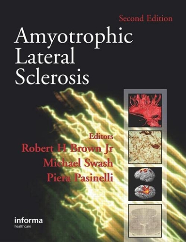
Support this website:
Order from Amazon
We will earn a commission.
Amyotrophic Lateral Sclerosis, or ALS, is a rare, incurable neuro-degenerative disease, of unknown etiology. With this disease, both upper (brain) and lower (spinal cord) motor neurons progressively degenerate and die, rendering immobile the muscles that they innervated. For anyone with a need or desire to appreciate what is known about ALS, this book provides a good foundation. R. Robbins
RJR Picks from Around the Web (updated 11 MAY 2018 )
Old Science
Weird Science
Treating Disease with Fecal Transplantation
Fossils of miniature humans (hobbits) discovered in Indonesia
Paleontology
Dinosaur tail, complete with feathers, found preserved in amber.
Astronomy

Mysterious fast radio burst (FRB) detected in the distant universe.
Big Data & Informatics

Big Data: Buzzword or Big Deal?
Hacking the genome: Identifying anonymized human subjects using publicly available data.